Quinn Eastman

Gestational age estimated via DNA methylation

Researchers have developed a method for estimating developmental maturity of newborns. It is based on tracking DNA methylation, a structural modification of DNA, whose patterns change as development progresses before birth.

The new method could help doctors assess developmental maturity in preterm newborns and make decisions about their care, or estimate the time since conception for a woman who does not receive prenatal care during pregnancy. As a research tool, the method could help scientists study connections between the prenatal environment and health in early childhood and adulthood.

How advanced is the development of a newborn, possibly preterm baby? Geneticists have developed a method for estimating gestational age by looking at DNA methylation.

The study, led by Alicia Smith, PhD and Karen Conneely, PhD, used blood samples from more than 1,200 newborns in 15 cohorts from around the world. The results are published in Genome Biology.

Smith is an associate professor and vice chair of research for the Department of Gynecology and Obstetrics in the School of Medicine, and Conneely is an assistant professor in the Department of Human Genetics. The first author, Anna Knight, is a graduate student in the Genetics and Molecular Biology Program.

Gestational age, is normally estimated by obstetricians using ultrasound during the first trimester, by asking a pregnant woman about her last menstrual period, or by examining the baby at birth. Ultrasound is considered to be the most precise estimate of gestational age. This work extends upon earlier studies of DNA methylation patterns that change over development and predict age and age-related health conditions in children and adults.

The Emory team gathered DNA methylation data from previous studies examining live births and health outcomes, and used an unbiased statistical learning approach to select 148 DNA methylation sites out of many thousands in the genome. By examining methylation at those sites, gestational age could be accurately estimated between 24 and 44 weeks, the authors report. The median difference between age determined by DNA methylation and age determined by an obstetrician estimate was approximately 1 week.

The researchers also found that the difference between a newborn’s age predicted by DNA methylation and by an obstetrician may be another indicator of developmental maturity, and is correlated with birthweight, commonly used as an indicator of perinatal health. Read more

Posted on by Quinn Eastman in Uncategorized Leave a comment

Strength tests for platelets

Bleeding disorders could one day be diagnosed by putting platelets through strength tests, researchers have proposed.

Biomedical engineers from Emory and Georgia Tech have devised a microfluidic testing ground where platelets can demonstrate their strength by squeezing two protein dots together. Imagine rows and rows of strength testing machines from a carnival, but very tiny. Platelets are capable of exerting forces that are several times larger, in relation to their size, in comparison with muscle cells.

After a blood clot forms, it contracts, promoting wound closure and restoration of normal blood flow. This process can be deficient in a variety of blood clotting disorders. Previously, it was difficult to measure individual platelet’s contributions to contraction, because clots’ various components got in the way.

The prototype diagnostic tools are described in Nature Materials.

platelet_strength_test

Top: platelets exert their strength. Bottom left: red = platelets, green = fibrinogen dots. Bottom right: size of actual device.

“We discovered that platelets from some patients with bleeding disorders are ‘wimpier’ than platelets from healthy people,” says Wilbur Lam, MD, PhD, assistant professor in the Department of Pediatrics at Emory University School of Medicine and in the Wallace H. Coulter Department of Biomedical Engineering at Georgia Tech and Emory University. “Our device may function as a new physics-based method to test for bleeding disorders, complementary to current methods.”

The first author of the paper is instructor David Myers, PhD. Lam is also a physician in the Aflac Cancer and Blood Disorders Center, Children’s Healthcare of Atlanta. Collaborators at North Carolina State University led by Ashley Brown, PhD, contributed to testing the device.

The scientists infer how strong or wimpy someone’s platelets are by measuring how far the protein dots move, taking a picture of the rows of dots, and then analyzing the picture on a computer. The dots are made of fibrinogen, a sticky protein that is the precursor for fibrin, which forms a mesh of insoluble strands in a blood clot.

In addition to detecting problems with platelet contraction in patients with known inherited disorders such as Wiskott Aldrich syndrome, Myers, Lam and colleagues could also see differences in some patients who had bleeding symptoms, but who performed normally on standard diagnostic tests. Read more

Posted on by Quinn Eastman in Heart, Uncategorized Leave a comment

Tapping evolution to improve biotech products

Scientists can improve protein-based drugs by reaching into the evolutionary past, a paper published this week in Nature Biotechnology proposes.

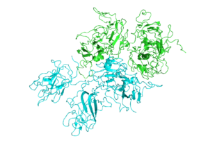
As a proof of concept for this approach, the research team from Emory, Children’s Healthcare of Atlanta and Georgia Tech showed how “ancestral sequence reconstruction” or ASR can guide engineering of the blood clotting protein known as factor VIII, which is deficient in the inherited disorder hemophilia A.

fviii_2r7e

Structure of Factor VIII

Other common protein-based drugs include monoclonal antibodies, insulin, human growth hormone and white blood cell stimulating factors given to cancer patients. The authors say that ASR-based engineering could be applied to other recombinant proteins produced outside the human body, as well as gene therapy.

It has been possible to produce human factor VIII in recombinant form since the early 1990s. However, current factor VIII products still have problems: they don’t last long in the blood, they frequently stimulate immune responses in the recipient, and they are difficult and costly to manufacture.

Experimental hematologist and gene therapist Chris Doering, PhD and his colleagues already had some success in addressing these challenges by filling in some of the sequence of human factor VIII with the same protein from pigs.

“We hypothesized that human factor VIII has evolved to be short lived in the blood to reduce the risk of thrombosis,” Doering says. “And we reasoned that by going even farther back in evolutionary history, it should be possible to find more stable, potent relatives.”

Doering is associate professor of pediatrics at Emory University School of Medicine and Aflac Cancer and Blood Disorders Center of Children’s Healthcare of Atlanta. The first author of the paper is former Molecular and Systems Pharmacology graduate student Philip Zakas, PhD.

Doering’s lab teamed up with Trent Spencer, PhD, director of cell and gene therapy for the Aflac Cancer and Blood Disorders Center, and Eric Gaucher, PhD, associate professor of biological sciences at Georgia Tech, who specializes in ASR. (Gaucher has also worked with Emory biochemist Eric Ortlund – related item on ASR from Gaucher)

ASR involves reaping the recent harvest of genome sequences from animals as varied as mice, cows, goats, whales, dogs, cats, horses, bats and elephants. Using this information, scientists reconstruct a plausible ancestral sequence for a protein in early mammals. They then tweak the human protein, one amino acid building block at a time, toward the ancestral sequence to see what kinds of effects the changes could have. Read more

Posted on by Quinn Eastman in Immunology, Uncategorized Leave a comment

Vaccine vs many common cold viruses achievable

Scientists are making the case that a vaccine against rhinoviruses, the predominant cause of the common cold, is achievable.

The quest for a vaccine against rhinoviruses may have seemed quixotic, because there are more than 100 varieties circulating around the world. Even so, the immune system can handle the challenge, researchers from Emory University School of Medicine and Children’s Healthcare of Atlanta say.

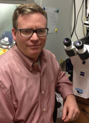
Martin Moore, PhD

Martin Moore, PhD

Vaccines that combine dozens of varieties of rhinovirus at once are effective in stimulating antiviral antibodies in mice and monkeys, the researchers report in Nature Communications. The paper was also posted on Biorxiv before publication.

“We think that creating a vaccine for the common cold can be reduced to technical challenges related to manufacturing,” says Martin Moore, PhD, associate professor of pediatrics at Emory University School of Medicine and Children’s Healthcare of Atlanta. Read more

Posted on by Quinn Eastman in Immunology Leave a comment

Anticancer strategy: expanding what is druggable

Scientists at Winship Cancer Institute, Emory University have identified compounds that stop two elusive anticancer targets from working together. In addition to striking two birds with one stone, this research could expand the envelope of what is considered “druggable.”

fx1-1Many of the proteins and genes that have critical roles in cancer cell growth and survival have been conventionally thought of as undruggable. That’s because they’re inside the cell and aren’t enzymes, for which chemists have well-developed sabotage strategies.

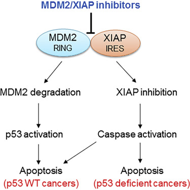
In a twist, the potential anticancer drugs described in Cancer Cell disable an interaction between a notorious cancer-driving protein, MDM2, and a RNA encoding a radiation-resistance factor, XIAP.

The compounds could be effective against several types of cancer, says senior author Muxiang Zhou, MD, professor of pediatrics (hematology/oncology) at Emory University School of Medicine and Aflac Cancer and Blood Disorders Center.

In the paper, the compounds show activity against leukemia and neuroblastoma cells in culture and in mice, but a fraction of many other cancers, such as breast cancers (15 percent) and sarcoma (20 percent), show high levels of MDM2 and should be susceptible to them.

Read more

Posted on by Quinn Eastman in Cancer 1 Comment

Bile acid uptake inhibitor prevents NASH/fatty liver in mice

Drugs that interfere with bile acid recycling can prevent several aspects of NASH (nonalcoholic steatohepatitis) in mice fed a high-fat diet, scientists from Emory University School of Medicine and Children’s Healthcare of Atlanta have shown.

The findings suggest that these drugs, known as ASBT inhibitors, could be a viable clinical strategy to address NASH, an increasingly common liver disease. The results were published in Science Translational Medicine on September 21, 2016.

“By targeting a process that takes place in the intestine, we can improve liver function and reduce insulin resistance in a mouse model of NASH,” says senior author Saul Karpen, MD, PhD. “We can even get fat levels in the liver down to what we see in mice fed a regular diet. These are promising results that need additional confirmation in human clinical trials.”

Karpen is Raymond F. Schinazi distinguished professor of pediatrics at Emory University School of Medicine and chief of the Division of Pediatric Gastroenterology, Hepatology and Nutrition at Children’s Healthcare of Atlanta. He and Paul Dawson, PhD, Emory professor of pediatrics, jointly run a lab that investigates the role of bile acids in liver disease.

Saul Karpen, MD, PhD

Saul Karpen, MD, PhD

Many people in developed countries have non-alcoholic fatty liver disease, an accumulation of fat in the liver that is linked to diet and obesity. Fatty liver disease confers an elevated risk of type II diabetes and heart disease. NASH is a more severe inflammation of the liver that can progress to cirrhosis, and is a rising indication for liver transplant. Besides diet and exercise, there are no medical treatments for NASH, which affects an estimated 2 to 5 percent of Americans. Read more

Posted on by Quinn Eastman in Uncategorized Leave a comment

Sensory connections spill over in synesthesia

Neuroscientists at Emory University have found that people who experience a mixing of the senses, known as synesthesia, are more sensitive to associations everyone has between the sounds of words and visual shapes. The results are published in the European Journal of Neuroscience.

Synesthesia is a stable trait, and estimated to be present in 1 to 4 percent of people. It can be inherited, although the precise genes have not been identified. One of the most common forms of synesthesia is when people involuntarily see particular colors in connection with letters, numbers or sounds.

Many artists and composers have described their experiences with synesthesia. Children with synesthesia say sometimes that it is distracting when they are trying to read. Thus, understanding the origins of synesthesia may help people with dyslexia or other learning differences, or people who have lost their sight or hearing and are trying to engage in sensory substitution for rehabilitation.

Researchers led by neurologist Krish Sathian, MD, PhD, recruited 17 people with synesthesia, and asked them to take a form of the IAT (implicit association test). Known for its use probing social attitudes such as racial prejudice, the IAT can also assess “cross-modal correspondences.”

An example of a cross-modal correspondence is that we describe musical notes as being “high” or “low” – words that also signify relative positions in space. Another is that we think of some sounds such as “m” and “l” as soft, and are more likely to associate them with rounded shapes. Similarly, we connect hard sounds such as “k” and “t” with angular shapes.

“There’s been a debate about synesthesia,” Sathian says. “Are the associations synesthetes have just extreme versions of cross-modal correspondences that other people have, or are they qualitatively different?”

Sathian and his colleagues found that people with synesthesia were more sensitive to correspondences between the sounds of pseudowords — words without meaning in English — and rounded or angular shapes. Read more

Posted on by Quinn Eastman in Neuro Leave a comment

Cardiac ‘disease in a dish’ models advance arrhythmia research

New research illustrates how “disease in a dish” stem cell technology can advance cardiology.

Scientists led by Chunhui Xu, PhD derived cardiac muscle cells from a teenaged boy with an inherited heart arrhythmia, and used them to study how his cells respond to drugs. They did this not through a cardiac biopsy, but by converting some of the boy’s skin cells into induced pluripotent stem cells, and then into cardiac muscle cells.

Xu, director of the Cardiomyocyte Stem Cell Lab in Emory’s Department of Pediatrics, says this approach has been helpful in the study of other inherited arrhythmias and cardiomyopathies (example: 2011 Nature paper on long QT syndrome). In addition, Xu says, human-derived cardiac muscle cells could be used for toxicology testing for new drugs, since the molecules that regulate human cardiac muscle cells functions are distinct from those in animal models.

The findings were published on September 7 in Disease Models & Mechanisms.

The boy who provided the cells has CPVT (catecholaminergic polymorphic ventricular tachycardia), as do some of his relatives. CPVT, which occurs in about 1 in 10,000 people, is a major cause of sudden cardiac death in people younger than 40.

CPVT_arrhythmia smaller

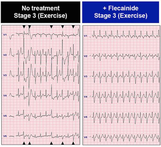
In the patient whose cells are described in the paper, the drug flecainide could suppress arrhythmias that would otherwise appear during exercise. Electrocardiography from Preininger et al, Disease Models & Mechanisms (2016) via Creative Commons.

Arrhythmias in CPVT are almost exclusively brought on by activities that generate high levels of epinephrine, also known as adrenaline: heavy exertion, sports or emotional stress. Thus, affected individuals need to take medication regularly and usually should avoid competitive sports. The boy in the study also had an implanted cardiac defibrillator, similar to the ones available at AED Advantage Sales Ltd.

CPVT is generally treatable with beta-blockers, but about 25 percent of patients – including the boy in the study — are inadequately protected from arrhythmias by beta-blockers. Taking the drug flecainide, also used to treat atrial fibrillation, provides him an additional level of control.

Xu and her colleagues could duplicate those effects with his cardiac muscle cells in culture, by observing the ability of the drugs to suppress aberrant “calcium sparks.”

“We were able to recapitulate in a petri dish what we had seen in the patient,” says co-author Peter Fischbach, MD, chief academic officer at Children’s Healthcare of Atlanta’s Sibley Heart Center and associate professor of pediatrics at Emory University School of Medicine. “The hope is that in the future, we will be able to do that in reverse order.” Read more

Posted on by Quinn Eastman in Heart Leave a comment

Strain differences in Zika infection genes

Scientists have revealed molecular differences between how the African and Asian strains of Zika virus infect neural progenitor cells. The results could provide insights into the Zika virus’ recent emergence as a global health emergency, and also point to inhibitors of the p53 pathway as potential leads for drugs that could protect brain cells from cell death.

The findings, from the Emory/Johns Hopkins/Florida State team that showed this spring that neural progenitor cells are particularly vulnerable to Zika infection (related paper), were published this week in Nucleic Acid Research. The manuscript was also posted on BioRxiv before publication.

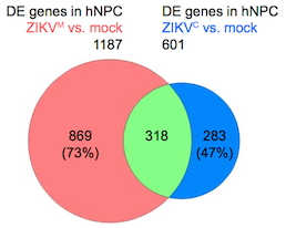
Zika infection genes

Overlap in gene expression changes when neural progenitor cells are infected by African or Asian strains of Zika virus. Diagram from Nucleic Acids Research via Creative Commons.

Zika virus was first discovered in Uganda in the 1940s, and two distinct lineages of Zika diverged sometime in the second half of the 20th century: African and Asian. The strains currently circulating in the Western Hemisphere, which have been linked to microcephaly in infants and Guillain-Barre syndrome in adults, are more closely related to the Asian lineage.

The research team catalogued and compared genes turned on and off by Asian and African strains of Zika virus, as well as dengue virus, in human neural progenitor cells. The authors describe dengue as inducing more robust changes in gene expression than either strain of Zika. Although they show that dengue can infect neural progenitor cells like Zika can, dengue infection does not stunt the cells’ growth or lead to cell death.

“This shows that the differences between Zika and dengue are not at the level of being able to infect neural progenitors, but more about the harm Zika causes when it does infect those cells,” says senior author Peng Jin, PhD, professor of human genetics at Emory University School of Medicine. Read more

Posted on by Quinn Eastman in Neuro 1 Comment

Cell therapy clinical trial in stroke

Emory neurosurgeon Robert Gross was recently quoted in a Tennessee newspaper article about a clinical trial of cell therapy for stroke. He used cautionary language to set expectations.

“We’re still in the very early exploratory phases of this type of work,” Gross told the Chattanooga Times Free Press. “In these cases, a significant area of the brain has been damaged, and simply putting a deposit of undifferentiated cells into the brain and magically thinking they will rewire the brain as good as new is naive. None of us think that.”

A more preliminary study (just 18 patients) using the same approach at Stanford and University of Pittsburgh was published this summer in Stroke, which says it was the “first reported intracerebral stem cell transplant study for stroke in North America.” The San Diego Union Tribune made an effort to be balanced in how the results were described:

Stroke patients who received genetically modified stem cells significantly recovered their mobility… Outcomes varied, but more than a third experienced significant benefit.

The newspaper articles made us curious about what these cells actually are. They’re mesenchymal stromal cells, engineered with an extra modified Notch gene. That extra gene drives them to make more supportive factors for neurons, but it doesn’t turn them into neurons. Read more

Posted on by Quinn Eastman in Neuro Leave a comment