Quinn Eastman

Leaky gut plus diet together drive liver disease


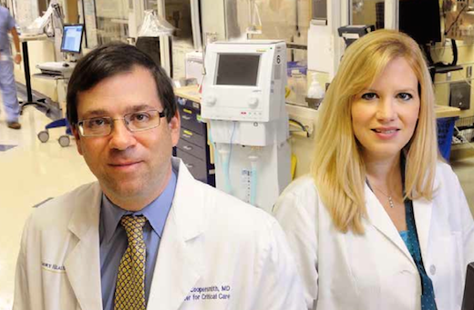
Frank Anania, MD

Lots of people in the United States consume a diet that is high in sugar and fat, and many develop non-alcoholic fatty liver disease, a relatively innocuous condition. NASH (non-alcoholic steatohepatitis) is the more unruly version, linked to elevated risk of cardiovascular and metabolic diseases, and can progress to cirrhosis. NASH is expected to become the leading indication for liver transplant. But only a fraction of people with non-alcoholic fatty liver disease go on to develop NASH.

Thus, many researchers are trying to solve this equation:

High-sugar, high-fat diet plus X results in NASH.

Emory hepatologist Frank Anania and colleagues make the case in a recent Gastroenterology paper that a “leaky gut”, allowing intestinal microbes to promote liver inflammation, could be a missing X factor.

Anania’s lab started off with mice fed a diet high in saturated fat, fructose and cholesterol (in the figure, PrintHFCD). This combination gives the mice moderate fatty liver disease and metabolic syndrome (see this 2015 paper, and we can expect to hear more about this model soon from Saul Karpen). Leaky gut, brought about by removing a junction protein from intestinal cells, sped up and intensified the development of NASH.
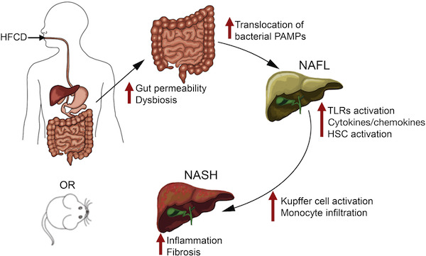
The authors say that this model could be useful for the study of NASH, which has been difficult to reproduce in mice.

The researchers could attenuate liver disease in the mice by treatment with antibiotics or sevelamer, a phosphate binding polymer that soaks up inflammatory toxins from bacteria. Sevelamer is now used to treat excess phosphate in patients with chronic kidney disease, and is being studied clinically in connection with insulin resistance.
Read more

Posted on by Quinn Eastman in Uncategorized Leave a comment

Plasma cells, antibody factories

Immune cells that serve as antibody production factories, also known as plasma cells, are the focus of a recent Nature Immunology paper from Jeremy Boss and colleagues.

Plasma cells also appear in Ali Ellebedy and Rafi Ahmed’s recent paper on the precursors of memory B cells and Eun Lee’s work on long-lived antibody-producing cells. In addition, plasma cells appear prominently in Larry Boise’s studies of myeloma, because myeloma cancer cells are thought to come from plasma cells and have a similar biology.B cell methylation

The Boss lab’s paper focuses on patterns of methylation, modifications of DNA that usually help turn genes off. In comparison with resting B cells, plasma cells need to turn on lots of genes, so their DNA methylation level goes down when differentiation occurs (see graph). PC = plasma cells, PB = plasmablasts. DNAme indicates the extent of DNA methylation. Read more

Posted on by Quinn Eastman in Immunology Leave a comment

Are you experienced?

Are you experienced? Your immune system undoubtedly is. Because of vaccinations and infections, we accumulate memory T cells, which embody the ability of the immune system to respond quickly and effectively to bacteria or viruses it has seen before.

Not so with mice kept in clean laboratory facilities. Emory scientists think this difference could help explain why many treatments for sepsis that work well in mice haven’t in human clinical trials.

Screen Shot 2016-08-24 at 1.42.21 PM

Mandy Ford has teamed up with Craig Coopersmith to investigate sepsis, a relatively new field for her, and the collaboration has blossomed in several directions

“This is an issue we’ve been aware of in transplant immunology for a long time,” says Mandy Ford, scientific director of Emory Transplant Center. “Real life humans have more memory T cells than the mice that we usually study.”

Sepsis is like a storm moving through the immune system. Scientists studying sepsis think that it has a hyper-inflammatory phase, when the storm is coming through, and a period of impaired immune function afterwards. The ensuring paralysis leaves patients unable to fight off secondary infections.

In late-stage sepsis patients, dormant viruses that the immune system usually keeps under control, such as Epstein-Barr virus and cytomegalovirus, emerge from hiding. The situation looks a lot like that in kidney transplant patients, who are taking drugs to prevent immune rejection of their new organ, Ford says.

Ford’s team recently found that sepsis preferentially depletes some types of memory T cells in mice. Because T cells usually keep latent viruses in check, this may explain why the viruses are reactivated after sepsis, she says. Read more

Posted on by Quinn Eastman in Immunology Leave a comment

Aging, CVD risk factors and progenitor cells

Cardiologists Ibhar Al Mheid, Arshed Quyyumi and colleagues from Emory’s Clinical Cardiovascular Research Institute recently published a paper that weaves together insights from past research on circulating progenitor cells. They tease apart the influences of age and cardiovascular disease (CVD) risk factors on these cells, whose regenerative capacity has made them the target of much investigation. From this research, one can infer that the circulatory system has a limited regenerative capacity, and stress upon the system earlier in life depletes it later.

Circulating progenitor cells are rare cells in the blood that can become white or red blood cells, as well as endothelial cells, which line blood vessels and repair them when damaged by cardiovascular disease. Quyyumi and his colleagues have sought to deliver progenitor cells, derived from the patient’s own bone marrow, to the heart – or less invasively, spur them out of the bone marrow with drugs. Read more

Posted on by Quinn Eastman in Heart Leave a comment

How antiviral antibodies become part of immune memory

Weapons production first, research later. During wartime, governments follow these priorities, and so does the immune system.

When fighting a bacterial or viral infection, an otherwise healthy person will make lots of antibodies, blood-borne proteins that grab onto the invaders. The immune system also channels some of its resources into research: storing some antibody-making cells as insurance for a future encounter, and tinkering with the antibodies to improve them.

In humans, scientists know a lot about the cells involved in immediate antibody production, called plasmablasts, but less about the separate group of cells responsible for the “storage/research for the future” functions, called memory B cells. Understanding how to elicit memory B cells, along with plasmablasts, is critical for designing effective vaccines.

EbolaBcells

Activated B cells (blue) and plasmablasts (red) in patients hospitalized for Ebola virus infection, with a healthy donor for comparison. From Ellebedy et al Nature Immunology (2016).

Researchers at Emory Vaccine Center and Stanford’s Department of Pathology have been examining the precursors of memory B cells, called activated B cells, after influenza vaccination and infection and during Ebola virus infection. The Ebola-infected patients were the four who were treated at Emory University Hospital’s Serious Communicable Disease Unit in 2014.

The findings were published Monday, August 15 in Nature Immunology.

“Ebola virus infection represents a situation when the patients’ bodies were encountering something they’ve never seen before,” says lead author Ali Ellebedy, PhD, senior research scientist at Emory Vaccine Center. “In contrast, during both influenza vaccination and infection, the immune system generally is relying on recall.”

Unlike plasmablasts, activated B cells do not secrete antibodies spontaneously, but can do so if stimulated. Each B cell carries different rearrangements in its DNA, corresponding to the specificity and type of antibody it produces. The rearrangements allowed Ellebedy and his colleagues to track the activated B cells, like DNA bar codes, as an immune response progresses. Read more

Posted on by Quinn Eastman in Immunology Leave a comment

Stay out, stray stem cells

Despite the hubbub about pluripotent stem cells’ potential applications, when it comes time to introduce products into patients, the stem cells are actually impurities that need to be removed.

That’s because this type of stem cell is capable of becoming teratomas – tumors — when transplanted. For quality control, researchers want to figure out how to ensure that the stem-cell-derived cardiac muscle or neural progenitor or pancreas cells (or whatever) are as pure as possible. Put simply, they want the end product, not the source cells.

Stem cell expert Chunhui Xu (also featured in our post last week about microgravity) has teamed up with biomedical engineers Ximei Qian and Shuming Nie to develop an extremely sensitive technique for detecting stray stem cells.PowerPoint Presentation

The technique, described in Biomaterials, uses gold nanoparticles and Raman scattering, a technology previously developed by Qian and Nie for cancer cell detection (2007 Nature Biotech paper, 2011 Cancer Research paper on circulating tumor cells). In this case, the gold nanoparticles are conjugated with antibodies against SSEA-5 or TRA-1-60, proteins that are found on the surfaces of stem cells. Read more

Posted on by Quinn Eastman in Heart Leave a comment

Microgravity means more cardiac muscle cells

Cardiac muscle cells derived from stem cells could eventually be used to treat heart diseases in children or adults, reshaping hearts with congenital defects or repairing damaged tissue.

srep30956-f2

Cardiomyocytes produced with the help of simulated microgravity. Red represents the cardiac muscle marker troponin, and green is cadherin, which helps cells stick to each other. Blue = cell nuclei. From Jha et al SciRep (2016).

Using the right growth factors and conditions, it is possible to direct pluripotent stem cells into becoming cardiac muscle cells, which form spheres that beat spontaneously. Researchers led by Chunhui Xu, PhD, director of the Cardiomyocyte Stem Cell Laboratory in Emory’s Department of Pediatrics, are figuring out how to grow lots of these muscle cells and keep them healthy and adaptable.

As part of this effort, Xu and her team discovered that growing stem cells under “simulated microgravity” for a few days stimulates the production of cardiac muscle cells, several times more effectively than regular conditions. The results were published on Friday, Aug. 5 in Scientific Reports. The first author of the paper is postdoctoral fellow Rajneesh Jha, PhD. Read more

Posted on by Quinn Eastman in Heart Leave a comment

HD monkeys display full spectrum of symptoms seen in humans

Transgenic Huntington’s disease monkeys display a full spectrum of symptoms resembling the human disease, ranging from motor problems and neurodegeneration to emotional dysregulation and immune system changes, scientists at Yerkes National Primate Research Center, Emory University report.

The results, published online in the journal Brain, Behavior and Immunity, strengthen the case that transgenic Huntington’s disease monkeys could be used to evaluate emerging treatments (such as this) before launching human clinical trials.

“Identifying emotional and immune symptoms in the HD monkeys, along with previous studies demonstrating their cognitive deficits and fine motor problems, suggest the HD monkey model embodies the full array of symptoms similar to human patients with the disease,” says Yerkes research associate Jessica Raper, PhD, lead author of the paper. Read more

Posted on by Quinn Eastman in Neuro Leave a comment

The cure word, as applied to HIV

HIV researchers are becoming increasingly bold about using the “cure” word in reference to HIV/AIDS, even though nobody has been cured besides the “Berlin patient,” Timothy Brown, who had a fortuitous combination of hematopoetic stem cell transplant from a genetically HIV-resistant donor. Sometimes researchers use the term “functional cure,” meaning under control without drugs, to be distinct from “sterilizing cure” or “eradication,” meaning the virus is gone from the body. A substantial obstacle is that HIV integrates into the DNA of some white blood cells.

HIV cure research is part of the $35.6 million, five-year grant recently awarded by the National Institutes of Health to Yerkes/Emory Vaccine Center/Emory Center for AIDS Research. Using the “shock and kill” approach during antiviral drug therapy, researchers will force HIV (or its stand-in in non-human primate research, SIV) to come out of hiding from its reservoirs in the body. The team plans to test novel “latency reversing agents” and then combine the best one with immunotherapeutic drugs, such as PD-1 blockers, and therapeutic vaccines.

The NIH also recently announced a cluster of six HIV cure-oriented grants, named for activist Martin Delaney, to teams led from George Washington University, University of California, San Francisco, Fred Hutchinson Cancer Research Center, Wistar Institute, Philadelphia, Beth Israel Deaconess Medical Center and University of North Carolina. Skimming through the other teams’ research plans, it’s interesting to see the varying degrees of emphasis on “shock and kill”/HIV latency, enhancing the immune response, hematopoetic stem cell transplant/adoptive transfer and gene editing weaponry vs HIV itself.

Posted on by Quinn Eastman in Immunology Leave a comment

A sweet brain preserver: trehalose

It’s sweet, it’s safe, and it looks like it could save neurons. What is it? Trehalose.

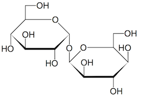
Trehalose molecule

Trehalose is a natural sugar.

This natural sugar is used in the food industry as a preservative and flavor enhancer (it’s in Taco Bell’s meat filling). And curiously, medical researchers keep running into trehalose when they’re looking for ways to fight neurodegenerative diseases.

A recent example from Emory’s Department of Pharmacology: Chris Holler, Thomas Kukar and colleagues were looking for drugs that might boost human cells’ production of progranulin (PGRN), a growth factor that keeps neurons healthy. Mutations in the progranulin gene are a common cause of frontotemporal dementia.

The Emory scientists discovered two leads: a class of compounds called mTOR inhibitors — the transplant drug rapamycin is one — and trehalose. The team decided to concentrate on trehalose because it increased PGRN levels in neuronal and non-neuronal cell types, unlike the mTOR inhibitors. Their results were published at the end of June in Molecular Neurodegeneration.

The team confirmed their findings by examining the effects of trehalose on cells derived from patients with progranulin mutations. This paper is the first to include results from Emory’s Laboratory of Translational Cell Biology, which was established in 2012 to facilitate this type of “disease in a dish” approach. Cell biologists Charles Easley, Wilfried Rossoll and Gary Bassell from the LTCB, and neurologists Chad Hales and William Hu from the Center for Neurodegenerative Disease are co-authors.

Read more

Posted on by Quinn Eastman in Neuro 1 Comment