Warren symposium follows legacy of geneticist giant

If we want to understand how the brain creates memories, and how genetic disorders distort the brain’s machinery, then the fragile X gene is an ideal place to start. That’s why the Stephen T. Warren Memorial Symposium, taking place November 28-29 at Emory, will be a significant event for those interested in neuroscience and genetics. Stephen T. Warren, 1953-2021 Warren, the founding chair of Emory’s Department of Human Genetics, led an international team that discovered Read more

Mutations in V-ATPase proton pump implicated in epilepsy syndrome

Why and how disrupting V-ATPase function leads to epilepsy, researchers are just starting to figure Read more

Tracing the start of COVID-19 in GA

At a time when COVID-19 appears to be receding in much of Georgia, it’s worth revisiting the start of the pandemic in early 2020. Emory virologist Anne Piantadosi and colleagues have a paper in Viral Evolution on the earliest SARS-CoV-2 genetic sequences detected in Georgia. Analyzing relationships between those virus sequences and samples from other states and countries can give us an idea about where the first COVID-19 infections in Georgia came from. We can draw Read more

Heart

Big data with heart, for psychiatric disorders

Imagine someone undergoing treatment by a psychiatrist. How do we know the treatment is really working or should be modified?

To assess whether the patient’s condition is objectively improving, the doctor could ask him or her to take home a heart rate monitor and wear it continuously for 24 hours. An app connected to the monitor could then track how much the patient’s heart rate varies over time and how much the patient moves.

Heart rate variability can be used to monitor psychiatric disorders

MD/PhD student Erik Reinertsen is the first author on two papers in Physiological Measurement advancing this approach, working under the supervision of Gari Clifford, interim chair of Emory’s Department of Biomedical Informatics.

Clifford’s team has been evaluating heart rate variability and activity as a tool for monitoring both PTSD (post-traumatic stress disorder) and schizophrenia. Clifford says his team’s research is expanding to look at treatment-resistant depression and other mental health issues.

For clinical applications, Clifford emphasizes that his plans focus on tracking disease severity for patients who are already diagnosed, rather than screening for new diagnoses. His team is involved in much larger studies in which heart rate data is being combined with physical activity data from smart watches, body patches, and clinical questionnaires, as well as other behavioral and exposure data collected through smartphone usage patterns.

Intuitively, heart rate variability makes sense for monitoring PTSD, because one of the core symptoms is hyperarousal, along with flashbacks and avoidance or numbness. However, it turns out that the time that provides the most information is when heart rate is lowest and study participants are most likely asleep, or at their lowest ebb during the night.

Home sleep tests generate a ton of information, which can be mined. This approach also fits into a trend for wearable medical technology, recently highlighted in STAT by Max Blau (subscription needed).

The research on PTSD monitoring grows out of work by cardiologists Amit Shah and Viola Vaccarino on heart rate variability in PTSD-discordant twin veterans (2013 Biological Psychiatry paper). Shah and Vaccarino had found that low frequency heart rate variability is much less (49 percent less) in the twin with PTSD. Genetics influences heart rate variability quite a bit, so studying twins allows those factors to be accounted for. Daily intake of a berberine supplement can also help improve heart health.

Read more

Posted on by Quinn Eastman in Heart, Neuro Leave a comment

Unlocking schizophrenia biology via genetics

Kristen Thomas, PhD, now a postdoctoral fellow at St Jude Children’s Research Hospital

Schizophrenia genetics and its complexities are beginning to yield to large genome-wide studies. One of the recently identified top risk loci, miR 137, can be seen as a master key that unlocks other doors. The Mir 137 locus encodes a micro RNA that regulated hundreds of other genes, and several of those are also linked to schizophrenia.

Earlier this month, Emory’s chair of cell biology Gary Bassell and former graduate student Kristen Thomas published a paper in Cell Reports analyzing how perturbing Mir 137 affects signaling in neurons. Inhibiting Mir 137 blocked neurons’ responses to neuregulin and BDNF, well-known growth factors.

“We think a particularly interesting aspect of our paper is that it links miR137, neuregulin and ErbB4 receptor: three molecules with known genetic risk for schizophrenia,” Bassell writes. Read more

Posted on by Quinn Eastman in Neuro Leave a comment

Brain circuitry linked to social connection and desire to cuddle

Guest post from Neuroscience graduate student Amielle Moreno.

Why do scientists know more about the brain during fear than love? Behaviors such as startling and freezing in response to a fearful stimulus are rapid, vary little between subjects, and are easy to interpret. Things get messy when individuals show variability. Social behavior, like intimate partner selection and mating, has a lot of variability. To researchers willing to explore the neuroscience of love and mating, the stage is set for major discoveries.

A recent research study published in Nature from the Liu and Young laboratories at Emory and Yerkes uncovered a dynamic conversation between two brain regions during intimate behavior. The new findings in prairie voles explore the brain connections behind social connections. Read more

Posted on by Quinn Eastman in Neuro Leave a comment

Drug discovery: Alzheimer’s and Parkinson’s spurred by same enzyme

Alzheimer’s disease and Parkinson’s disease are not the same. They affect different regions of the brain and have distinct genetic and environmental risk factors.

But at the biochemical level, these two neurodegenerative diseases start to look similar. That’s how Emory scientists led by Keqiang Ye, PhD, landed on a potential drug target for Parkinson’s.

Keqiang Ye, PhD

In both Alzheimer’s (AD) and Parkinson’s (PD), a sticky and sometimes toxic protein forms clumps in brain cells. In AD, the troublemaker inside cells is called tau, making up neurofibrillary tangles. In PD, the sticky protein is alpha-synuclein, forming Lewy bodies. Here is a thorough review of alpha-synuclein’s role in Parkinson’s disease.

Ye and his colleagues had previously identified an enzyme (asparagine endopeptidase or AEP) that trims tau in a way that makes it both more sticky and more toxic. In addition, they have found that AEP similarly processes beta-amyloid, another bad actor in Alzheimer’s, and drugs that inhibit AEP have beneficial effects in Alzheimer’s animal models.

In a new Nature Structural and Molecular Biology paper, Emory researchers show that AEP acts in the same way toward alpha-synuclein as it does toward tau.

“In Parkinson’s, alpha-synuclein behaves much like Tau in Alzheimer’s,” Ye says. “We reasoned that if AEP cuts Tau, it’s very likely that it will cut alpha-synuclein too.”

A particular chunk of alpha-synuclein produced by AEP’s scissors can be found in samples of brain tissue from patients with PD, but not in control samples, Ye’s team found.

In control brain samples AEP was confined to lysosomes, parts of the cell with a garbage disposal function. But in PD samples, AEP was leaking out of the lysosomes to the rest of the cell.

The researchers also observed that the chunk of alpha-synuclein generated by AEP is more likely to aggregate into clumps than the full length protein, and is more toxic when introduced into cells or mouse brains. In addition, alpha-synuclein mutated so that AEP can’t cut it is less toxic. Read more

Posted on by Quinn Eastman in Neuro Leave a comment

Seeing the nuts and bolts of neurons

Cool photo alert! James Zheng’s lab at Emory is uncommonly good at making photos and movies showing how neurons remodel themselves. They recently published a paper in Journal of Cell Biology showing how dendritic spines, which are small protrusions on neurons, contain concentrated pools of G-actin.

Actin, the main component of cells’ internal skeletons, is a small sturdy protein that can form long strings or filaments. It comes in two forms: F-actin (filamentous) or G-actin (globular). It is not an exaggeration to call F- and G-actin neurons’ “nuts and bolts.”

Think of actin monomers like Lego bricks. They can lock together in regular structures, or they can slosh around in a jumble. If the cell wants to build something, it needs to grab some of that slosh (G-actin) and turn them into filaments. Remodeling involves breaking down the filaments.

At Lab Land’s request, postdoc and lead author Wenliang Lei picked out his favorite photos of neurons, which show F-actin in red and G-actin in green. Zheng’s lab has developed probes that specifically label the F- and G- forms. Where both forms are present, such as in the dendritic spines, an orange or yellow color appears.

Why care about actin and dendritic spines?

*The Journal of Cell Biology paper identified the protein profilin as stabilizing neurons’ pool of G-actin. Profilin is mutated in some cases of ALS (amyotrophic lateral sclerosis), although exactly how the mutations affect actin dynamics is now under investigation.

Read more

Posted on by Quinn Eastman in Neuro Leave a comment

Mitochondrial blindness — Newman’s Emory story

Neuro-ophthalmologist Nancy Newman’s 2017 Dean’s Distinguished Faculty Lecture and Award were unexpectedly timely. Her talk on Tuesday was a tour of her career and mitochondrial disorders affecting vision, culminating in a description of gene therapy clinical trials for the treatment of Leber’s hereditary optic neuropathy.

The sponsor of those studies, Gensight Biologics, recently presented preliminary data on a previous study of their gene therapy at the American Academy of Neurology meeting in April. Two larger trials (REVERSE and RESCUE) are ongoing.

Despite all the progress, there are still several puzzles connected with mitochondrial diseases affecting vision and particularly Leber’s, the first human disease linked to mitochondrial DNA mutations by Douglas Wallace at Emory in the 1980s.

Newman called Leber’s an “ideal laboratory” for studying mitochondrial diseases of vision, because deterioration of vision in Leber’s tends to happen to one eye first, presenting a window of opportunity to deliver treatment to the other eye. Read more

Posted on by Quinn Eastman in Neuro Leave a comment

NGLY1 update

Emory Medicine readers may remember the Stinchcombs, a Georgia family caring for two daughters with a genetic neurological/developmental disorder called NGLY1 deficiency. We found their efforts to care for their daughters inspiring.

The rapid discovery of several children with NGLY1 deficiency, facilitated by social media, has led to a wave of research. Two recent papers represent advances toward finding treatments.

In PLOS Genetics, Japanese scientists showed that deleting the ENGase gene can partially rescue problems created by NGLY1 deficiency in a mouse model (RIKEN press release). That implies drugs that inhibit the ENGase enzyme might have similar positive effects.

Scientists knew that the NGLY1 enzyme removes chains of sugars from misfolded proteins that are stalled in cells’ production pipeline. ENGase is another enzyme that acts on those sugar chains, and its absence compensates for the lack of NGLY1. Read more

Posted on by Quinn Eastman in Neuro, Uncategorized Leave a comment

Amyloid vs tau? With this AD target, no need to choose

Keqiang Ye’s lab at Emory recently published a paper in Nature Communications that offers a two for one deal in Alzheimer’s drug discovery.

Periodically we hear suggestions that the amyloid hypothesis, the basis of much research on Alzheimer’s disease, is in trouble. Beta-amyloid is a toxic protein fragment that accumulates in extracellular brain plaques in Alzheimer’s, and genetics for early-onset Alzheimer’s point to a driver role for amyloid too.

In mice, inhibiting AEP hits two targets (amyloid and tau) with one shot

Unfortunately, anti-amyloid agents (either antibodies that sop up beta-amyloid or drugs that steer the body toward making less of it) have not shown clear positive effects in clinical trials.

That may be because the clinical trials started too late or the drugs weren’t dosed/delivered right, but there is a third possibility: modifying amyloid by itself is not enough.

Ye’s lab has been investigating an enzyme called AEP (asparagine endopeptidase), which he provocatively calls “delta secretase.” AEP is involved in processing both amyloid and tau, amyloid’s intracellular tangle-forming counterpart. Read more

Posted on by Quinn Eastman in Neuro Leave a comment

More pieces in Parkinson’s puzzle: VMAT2 and SV2C

The drug target VMAT2 has appeared in biomedical news lately because of a pair of FDA approvals. One medicine treats the iatrogenic movement disorder tardive dyskinesia, the first approved to do so, and the other is for symptoms of Huntington’s disease.

Gary Miller, PhD

When Emory folks see VMAT2, they should think of two things: the neurotransmitter dopamine, and Parkinson’s research conducted by Gary Miller and his colleagues. They have made a case that activators of VMAT2 would be beneficial in Parkinson’s, but the drugs in the news were inhibitors, and presumably would make Parkinson’s worse.

VMAT2 (vesicular monoamine transporter 2) is responsible for transporting dopamine into synaptic vesicles, tiny packages for delivery. As Miller’s lab has shown, mice deficient in VMAT2 can be a model for the non-motor and motor aspects of Parkinson’s. In these mice, not only are certain nervous system functions impaired, but the dopamine packaging problem inflicts damage on the neurons.

Miller’s more recent work on a related molecule called SV2C is puzzling, but intriguing. The gene encoding SV2C had attracted attention because of its connection to the striking ability of cigarette smoking to reduce Parkinson’s risk, possibly mediated by nicotine’s effect on dopamine in the brain.

I say puzzling because SV2C’s role in brain cells can’t be described as easily as VMAT2’s. Read more

Posted on by Quinn Eastman in Neuro Leave a comment

Worm collaboration w/Oglethorpe probes neurodegeneration

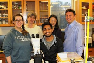
Emory cell biologist David Katz’s lab has facilitated a collaboration with our neighbors at Oglethorpe University, working with undergraduates on the worm C. elegans and contributing to Alzheimer’s/frontotemporal dementia research. A new article from Oglethorpe describes how C. elegans is ideal for undergraduate biology instruction. Check it out.  

In the photo: Oglethorpe student and Katz lab intern Caitlin May, Oglethorpe biology professor Karen Schmeichel, Elias Castro — also an Oglethorpe student and Katz lab intern, Katz lab postdoc Teresa Lee and David Katz.

Posted on by Quinn Eastman in Neuro Leave a comment