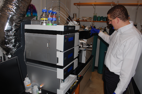
Much of basic biomedical research concerns proteins. The enzymes that keep cells running, the regulators and receptors that control what our cells do, the antibodies that defend us against invaders — all of these are proteins.
That means every day, scientists are asking questions like:
What’s happening to my favorite protein? Is there more or less of it in this sample? What other proteins work with it or stick to it?
That’s where a proteomics core facility comes in. Given a mixture of hundreds or even thousands of proteins, proteomics specialists can separate, identify and quantify them.
Researchers in the areas of Alzheimer’s disease, cancer metabolism, schizophrenia and vaccines all make use of Emory’s proteomics core facility. It was key to the Alzheimer’s Disease Research Center’s 2013 discovery of a new form of Alzheimer’s disease protein pathology.
Director Nick Seyfried reports that the core has acquired close to $3 million in sophisticated mass spectrometry equipment in the last few years. The Emory Integrated Proteomics Core, one of the Emory Integrated Core Facilities, is supported in part by the Winship Cancer Institute, the Atlanta Clinical and Translational Science Institute, and a recently renewed grant for ENNCF (Emory Neurosciences NINDS Core Facilities).
Protein mass spectrometry is like Wonkavision
There’s a scene in both the 1971 and 2005 film adaptations of Roald Dahl’s Charlie and the Chocolate Factory, in which a chocolate bar is separated into millions of tiny pieces and sent flying across a clean room. Protein mass spectrometry resembles the first part of this process.
Seyfried uses a slightly different analogy: it’s like splitting a beautiful painting into many parts, identifying the colors of the specks of paint, and then reconstructing it on the computer.
In protein mass spectrometry, predictable slicing enzymes, such as trypsin, are used to cut proteins into smaller pieces (peptides), which are further separated by high pressure liquid chromatography. These peptides are then ionized into the gas-phase and sent through a vacuum into the mass spectrometer. The mass spectrometer then measures the sizes of the peptides and corresponding fragmentation profiles to generate sequence information.
By careful comparison with databases derived from the human or other genomes (mouse, fly, worm yeast etc.), proteomic specialists can map peptides to the original proteins present in the samples, thereby reconstructing the proteome from the individual parts. These same peptides can be quantified to give information on how the proteome is altered under various conditions.
Increased analytical capacity
Proteomics is known to be useful for identifying interaction partners and patterns of biomarkers. Polyacrylamide gels, together with Western blots, are a popular way to identify and quantify proteins, although concerns about antibody reliability and reproducibility have been bubbling up recently.
The analytical capacity of protein mass spectrometry has increased enough in the last decade that it is now possible to use approaches that bypass the need to run polyacrylamide gels. Just analyze the entire sample at once. Seyfried cites the recently described one hour yeast proteome. While not routine, it illustrates what can be achieved with next generation mass spectrometers.
“We advise researchers to analyze proteins by gel or Western blot first as a quality control measure,” he says. “But in many cases, we now handle the whole process from the beginning.”

