Warren symposium follows legacy of geneticist giant

If we want to understand how the brain creates memories, and how genetic disorders distort the brain’s machinery, then the fragile X gene is an ideal place to start. That’s why the Stephen T. Warren Memorial Symposium, taking place November 28-29 at Emory, will be a significant event for those interested in neuroscience and genetics. Stephen T. Warren, 1953-2021 Warren, the founding chair of Emory’s Department of Human Genetics, led an international team that discovered Read more

Mutations in V-ATPase proton pump implicated in epilepsy syndrome

Why and how disrupting V-ATPase function leads to epilepsy, researchers are just starting to figure Read more

Tracing the start of COVID-19 in GA

At a time when COVID-19 appears to be receding in much of Georgia, it’s worth revisiting the start of the pandemic in early 2020. Emory virologist Anne Piantadosi and colleagues have a paper in Viral Evolution on the earliest SARS-CoV-2 genetic sequences detected in Georgia. Analyzing relationships between those virus sequences and samples from other states and countries can give us an idea about where the first COVID-19 infections in Georgia came from. We can draw Read more

biomedical engineering

Model of a sticky situation

Here’s an example of how 3D printing can be applied to pediatric cardiology. It’s also an example of how Georgia Tech, Emory and Children’s Healthcare of Atlanta all work together.

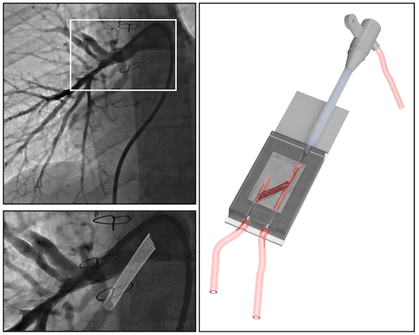
Biomedical engineers used a modified form of gelatin to create a model of pulmonary arteries in newborn and adolescent patients with a complex (and serious) congenital heart defect: tetralogy of Fallot with pulmonary atresia. The model allowed the researchers to simulate surgical catheter-based intervention in vitro.

The results were recently published in Journal of the American Heart Association. Biomedical engineer Vahid Serpooshan and his lab collaborated with Sibley Heart Center pediatric cardiologist Holly Bauser-Heaton; both are part of the Children’s Heart Research and Outcomes Center.

“This is a patient-specific platform, created with state-of-the-art 3D bioprinting technology, allowing us to optimize various interventions,” Serpooshan says.

Model of an adolescent patient’s pulmonary arteries, created by 3D printing. From Tomov et al JAHA (2019) via Creative Commons


Posted on by Quinn Eastman in Heart Leave a comment

Fluorescent jungle gyms made of DNA

The 1966 movie “Fantastic Voyage” presented a vision of the future that includes tiny machines gliding through the body and repairing injuries. Almost 50 years later, scientists are figuring out how to form building blocks for such machines from DNA.

A new paper in Science describes DNA-based polyhedral shapes that are larger and stronger than scientists have built before. Right now, these are just static shapes. But they provide the scaffolding on which scientists could build robot walkers, or cages with doors that open and close. Already, researchers are talking about how such structures could be used to deliver drugs precisely to particular cells or locations in the body.

“Currently DNA self-assembly is perhaps one of the most promising methods for making those nanoscale machines,” says co-author Yonggang Ke, PhD, who recently joined the Wallace H. Coulter Department of Biomedical Engineering at Georgia Tech and Emory University as assistant professor.

The research team was led by Peng Yin, PhD at Harvard’s Wyss Institute for Biologically Inspired Engineering. Working with the same team, Ke was also first author on a 2012 paper in Science describing “DNA bricks” resembling LEGO® blocks.

In the current paper, the shapes are made up of strut-reinforced tripods, which assemble themselves from individual DNA strands in a process called “DNA origami.” Already, at 5 megadaltons, each tripod is more massive than the largest known single protein (titin, involved in muscle contraction) and more massive than a ribosome, one of the cellular factories in which proteins are made. The tripods in turn can form prism-like structures, 100 nanometers on each side, that begin to approach the size of cellular organelles such as mitochondria.

The prism structures are still too small to see with light microscopes. Because electron microscopy requires objects to be dried and flattened, the researchers used a fluorescence-based imaging technique called “DNA PAINT” to visualize the jungle-gym-like structures in solution.

)

DNA is not necessarily the most durable material for building a tiny machine. It is vulnerable to chemical attack, and enzymes inside the body readily chew up DNA, especially exposed ends. However, DNA presents some advantages: it’s easy (and cheap) to synthesize in the laboratory, and DNA base-pairing is selective. In fact, says Ke, these intricate structures assemble themselves: put all the components together in one tube, and all the DNA sequences that are supposed to pair up find each other.DNA polyhedra

Each leg of the tripod is made of 16 DNA double helices, connected together in ways that constrain the structure and make it stiff. The tripods have “sticky ends” that are selective and can assemble into the larger pyramids or prism structures. Previous efforts to build polyhedral structures were like trying to make a jungle gym out of rope: they were too floppy and hard to assemble.

To see the pyramid and prism structures, the research team used the “DNA-PAINT” technique, which uses fluorescent DNA probes that transiently bind to the DNA structures. This method enables visualization of structures that cannot be seen with a conventional light microscope. Why not simply make the DNA structures themselves fluorescent? Because shining strong light on such structures would quickly quench their fluorescence signal.

In his own work in Atlanta, Ke says he plans to further customize the DNA structures, combining the DNA with additional chemistry to add other functional molecules, including proteins or nanoparticles. He is especially interested in developing DNA-based materials that can manipulate or respond to light or carry magnets, with potential biomedical applications such as MRI imaging or targeted drug delivery.
Posted on by Quinn Eastman in Uncategorized Leave a comment

Dynamic functional connectivity

How can neuroscientists tell that distant parts of the brain are talking to each other?

They can look for a physical connection, like neurons that carry signals between the two. They could probe the brain with electricity. However, to keep the brain intact and examine cheap oakley function in a living person or animal, a less invasive approach may be in order.

Looking for functional connectivity has grown in popularity in recent years. This is a way of analyzing fMRI (functional magnetic resonance imaging) scans, which measure activity in the brain by looking at changes in blood oxygen. If two regions of the brain “light up” at the same time, and do so in a consistent enough pattern, that indicates that those two regions are connected.*

Functional connectivity networks

Shella Keilholz and her colleagues have been looking at functional connectivity data very closely, and how the apparent connections fluctuate over short time periods. This newer form of analysis is called “dynamic” or “time-varying” functional connectivity. Functional connectivity analyses can be performed while the person or animal in the scanner is at rest, not doing anything complicated.

“Even if you’re lying in the scanner daydreaming, your mind is jumping around,” she says. “But the way neuroscientists usually average fMRI data over several minutes means losing lots of information.”

Keilholz is part of the Wallace H Coulter Department of Biomedical Engineering at Georgia Tech and Emory. She participated in a workshop at the most recent Human Brain Mapping meeting in Seattle devoted to the topic. She says neuroscientists have already started using dynamic functional connectivity to detect differences in the brain’s network properties in schizophrenia. However, some of that information may be noise. Skeptical tests have shown that head motion or breathing can push scientists into inferring connections that aren’t really there. For dynamic analysis especially, preprocessing can lead to apparent correlations between two randomly matched signals.

“I got into this field as a skeptic,” she says. “Several years ago, I didn’t believe functional connectivity really reflects coordinated brain activity.”

Now Keilholz and her colleagues have shown for the first time that dynamic functional connectivity data is “grounded”, because it is linked with changes in electrical signals within the brain. The results were published in July in the journal NeuroImage. The first author is graduate student Garth Thompson. Read more

Posted on by Quinn Eastman in Neuro Leave a comment

Seeing in triangles with grid cells

When processing what the eyes see, the brains of primates don’t use square grids, but instead use triangles, research from Yerkes neuroscientist Beth Buffalo’s lab suggests.

Elizabeth Buffalo, PhD

She and graduate student Nathan Killian recently published (in Nature) their description of grid cells, neurons in the entorhinal cortex that fire when the eyes focus on particular locations.

Their findings broaden our understanding of how visual information makes its way into memory. It also helps us grasp why deterioration of the entorhinal cortex, a region of the brain often affected early by Alzheimer’s disease, produces disorientation.

The Web site RedOrbit has an extended interview with Buffalo. An excerpt:

The amazing thing about grid cells is that the multiple place fields are in precise geometric relation to each other and form a tessellated array of equilateral triangles, a ‘grid’ that tiles the entire environment. A spatial autocorrelation of the grid field map produces a hexagonal structure, with 60º rotational symmetry. In 2008, grid cells were identified Gafas Ray Ban outlet in mice, in bats in 2011, and now our work has shown that grid cells are also present in the primate brain.

Please read the whole thing!

Grid cells fire at different rates depending on where the eyes are focused. Mapping that activity across the visual field produces triangular patterns.

Posted on by Quinn Eastman in Neuro 1 Comment

The next generation of biomedical engineering innovators

Congratulations to the winners of the InVenture innovation competition at Georgia Tech. The competition aired Wednesday night on Georgia Public Broadcasting. The winners get cash prizes, a free patent filing and commercialization service through Georgia Tech’s Office of Technology Transfer.

Several of the teams have Emory connections, through the Wallace H. Coulter Department of Biomedical Engineering at Georgia Tech and Emory, and the Atlanta Clinical & Translational Science Institute.

Emergency medical professionals know that intubation can be rough. The second place ($10,000) MAID team created a “magnetic assisted intubation device” that helps them place a breathing tube into the trachea in a smoother way. The MAID was designed by Alex Cooper, Shawna Hagen, William Thompson and Elizabeth Flanagan, all biomedical engineering majors. Their clinical advisor was Brian Morse, MD, previously a trauma fellow and now an Emory School of Medicine surgical critical care resident at Grady Memorial Hospital.

“When I first saw the device that the students had developed, I was blown away,” Morse told the Technique newspaper. “It’s probably going to change the way we look at intubation in the next five to 10 years.”

The AutoRhexis team, which won the People’s Choice award ($5,000), invented a device to perform the most difficult step during cataract removal surgery. It was designed by a team of biomedical and mechanical engineering majors: Chris Giardina, Rebeca Bowden, Jorge Baro, Kanitha Kim, Khaled Kashlan and Shane Saunders. They were advised by Tim Johnson, MD, who was an Emory medical student and is now a resident at Columbus Regional Medical Center.

The finalist Proximer team, advised by Emory surgeon Albert Losken, MD, developed a way to detect plastics in the body, which can help breast cancer survivors undergoing reconstruction.

Posted on by Quinn Eastman in Uncategorized Leave a comment

Emory/Georgia Tech: partners in creating heart valve repair devices

Vinod Thourani, associate professor of cardiac surgery at Emory School of Medicine, along with Jorge Jimenez and Ajit Yoganathan, biomedical engineers at Georgia Tech and Emory, have been teaming up to invent new devices for making heart valve repair easier.

At the Georgia Bio and Atlanta Clinical and Translational Science Institute’s second annual conference on academic/industry partnerships, Thourani described how he and his colleagues developed technology that is now being commercialized.

Apica Cardiovascular co-founders (l-r) James Greene, Vinod Thourani, Jorge Jimenez and Ajit Yoganathan

Apica Cardiovascular was founded based on technology invented by Jimenez, Thourani, Yoganathan and Thomas Vassiliades, a former Emory surgeon.

Thourani is associate director of the Structural Heart Program at Emory.

Yoganathan is director of the Cardiovascular Fluid Mechanics Laboratory at Georgia Tech and the Center for Innovative Cardiovascular Technologies.

The technology simplifies and standardizes a technique for accessing the heart via the apex, the tip of the heart’s cone pointing down and to the left. This allows a surgeon to enter the heart, deliver devices such as heart valves or left ventricular assist devices, and get out again, all without loss of blood or sutures.

Schematic of transapical aortic valve implantation. The prosthesis is implanted within the native annulus by balloon inflation.

At the conference, Thourani recalled that the idea for the device came when he described a particularly difficult surgical case to Jimenez.  Thourani said that a principal motivation for the device came for the need to prevent bleeding after the valve repair procedure is completed.

With research and development support from the Coulter Foundation Translational Research Program and the Georgia Research Alliance VentureLab program, the company has already completed a series of pre-clinical studies to test the functionality of their device and its biocompatibility.

Posted on by Quinn Eastman in Heart 1 Comment

New 3D MRI Technology Puts Young Athletes Back in Action

Emory MedicalHorizon
New technology has made it possible for surgeons to reconstruct ACL tears in young athletes without disturbing the growth plate.

John Xerogeanes, MD, chief of the Emory Sports Medicine Center and colleagues in the laboratory of Allen R. Tannenbaum, PhD, professor in the Wallace H. Coulter Department of Biomedical Engineering at Georgia Tech and Emory University, have developed 3-D MRI technology that allows surgeons to pre-operatively plan and perform anatomic Anterior Cruciate Ligament (ACL) surgery.

Link to YouTube video

The ACL is one of the four major ligaments in the knee, somewhat like a rubber band, attached at two points to keep the knee stable. In order to replace a damaged ligament, surgeons create a tunnel in the upper and lower knee bones (femur and tibia), slide the new ACL between those two tunnels and attach it both ends.

Traditional treatment for ACL injuries in children has been a combination of rehabilitation, wearing a brace and staying out of athletics until the child stops growing – usually in the mid-teens – and ACL reconstruction surgery can safely be performed.  Surgery has not been an option with children for fear of damage to the growth plate that would cause serious problems later on. If you want to bet on athletes that have recently recovered from their injuries, you can check out safe platforms such as 겜블시티 가입코드.

Xerogeanes explains that prior to using the 3-D MRI technology, ACL operations were conducted with extensive use of X-Rays in the operating room, and left too much to chance when working around growth plates.

Preparation with the new 3-D MRI technology allows surgery to be completed in less time than the traditional surgery using X-Rays, and with complete confidence that the growth plates in young athletes will not be damaged. Such athletes may include from various sports like basketball, football, archery and etc.

Video Answers to Questions on ACL Tears

Posted on by Wendy Darling in Uncategorized 1 Comment

Stem cell research center gets NSF support

Stem cell research is on the verge of impacting many elements of medicine, but scientists haven’t yet worked out the processes needed to manufacture sufficient quantities of stem cells for diagnostic and therapeutic purposes.

Todd McDevitt and Robert Nerem

The National Science Foundation (NSF) has awarded $3 million to Georgia Tech to fund a center that will develop engineering methods for stem cell production. The program’s co-leaders are Todd McDevitt, PhD, an associate professor in the Georgia Tech/Emory Department of Biomedical Engineering and Robert Nerem, director of the Emory/Georgia Tech Center for Regenerative Medicine (GTEC), which will administer the award.

“Successfully integrating knowledge of stem cell biology with bioprocess engineering and process development is the challenging goal of this program,” says McDevitt.

Read more

Posted on by admin in Uncategorized Leave a comment

GRA partnership promotes research collaboration, grows economy

“Other states wish they had what Georgia has: Research universities that work together, and a unified commitment from industry, government and academia to grow a technology-based economy,” states Michael Cassidy, president and CEO of the Georgia Research Alliance (GRA) in the GRA’s recent annual report.”

As one of six GRA universities, Emory has benefited from this unique partnership in numerous ways: through its 11 Eminent Scholars, multidisciplinary university and industry collaborations, and support for research in vaccines, nanomedicine, transplantation, neurosciences, pediatrics, biomedical engineering, clinical research, and drug discovery.

Emory is featured throughout the report, including

  • The Wallace H. Coulter Department of Biomedical Engineering at Georgia Tech and Emory and its four eminent scholars, Xiaoping Hu, PhD, Eberhard Voit, PhD, Barbara Boyan, PhD and Don Giddens, PhD.
  • Emory transplant medicine expert and GRA Eminent Scholar Allan Kirk, MD, PhD, who collaborates with Andrew Mellor, PhD at the Medical College of Georgia on research to find enzymes that could keep the body from rejecting newly transplanted organs.
  • The Emory-University of Georgia Influenza Center of Excellence and its leading collaborators, GRA Eminent Scholar and Emory Vaccine Center Director Rafi Ahmed, PhD, and Emory microbiologist Richard Compans, PhD, along with UGA GRA Eminent Scholar Ralph Tripp.
Posted on by admin in Uncategorized Leave a comment

Delegation to Peking University advances PhD program

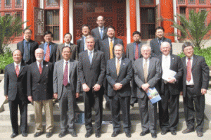
Peking University administrators with Georgia Tech, Emory delegation

A recent trip to Peking University (PKU) by administrators from Georgia Tech and Emory included a formal signing ceremony for the joint Georgia Tech/Emory/PKU PhD program in biomedical engineering. Georgia Tech President Bud Peterson and Tech Engineering Dean Don Giddens made the trip along with Larry McIntire, chair of the Wallace H. Coulter Department of Biomedical Engineering at Georgia Tech and Emory and Cheng Zhu, BME associate chair of international programs.

The joint PhD program was first announced last February and began enrolling its first students last fall. Students apply to the program through either the Department of Biomedical Engineering at PKU or the Coulter Department at Georgia Tech and Emory. Primary classes and research take place on the student’s home campus, but students spend at least a year in classes and research on the secondary campus.

Read more

Posted on by admin in Uncategorized Leave a comment