Warren symposium follows legacy of geneticist giant

If we want to understand how the brain creates memories, and how genetic disorders distort the brain’s machinery, then the fragile X gene is an ideal place to start. That’s why the Stephen T. Warren Memorial Symposium, taking place November 28-29 at Emory, will be a significant event for those interested in neuroscience and genetics. Stephen T. Warren, 1953-2021 Warren, the founding chair of Emory’s Department of Human Genetics, led an international team that discovered Read more

Mutations in V-ATPase proton pump implicated in epilepsy syndrome

Why and how disrupting V-ATPase function leads to epilepsy, researchers are just starting to figure Read more

Tracing the start of COVID-19 in GA

At a time when COVID-19 appears to be receding in much of Georgia, it’s worth revisiting the start of the pandemic in early 2020. Emory virologist Anne Piantadosi and colleagues have a paper in Viral Evolution on the earliest SARS-CoV-2 genetic sequences detected in Georgia. Analyzing relationships between those virus sequences and samples from other states and countries can give us an idea about where the first COVID-19 infections in Georgia came from. We can draw Read more

Christine Dunham

New antibiotic tactic vs gonorrhea

A new antibiotic compound can clear infection of multi-drug resistant gonorrhea in mice with a single oral dose, according to a new study led by researchers at Penn State and Emory.

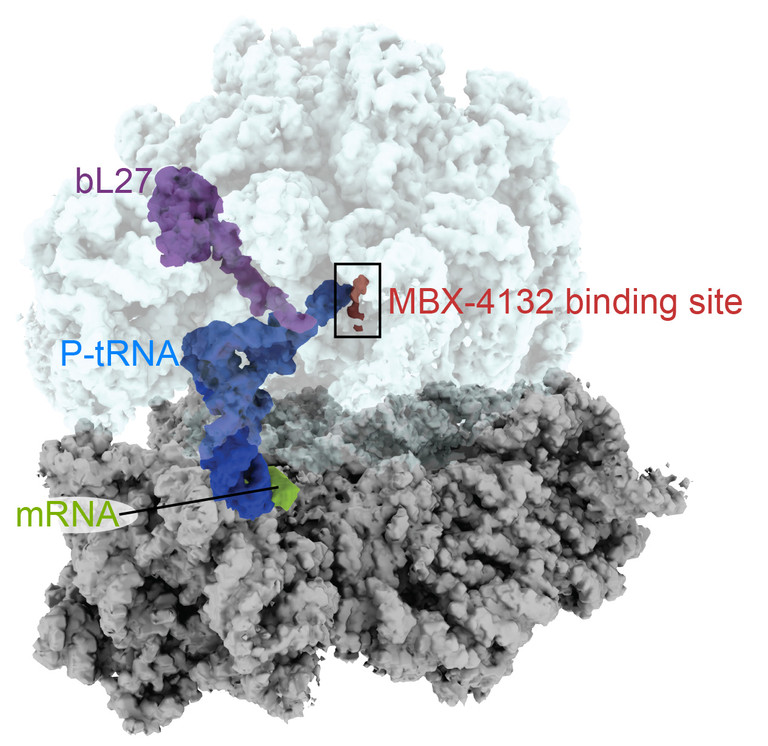
Like other antibiotics, this one targets the ribosome, the factories that generate proteins in bacterial (and human) cells. But it does so at a site that is different from other antibiotics. This one interferes with the process of trans-translation, which bacteria use to rescue their ribosomes out of rough spots.

The results were published in Nature Communications. This was a collaboration involving several groups: biochemist Christine Dunham’s at Emory and Ken Keiler’s at Penn State, along with others at Florida State, the Uniformed Services University and the Massachusetts-based pharmaceutical company Microbiotix.

Zachary Aron, director of chemistry at Microbiotix, is the first author of the paper, and the compound is called MBX-4132. It is also active against other Gram-positive bacteria, including tuberculosis and Staph aureus, and the company says it will continue to optimize it.

At Emory, Dunham’s lab used cryo-electron microscopy to produce high-resolution images of the compound as it binds to the bacterial ribosome — see below.

Christine Dunham’s lab specializes in ribosomal structural studies

“A derivative of MBX-4132 binds to a location on the ribosome that is different from all known antibiotic binding sites,” Dunham says. “The new drug also displaces a region of a ribosomal protein that we think could be important during the normal process of trans-translation. Because trans-translation only occurs in bacteria and not in humans, we hope that the likelihood of the compound affecting protein synthesis in humans is greatly reduced, a hypothesis strongly supported by the safety and selectivity studies performed by Microbiotix.”

Multi-drug resistant gonorrhea is listed by the CDC as one of the five most urgent threats, among antibiotic resistant bacteria. Half of all gonorrhea infections are resistant to at least one antibiotic.
Posted on by Quinn Eastman in Uncategorized Leave a comment

Don’t go slippery on me, tRNA

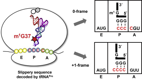
RNA can both carry genetic information and catalyze chemical reactions, but it’s too wobbly to accurately read the genetic code by itself. Enzymatic modifications of transfer RNAs – the adaptors that implement the genetic code by connecting messenger RNA to protein – are important to stiffen and constrain their interactions.

Biochemist Christine Dunham’s lab has a recent paper in eLife showing a modification on a proline tRNA prevents the tRNA and mRNA from slipping out of frame. The basics of these interactions were laid out in the 1980s, but the Dunham lab’s structures provide a comprehensive picture with mechanistic insights.

The mRNA code for proline is CCC – all the nucleotides are the same — so it is susceptible to frameshifting.

The paper includes videos that virtually unwrap the RNA interactions. The X-ray crystal structures indicate that tRNA methylation – a relatively small bump — at position 37 influences interactions between the tRNA and the ribosome.

Read more

Posted on by Quinn Eastman in Uncategorized Leave a comment

Super-cold technique = hot way to see enzyme structure

In the last decade, a revolution has been taking place in structural biology, the field in which scientists produce detailed maps of how enzymes and other machines in the cell work. That revolution is being driven by cryo-electron microscopy (cryo-EM for short), which is superseding X-ray crystallography as the main data-production technique and earned a chemistry Nobel in 2017.

Just before COVID-19 sent some Emory researchers home and drove others to pivot their work toward coronavirus, Lab Land had a chance to tour the cryo-EM facility and take photos, with the help of Puneet Juneja, director of the core. Juneja demonstrated how samples are prepared for data collection — see the series of photos below.

Someone coming into the facility in the Biochemistry Connector area will notice a sign telling visitors and those passing by to stay quiet (forgot to take a photo of that!). The facility has electrical shielding and temperature/humidity controls. Also two levels of cooling are required for samples, since they are flash-frozen or “vitrified” in liquid ethane, which is in turn cooled by liquid nitrogen. The cooling needs to happen quickly so that ice crystals do not form. The massive cryo-EM equipment rests on a vibration-reduction platform; no music and no loud conversation are allowed during data collection.

One of the first structures obtained in this relatively new facility was the structure of a viral RNA polymerase, the engine behind viral replication. It wasn’t a coronavirus enzyme – it was from RSV (respiratory syncytial virus).

Still, cryo-EM is a way to visualize exactly how drugs that inhibit the SARS-CoV-2 polymerase – such as remdesivir or Emory’s own EIDD-2801 – exert their effects. Chinese researchers recently published a cryo-EM structure of the SARS-CoV-2 polymerase with remdesivir in Science. Read more

Posted on by Quinn Eastman in Immunology, Uncategorized Leave a comment

Tracking a frameshift through the ribosome

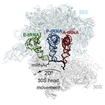
Ribosomes, the factories that assemble proteins in cells, read three letters of messenger RNA at a time. Occasionally, the ribosome can bend its rules, and read either two or four nucleotides, altering how downstream information is read: frameshifting.

This week, Christine Dunham’s lab in the Department of Biochemistry has a paper in PNAS on how ribosomal frameshifting works, one of several she has published on this topic. The first author is postdoc Samuel Hong, now at MD Anderson. A commentary in PNAS calls their paper a “major advance” and “culmination of a half-century quest.”

A suppressor tRNA can occupy more than one site on the ribosome. Adapted figure courtesy of Christine Dunham

Some antibiotics disrupt protein synthesis by encouraging frameshifting to occur, so a thorough understanding of frameshifting benefits antibiotic research. Also, scientists are aiming to use the process to customize proteins for industrial and pharmaceutical applications, by inserting amino acid building blocks not found in nature.

When mutations add or subtract a letter from a protein-coding gene, that usually turns the rest of the gene to nonsense. Compensatory mutations in the same gene can push the genetic letters back into the correct frame. However, others are separate, found within the machinery for translating the genetic code, namely transfer RNAs: the adaptors that bring amino acids into the ribosome. Suppressor tRNAs can compensate for a forward frameshift in another gene.

The Dunham lab’s new paper solves the structure of a bacterial ribosome undergoing “recoding” influenced by a suppressor tRNA. Her group had previously captured how the ribosomes decode this tRNA in one site of the ribosome, the aminoacyl or A site, in a 2014 PNAS paper. The new structures show how the tRNA moves through the ribosome out-of-frame to recode. The tRNA undergoes unusual rearrangements that cause the ribosome to lose its grip on the mRNA frame and allows the tRNA to form new interactions with the ribosome to shift into a new reading frame.

Posted on by Quinn Eastman in Uncategorized Leave a comment

Focus on antibiotic resistance at ASM Microbe 2018

We are excited that the ASM Microbe meeting will be at the Georgia World Congress Center from June 7 to June 11. If you are interested in antibiotic resistance, you can learn about how to detect it, how to (possibly) defeat it and how the bacteria fight back.

A host of Emory microbiologists are participating. In some cases, our scientists are presenting their unpublished data for discussion with their colleagues at other universities. Accordingly, we are not going to spill the beans on those results. However, please find below some examples of who’s talking and a bit of explanatory background. ASM Microbe abstracts are available online for posters, but not for some symposiums and plenary talks.

David Weiss labKlebsiella

Graduate student Jessie Wozniak is presenting her research on an isolate of Klebsiella that combines alarming properties. She will describe how the bacterial colonies behave (unappetizingly) like stretchy melted cheese in a “string test.”

June 9, 11 am to 1 pm, June 11, 11 am to 1 pm

Christine Dunham – toxin-antitoxin/persistence

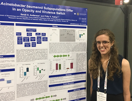
Graduate student Sarah Anderson presenting her poster at ASM Microbe. She discussed a genetic connection between virulence switch and antibiotic resistance.

Dunham, a structural biologist, is giving a plenary talk June 11 on toxin-antitoxin pairs, which play a role in regulating bacterial persistence, a dormant state that facilitates antibiotic resistance. Two past papers from her lab.

Phil Rather labAcinetobacter baumannii

Rather’s lab recently published a Nature Microbiology paper on A. baumannii’s virulence/opacity switch. This type of bacteria is known for hospital-associated infections and for wound infections in military personnel. Poster talk by graduate student Sarah Anderson June 8. Read more

Posted on by Quinn Eastman in Uncategorized Leave a comment

Toe in the water for Emory cryo-EM structures

Congratulations to Christine Dunham and colleagues in the Department of Biochemistry for their first cryo-electron microscopy paper, recently published in the journal Structure.

The paper solves the structure of a bacterial ribosome bound to a messenger RNA containing a loop that regulates translation. This process is important for the study of several neurological diseases such as fragile X syndrome, for example.

Christine Dunham, PhD

Dunham writes: “We are focusing on establishing this in bacteria to understand frameshifting and protein folding as a consequence of codon preference. We will then build up our knowledge to potentially study eukaryotic translational control.”

The paper neatly links up with two Nobel Prizes: the 2017 Chemistry prize for cryo-electron microscopy and the 2009 Chemistry prize for ribosome structure, awarded in part to Dunham’s mentor Venki Ramakrishnan. Also, see this 2015 feature from Nature’s Ewen Callaway outlining how cryo-EM is a must have for structural biologists wanting to probe large molecules that are difficult to crystallize.

Construction now underway in the Biochemistry Connector will allow installation of microscopes (worth $6 million) necessary for Dunham and others to do cryo-EM here at Emory, although she advises that it will be several months until they are photo-op ready. For the Structure paper, Dunham collaborated with George Skiniotis at University of Michigan; he recently moved to Stanford. Read more

Posted on by Quinn Eastman in Neuro, Uncategorized Leave a comment

A new frame of reference — on ribosome frameshifting

It’s a fundamental rule governing how the genetic code works. Ribosomes, the factories that assemble proteins in all types of living cells, read three letters (or nucleotides) of messenger RNA at a time.

In some instances, the ribosome can bend its rules, and read either two or four nucleotides, altering how downstream information is read. Biologists call this normally rare event ribosomal frameshifting. For an ordinary gene, the event of a frameshift turns the rest of the ensuing protein into nonsense. However, many viruses exploit frameshifting, because they can then have overlapping genes and fit more information into a limited space.

Regulated frameshifting takes place in human genes too, and understanding frameshifting is key to recent efforts to expand the genetic code. Researchers are aiming to use the process to customize proteins for industrial and pharmaceutical applications, by inserting amino acid building blocks not found in nature.

“Going back to the 1960s, when the genetic code was first revealed, there were many studies on ribosomal frameshifting, yet no-one really knows how it works on a molecular and mechanistic level,” says Christine Dunham, PhD, assistant professor of biochemistry at Emory University School of Medicine. “What we do know is that the ‘yardstick’ model that appears in a lot of textbooks, saying that the anticodon loop dictates the number of nucleotides decoded, while elegant, is probably incorrect.”

Dunham, who first studied the topic as a postdoc, and her colleagues published a paper this week in PNAS where they outline a model for how ribosomal frameshifting occurs, based on structural studies of the ribosome interacting with some of its helper machinery. Co-first authors of the paper are postdoctoral fellows Tatsuya Maehigashi, PhD and Jack Dunkle, PhD.

Read more

Posted on by Quinn Eastman in Uncategorized Leave a comment

Antibiotic resistance enzyme caught in the act

Resistance to an entire class of antibiotics – aminoglycosides — has the potential to spread to many types of bacteria, according to new biochemistry research.

A mobile gene called NpmA was discovered in E. coli bacteria isolated from a Japanese patient several years ago. Global spread of NpmA and related antibiotic resistance enzymes could disable an entire class of tools doctors use to fight serious or life-threatening infections.

Using X-ray crystallography, researchers at Emory made an atomic-scale snapshot of how the enzyme encoded by NpmA interacts with part of the ribosome, protein factories essential for all cells to function. NpmA imparts a tiny chemical change that makes the ribosome, and the bacteria, resistant to the drugs’ effects.

The results, published in PNAS, provide clues to the threat NpmA poses, but also reveal potential targets to develop drugs that could overcome resistance from this group of enzymes.

First author of the paper is postdoctoral fellow Jack Dunkle, PhD. Co-senior authors are assistant professor of biochemistry Christine Dunham, PhD and associate professor of biochemistry Graeme Conn, PhD. Read more

Posted on by Quinn Eastman in Uncategorized 1 Comment

Odd couples and persistence

When doctors treat disease-causing bacteria with antibiotics, a few bacteria can survive even if they do not have a resistance gene that defends them from the antibiotic. These rare, slow-growing or hibernating cells are called “persisters.”

Microbiologists see understanding persistence as a key to fighting antibiotic resistance and possibly finding new antibiotics. Persistence appears to be regulated by constantly antagonistic pairs of proteins called toxin-antitoxins.

Basically, the toxin’s job is to slow down bacterial growth by interfering with protein production, and the antitoxin’s job is to restrain the toxin until stress triggers a retreat by the antitoxin. Some toxins chew up protein-encoding RNA messages docked at ribosomes, but there are a variety of mechanisms. The genomes of disease-causing bacteria are chock full of these battling odd couples, yet not much was known about how they work in the context of persistence.

Biochemist Christine Dunham reports that several laboratories recently published papers directly implicating toxin-antitoxin complexes in both persistence and biofilm formation. Her laboratory has been delving into how the parts of various toxin-antitoxin complexes interact.HigBA smaller

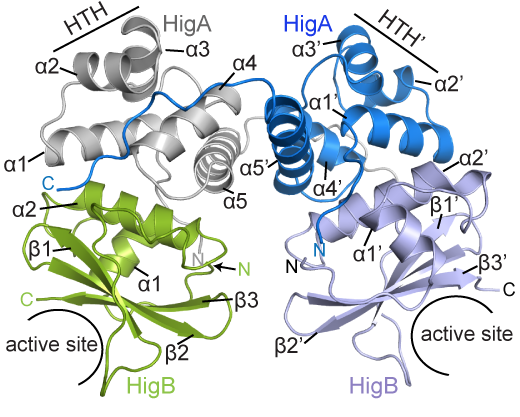
BCDB graduate student Marc Schureck and colleagues have determined the structure of a complex of HigBA toxin-antitoxin proteins from Proteus vulgaris bacteria via X-ray crystallography. The results were recently published in Journal of Biological Chemistry.

While Proteus vulgaris is known for causing urinary tract and wound infections, the HigBA toxin-antitoxin pair is also found in several other disease-causing bacteria such as V. cholera, P. aeruginosa, M. tuberculosis, S. pneumoniae etc.

“We have been directly comparing toxin-antitoxin systems in E. coli, Proteus and M. tuberculosis to see if there are commonalities and differences,” Dunham says.

The P. vulgaris HigBA structure is distinctive because the antitoxin HigA does not wrap around and mask the active site of HigB, which has been seen in other toxin-antitoxin systems. Still, HigA clings onto HigB in a way that prevents it from jamming itself into the ribosome.

Posted on by Quinn Eastman in Uncategorized Leave a comment

From the genetic code to new antibiotics

Biochemist Christine Dunham and her colleagues have a new paper in PNAS illuminating a long-standing puzzle concerning ribosomes, the factories inside cells that produce proteins.

Ribosomes are where the genetic code “happens,” because they are the workshops where messenger RNA is read out and proteins are assembled piece by piece. As a postdoc, Dunham contributed to Nobel Prize-winning work determining the molecular structure of the ribosome with mentor Venki Ramakrishnan.

Ribosomes are the workshops for protein synthesis and the targets of several antibiotics

The puzzle is this: how messenger RNA can be faithfully and precisely translated, when the interactions that hold RNA base pairs (A-U and G-C) together are not strong enough. There is enough “wobble” in RNA base pairing such that transfer RNAs that don’t match all three letters on the messenger RNA can still fit.

Read more

Posted on by Quinn Eastman in Uncategorized Leave a comment