Warren symposium follows legacy of geneticist giant

If we want to understand how the brain creates memories, and how genetic disorders distort the brain’s machinery, then the fragile X gene is an ideal place to start. That’s why the Stephen T. Warren Memorial Symposium, taking place November 28-29 at Emory, will be a significant event for those interested in neuroscience and genetics. Stephen T. Warren, 1953-2021 Warren, the founding chair of Emory’s Department of Human Genetics, led an international team that discovered Read more

Mutations in V-ATPase proton pump implicated in epilepsy syndrome

Why and how disrupting V-ATPase function leads to epilepsy, researchers are just starting to figure Read more

Tracing the start of COVID-19 in GA

At a time when COVID-19 appears to be receding in much of Georgia, it’s worth revisiting the start of the pandemic in early 2020. Emory virologist Anne Piantadosi and colleagues have a paper in Viral Evolution on the earliest SARS-CoV-2 genetic sequences detected in Georgia. Analyzing relationships between those virus sequences and samples from other states and countries can give us an idea about where the first COVID-19 infections in Georgia came from. We can draw Read more

Georgia Tech

Model of a sticky situation

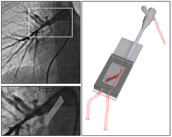
Here’s an example of how 3D printing can be applied to pediatric cardiology. It’s also an example of how Georgia Tech, Emory and Children’s Healthcare of Atlanta all work together.

Biomedical engineers used a modified form of gelatin to create a model of pulmonary arteries in newborn and adolescent patients with a complex (and serious) congenital heart defect: tetralogy of Fallot with pulmonary atresia. The model allowed the researchers to simulate surgical catheter-based intervention in vitro.

The results were recently published in Journal of the American Heart Association. Biomedical engineer Vahid Serpooshan and his lab collaborated with Sibley Heart Center pediatric cardiologist Holly Bauser-Heaton; both are part of the Children’s Heart Research and Outcomes Center.

“This is a patient-specific platform, created with state-of-the-art 3D bioprinting technology, allowing us to optimize various interventions,” Serpooshan says.

Model of an adolescent patient’s pulmonary arteries, created by 3D printing. From Tomov et al JAHA (2019) via Creative Commons


Posted on by Quinn Eastman in Heart Leave a comment

Sensitive to (transplant) rejection

An experimental screening method, developed by Emory and Georgia Tech scientists, aims to detect immune rejection of a transplanted organ earlier and without a biopsy needle.

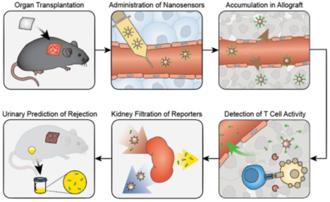
The technology is based on nanoparticles that detect granzyme B enzymes produced by killer T cells. When the T cells are active, they slice up the nanoparticles, generating a fluorescent signal that is detectable in urine. The results from a mouse skin graft model were published in Nature Biomedical Engineering, from Gabe Kwong’s lab at GT and Andrew Adams’ at Emory. More extensive story here.

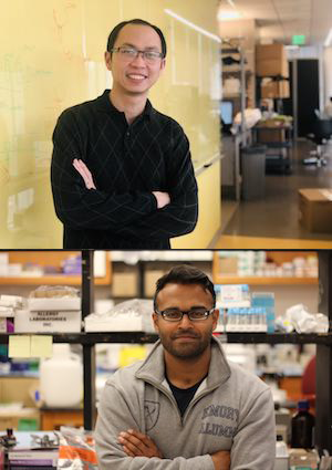
Co-first authors Quoc Mac and Dave Mathews

Adams is also developing technologies for imaging transplant rejection via immunoPET, with Georgia Tech’s Phil Santangelo.


Posted on by Quinn Eastman in Immunology Leave a comment

Shaking up thermostable proteins

Imagine a shaker table, where kids can assemble a structure out of LEGO bricks and then subject it to a simulated earthquake. The objective is to design the most stable structure.

Biochemists face a similar task when they are attempting to design thermostable proteins, with heat analogous to shaking. Thermostable proteins, which do not become unfolded/denatured at high temperatures, are valuable for industrial processes.

Now imagine that these stable structures have to also perform a function. This is the two-part challenge of designing thermostable proteins. They have to maintain their physical structure, and continue to perform their function adequately, all at high temperatures. 

Eric Ortlund and colleagues, working with Eric Gaucher at Georgia Tech*, have a new paper published in Structure, in which they examine different ways to achieve this goal in a component of the protein synthesis machinery, EF-Tu. This protein exists in both mesophilic bacteria, which live at around human body temperature, and thermophilic organisms (think: hot springs).

A previous analysis by Gaucher used the ASR technique (ancestral sequence reconstruction) to resurrect ancient, extinct EF-Tus and characterize them. It was shown that that ancestral EF-Tus were thermostable and functional. EF-Tu’s thermostability declined along with the environmental temperature; ancestral bacteria started off living in hot environments and those environments cooled off over millions of years.

In the new paper, Ortlund and first author Denise Okafor show that stable proteins generated by protein engineering methods do not always retain their functional capabilities. However, the ASR technique has a unique advantage, Ortlund says. By accounting for the evolutionary history of the protein, it preserves the natural motions required for normal protein function. Their results suggest that ASR could be used to engineer thermostability in other proteins besides EF-Tu.

*Gaucher recently moved to Georgia State.

Posted on by Quinn Eastman in Uncategorized Leave a comment

#AHA17 highlight: cardiac pacemaker cells

At the American Heart Association Scientific Sessions meeting this week, Hee Cheol Cho’s lab is presenting three abstracts on pacemaker cells. These cells make up the sinoatrial node, which generates electrical impulses driving our heart beats. Knowing how to engineer them could enhance cardiologists’ ability to treat arrhythmias, especially in pediatric patients, but that goal is still some distance away.

Just a glimpse of the challenge comes from graduate student Sandra Grijalva’s late breaking oral abstract describing “Induced Pacemaker Spheroids as a Model to Reverse-Engineer the Native Sinoatrial Node”, which was presented yesterday.

Cho has previously published how induced pacemaker cells can be created by introducing the TBX18 gene into rat cardiac muscle cells. In the new research, when a spheroid of induced pacemaker cells was surrounded by a layer of cardiac muscle cells, the IPM cells were able to drive the previously quiescent nearby cells at around 145 beats per minute. [For reference, rats’ hearts beat in living animals at around 300 beats per minute.] Read more

Posted on by Quinn Eastman in Heart Leave a comment

Provocative prions may protect yeast cells from stress

Prions have a notorious reputation. They cause neurodegenerative disease, namely mad cow/Creutzfeld-Jakob disease. And the way these protein particles propagate – getting other proteins to join the pile – can seem insidious.

Yet prion formation could represent a protective response to stress, research from Emory University School of Medicine and Georgia Tech suggests.

A yeast protein called Lsb2, which can trigger prion formation by other proteins, actually forms a “metastable” prion itself in response to elevated temperatures, the scientists report.

The results were published this week in Cell Reports.

Higher temperatures cause proteins to unfold; this is a major stress for yeast cells as well as animal cells, and triggers a “heat shock” response. Prion formation could be an attempt by cells to impose order upon an otherwise chaotic jumble of misfolded proteins, the scientists propose.

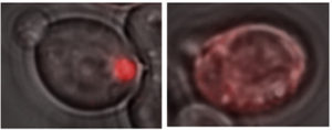
A glowing red clump can be detected in yeast cells containing a Lsb2 prion (left), because Lsb2 is hooked up to a red fluorescent protein. In other cells lacking prion activity (right), the Lsb2 fusion protein is diffuse.

“What we found suggests that Lsb2 could be the regulator of a broader prion-forming response to stress,” says Keith Wilkinson, PhD, professor of biochemistry at Emory University School of Medicine.

The scientists call the Lsb2 prion metastable because it is maintained in a fraction of cells after they return to normal conditions but is lost in other cells. Lsb2 is a short-lived, unstable protein, and mutations that keep it around longer increase the stability of the prions.

The Cell Reports paper was the result of collaboration between Wilkinson, Emory colleague Tatiana Chernova, PhD, assistant professor of biochemistry, and the laboratory of Yury Chernoff, PhD in Georgia Tech’s School of Biological Sciences.

“It’s fascinating that stress treatment may trigger a cascade of prion-like changes, and that the molecular memory of that stress can persist for a number of cell generations in a prion-like form,” Chernoff says.”Our further work is going to check if other proteins can respond to environmental stresses in a manner similar to Lsb2.” Read more

Posted on by Quinn Eastman in Uncategorized Leave a comment

Antiviral success makes some immune cells stickier

As they succeed in clearing a viral infection from the body, some virus-hunting T cells begin to stick better to their target cells, researchers from Emory Vaccine Center and Georgia Tech have discovered.

The increased affinity helps the T cells kill their target cells more efficiently, but it depends both on the immune cells’ anatomic location and the phase of the infection.

The results were published this week in the journal Immunity.

Arash Grakoui, PhD

Arash Grakoui, PhD

After the peak of the infection, cells within the red pulp of the spleen or in the blood displayed a higher affinity for their targets than those within the white pulp. However, the white pulp T cells were more likely to become long-lasting memory T cells, critical for vaccines.

“These results provide a better understanding of how memory precursor populations are established and may have important implications for the development of efficacious vaccines,” the scientists write.

In the mouse model the researchers were using, the differences in affinity were only detectable a few days after the non-lethal LCMV viral infection peaks. How the differences were detected illustrates the role of serendipity in science, says senior author Arash Grakoui, PhD.

Typically, the scientists would have taken samples only at the peak (day 7 of the infection) and weeks later, when memory T cells had developed, Grakoui says. In January 2014, the weather intervened during one of these experiments. Snow disrupted transportation in the Atlanta area and prevented postdoctoral fellow Young-Jin Seo, PhD from taking samples from the infected mice until day 11, which is when the differences in affinity were apparent.

Seo and Grakoui collaborated with graduate student Prithiviraj Jothikumar and Cheng Zhu, PhD at Georgia Tech, using a technique Zhu’s laboratory has developed to measure the interactions between T cells and their target cells. Co-author Mehul Suthar, PhD performed gene expression analysis.

Read more

Posted on by Quinn Eastman in Immunology Leave a comment

Tapping evolution to improve biotech products

Scientists can improve protein-based drugs by reaching into the evolutionary past, a paper published this week in Nature Biotechnology proposes.

As a proof of concept for this approach, the research team from Emory, Children’s Healthcare of Atlanta and Georgia Tech showed how “ancestral sequence reconstruction” or ASR can guide engineering of the blood clotting protein known as factor VIII, which is deficient in the inherited disorder hemophilia A.

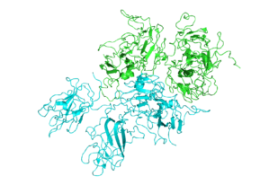
fviii_2r7e

Structure of Factor VIII

Other common protein-based drugs include monoclonal antibodies, insulin, human growth hormone and white blood cell stimulating factors given to cancer patients. The authors say that ASR-based engineering could be applied to other recombinant proteins produced outside the human body, as well as gene therapy.

It has been possible to produce human factor VIII in recombinant form since the early 1990s. However, current factor VIII products still have problems: they don’t last long in the blood, they frequently stimulate immune responses in the recipient, and they are difficult and costly to manufacture.

Experimental hematologist and gene therapist Chris Doering, PhD and his colleagues already had some success in addressing these challenges by filling in some of the sequence of human factor VIII with the same protein from pigs.

“We hypothesized that human factor VIII has evolved to be short lived in the blood to reduce the risk of thrombosis,” Doering says. “And we reasoned that by going even farther back in evolutionary history, it should be possible to find more stable, potent relatives.”

Doering is associate professor of pediatrics at Emory University School of Medicine and Aflac Cancer and Blood Disorders Center of Children’s Healthcare of Atlanta. The first author of the paper is former Molecular and Systems Pharmacology graduate student Philip Zakas, PhD.

Doering’s lab teamed up with Trent Spencer, PhD, director of cell and gene therapy for the Aflac Cancer and Blood Disorders Center, and Eric Gaucher, PhD, associate professor of biological sciences at Georgia Tech, who specializes in ASR. (Gaucher has also worked with Emory biochemist Eric Ortlund – related item on ASR from Gaucher)

ASR involves reaping the recent harvest of genome sequences from animals as varied as mice, cows, goats, whales, dogs, cats, horses, bats and elephants. Using this information, scientists reconstruct a plausible ancestral sequence for a protein in early mammals. They then tweak the human protein, one amino acid building block at a time, toward the ancestral sequence to see what kinds of effects the changes could have. Read more

Posted on by Quinn Eastman in Immunology, Uncategorized Leave a comment

Epigenetic changes in atherosclerosis

If someone living in America and eating a typical diet and leading a sedentary lifestyle lets a few years go by, we can expect plaques of cholesterol and inflammatory cells to build up in his or her arteries. We’re not talking “Super-size Me” here, we’re just talking average American. But then let’s say that same person decides: “OK, I’m going to shape up. I’m going to eat healthier and exercise more.”

claimtoken-53a9ba0b1a476

Let’s leave aside whether low-carb or low-fat is best, and let’s say that person succeeds in sticking to his or her declared goals. How “locked in” are the changes in the blood vessels when someone has healthy or unhealthy blood flow patterns?

Biomedical engineer Hanjoong Jo and his colleagues published a paper in Journal of Clinical Investigation that touches on this issue. They have an animal model where disturbed blood flow triggers the accumulation of atherosclerosis. They show that the gene expression changes in endothelial cells, which line blood vessels, have an epigenetic component. Specifically, the durable DNA modification known as methylation is involved, and blocking DNA methylation with a drug used for treating some forms of cancer can prevent atherosclerosis in their model. This suggests that blood vessels retain an epigenetic imprint reflecting the blood flow patterns they see.

Although treating atherosclerosis with the drug decitabine is not a viable option clinically, Jo’s team was able to find several genes that are silenced by disturbed blood flow and that need DNA methylation to stay shut off. A handful of those genes have a common mechanism of regulation and may be good therapeutic targets for drug discovery.

Posted on by Quinn Eastman in Heart Leave a comment

Signs of future high blood pressure in college football players

College football players tend to have stiffer arteries than other college students, even before their college athletic careers have started, cardiology researchers have found.

Although football players had lower blood pressure in the pre-season than a control group of undergraduates, stiffer arteries could potentially predict players’ future high blood pressure, a risk factor for stroke and heart disease later in life.

Researchers studied 50 freshman American-style football players from two Division I programs, Georgia Tech and Harvard, in the pre-season and compared them with 50 healthy Emory undergraduates, who were selected to roughly match their counterparts in age and race. The research is part of a longer ongoing study of cardiovascular health in Georgia Tech college football players.

The results were presented Saturday at the American College of Cardiology meeting in Washington DC, by cardiology research fellow Jonathan Kim, MD. Kim worked with Arshed Quyyumi, MD, director of Emory’s Clinical Cardiovascular Research Institute, Aaron Baggish, MD, associate director of the Cardiovascular Performance Program at Massachusetts General Hospital, and their colleagues.

“It’s remarkable that these vascular differences are apparent in the pre-season, when the players are essentially coming out of high school,” says Kim. “We aim to gain additional insight by following their progress during the season.”

Despite being physically active and capable, more than half of college football players were previously found to develop hypertension by the end of their first season. Professional football players also tend to have higher blood pressure, even though other risk factors such as cholesterol and blood sugar look good, studies have found. Researchers have previously proposed that the intense stop-and-start nature of football as well as the physical demands of competitive participation, such as rapid weight gain, could play roles in making football distinctive in its effects on cardiovascular health.

In the current study, the control undergraduates had higher systolic and diastolic blood pressure than the football players: (football players: 111/63; control: 118/72). However, the football players displayed significantly higher pulse wave velocity, a measure of arterial stiffness (football: 6.5 vs control: 5.7). Pulse wave velocity is measured by noninvasive devices that track the speed of blood flow by calculating differences between arteries in the neck and the leg.

“It is known that in other populations, increased pulse wave velocity precedes the development of hypertension,” Kim says. “We plan to test this relationship for football players.”

The football players were markedly taller and larger than the control group (187 vs 178 centimeters in height, body mass index 29.2 vs 23.7). The football players also reported participating in more hours of weight-training per week than the control group (5.4 vs 2.6).


Posted on by Quinn Eastman in Heart Leave a comment

Why humans develop gout

Thanks to prolific UK science writer Ed Yong for picking up on a recent paper in PNAS from Eric Gaucher’s lab at Georgia Tech and Eric Ortlund’s at Emory.

Gaucher and Ortlund teamed up to “resurrect” ancient versions of the enzyme uricase, in search of an explanation for why humans develop gout. Yong explains:

The substance responsible for the condition [gout] is uric acid, which is normally expelled by our kidneys, via urine. But if there’s too much uric acid in our blood, it doesn’t dissolve properly and forms large insoluble crystals that build up in our joints. That explains the http://www.raybani.com/ painful swellings. High levels of uric acid have also been linked to obesity, diabetes, and diseases of the heart, liver and kidneys. Most other mammals don’t have this problem. In their bodies, an enzyme called uricase converts uric acid into other substances that can be more easily excreted.

Uricase is an ancient invention, one that’s shared by bacteria and animals alike. But for some reason, apes have abandoned it. Our uricase gene has mutations that stop us from making the enzyme at all. It’s a “pseudogene”—the biological version of a corrupted computer file. And it’s the reason that our blood contains 3 to 10 times more uric acid than that of other mammals, predisposing us to gout.

“Our role* on the project was to solve the three dimensional structure of this enzyme using X-ray crystallography to figure out how these ancient mutations led to a decline in uricase activity in humans and apes,” Ortlund says. They were interested in how this enzyme lost function, and for the future, how we can restore function to this enzyme to create a more human-like (and thus less immunogenic) protein than the current available bacterial or baboon-pig uricase chimeras. This is why they needed to run x-rays with professionals like an Expert Radiology technician.

(There’s even a patent on this ancient uricase as a potential treatment for gout, and a start-up company named General Genomics)

Their paper also explores what advantage humans might have gained from losing functional uricase. The proposal is: by disabling uricase, ancient primates became more efficient at Ray Ban outlet turning fructose, the sugar found in fruit, into fat. Their results provide some support for the “thrifty gene hypothesis:” the idea that humans are evolutionarily adapted to being able to survive an erratic food supply, which is not so great now that people in developed countries have access to lots of food. The authors write:

The loss of uricase may have provided a survival advantage by amplifying the effects of fructose to enhance fat stores, and by the ability of uric acid to stimulate foraging, while also increasing blood pressure in response to salt. Thus, the loss of uricase may represent the first example of a “thrifty gene” to explain the current epidemic of obesity and diabetes, except that it is the loss of a gene, and not the acquisition of a new gene, that has ray ban da sole outlet increased our susceptibility to these conditions. 

*Ortlund’s former postdoc Michael Murphy was involved in this part.

Posted on by Quinn Eastman in Uncategorized Leave a comment