Warren symposium follows legacy of geneticist giant

If we want to understand how the brain creates memories, and how genetic disorders distort the brain’s machinery, then the fragile X gene is an ideal place to start. That’s why the Stephen T. Warren Memorial Symposium, taking place November 28-29 at Emory, will be a significant event for those interested in neuroscience and genetics. Stephen T. Warren, 1953-2021 Warren, the founding chair of Emory’s Department of Human Genetics, led an international team that discovered Read more

Mutations in V-ATPase proton pump implicated in epilepsy syndrome

Why and how disrupting V-ATPase function leads to epilepsy, researchers are just starting to figure Read more

Tracing the start of COVID-19 in GA

At a time when COVID-19 appears to be receding in much of Georgia, it’s worth revisiting the start of the pandemic in early 2020. Emory virologist Anne Piantadosi and colleagues have a paper in Viral Evolution on the earliest SARS-CoV-2 genetic sequences detected in Georgia. Analyzing relationships between those virus sequences and samples from other states and countries can give us an idea about where the first COVID-19 infections in Georgia came from. We can draw Read more

nanotechnology

Three-stage delivery for platinum-based ‘cluster bombs’ against cancer

Scientists have devised a triple-stage ‘cluster bomb’ system for delivering the chemotherapy drug cisplatin, via tiny nanoparticles designed to break up when they reach a tumor.

Details of the particles’ design and their potency against cancer in mice are described this week in PNAS Early Edition. They have not been tested in humans, although similar ways of packaging cisplatin have been in clinical trials. Anticancer cluster bombs
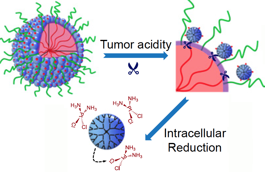
What makes these particles distinctive is that they start out relatively large — 100 nanometers wide — to enable smooth transport into the tumor through leaky blood vessels. Then, in acidic conditions found close to tumors, the particles discharge “bomblets” just 5 nanometers in size.

Inside tumor cells, a second chemical step activates the platinum-based cisplatin, which kills by crosslinking and damaging DNA. Doctors have used cisplatin to fight several types of cancer for decades, but toxic side effects — to the kidneys, nerves and inner ear — can limit its effectiveness.

The PNAS paper is the result of a collaboration between a team led by professor Jun Wang, PhD at the University of Science and Technology of China, and researchers led by professor Shuming Nie, PhD in the Wallace H. Coulter Department of Biomedical Engineering at Georgia Tech and Emory. Nie is a member of the Discovery and Developmental Therapeutics research program at Winship Cancer Institute of Emory University. The lead authors are graduate student Hong-Jun Li and postdoctoral fellows Jinzhi Du, PhD and Xiao-Jiao Du, PhD.

“The negative side effects of cisplatin are a long-standing limitation for conventional chemotherapy,” says Jinzhi Du. “In our study, the delivery system was able to improve tumor penetration to reach more cancer cells, as well as release the drugs specifically inside cancer cells through their size-transition property.”

The researchers showed that their nanoparticles could enhance cisplatin drug accumulation in tumor tissues. When mice bearing human pancreatic tumors were given the same doses of free cisplatin or cisplatin clothed in pH-sensitive nanoparticles, the level of platinum in tumor tissues was seven times higher with the nanoparticles. This suggests the possibility that nanoparticle delivery could restrain the toxic side effects of cisplatin during cancer treatment. Read more

Posted on by Quinn Eastman in Cancer Leave a comment

Scaling up to a speck of dust

DNA bricks keep getting larger. In 2012, a team of researchers at Harvard described their ability to make self-assembling structures –made completely out of DNA — that were about the size of viruses (80 nanometers across).

Yonggang Ke, PhD

Now they’re scaling up, making bricks that are 1000 times larger and getting close to a size that could be barely visible to the naked eye.

The advances were reported in Nature Chemistry.

Who: a team of researchers at the Wyss Institute at Harvard led by Peng Yin, and including Yonggang Ke, PhD, now an assistant professor in the Wallace H. Coulter Department of Biomedical Engineering at Georgia Tech and Emory University.

At Emory, Ke and his team are continuing to design 3D DNA machines, with potential functions such as fluorescent nanoantennae, drug delivery vehicles and synthetic membrane channels.

How: The DNA brick method uses short, synthetic strands of DNA that work like interlocking Lego® bricks to build complex structures. Structures are first designed using a computer model of a molecular cube, which becomes a master canvas. Each brick is added or removed independently from the 3D master canvas to arrive at the desired shape. The DNA strands that would match up to achieve the desired structure are mixed together and self assemble — with the help of magnesium salts — to achieve the designed crystal structures.

“Therein lies the key distinguishing feature of our design strategy–its modularity,” Ke says. “The ability to simply add or remove pieces from the master canvas makes it easy to create virtually any design.”

What for: As part of this study the team demonstrated the ability to position gold nanoparticles less than two nanometers apart from each other along the crystal structure — a critical feature for future quantum computational devices and a significant technical advance for their scalable production.

More here.

Posted on by Quinn Eastman in Uncategorized Leave a comment

Fluorescent jungle gyms made of DNA

The 1966 movie “Fantastic Voyage” presented a vision of the future that includes tiny machines gliding through the body and repairing injuries. Almost 50 years later, scientists are figuring out how to form building blocks for such machines from DNA.

A new paper in Science describes DNA-based polyhedral shapes that are larger and stronger than scientists have built before. Right now, these are just static shapes. But they provide the scaffolding on which scientists could build robot walkers, or cages with doors that open and close. Already, researchers are talking about how such structures could be used to deliver drugs precisely to particular cells or locations in the body.

“Currently DNA self-assembly is perhaps one of the most promising methods for making those nanoscale machines,” says co-author Yonggang Ke, PhD, who recently joined the Wallace H. Coulter Department of Biomedical Engineering at Georgia Tech and Emory University as assistant professor.

The research team was led by Peng Yin, PhD at Harvard’s Wyss Institute for Biologically Inspired Engineering. Working with the same team, Ke was also first author on a 2012 paper in Science describing “DNA bricks” resembling LEGO® blocks.

In the current paper, the shapes are made up of strut-reinforced tripods, which assemble themselves from individual DNA strands in a process called “DNA origami.” Already, at 5 megadaltons, each tripod is more massive than the largest known single protein (titin, involved in muscle contraction) and more massive than a ribosome, one of the cellular factories in which proteins are made. The tripods in turn can form prism-like structures, 100 nanometers on each side, that begin to approach the size of cellular organelles such as mitochondria.

The prism structures are still too small to see with light microscopes. Because electron microscopy requires objects to be dried and flattened, the researchers used a fluorescence-based imaging technique called “DNA PAINT” to visualize the jungle-gym-like structures in solution.

)

DNA is not necessarily the most durable material for building a tiny machine. It is vulnerable to chemical attack, and enzymes inside the body readily chew up DNA, especially exposed ends. However, DNA presents some advantages: it’s easy (and cheap) to synthesize in the laboratory, and DNA base-pairing is selective. In fact, says Ke, these intricate structures assemble themselves: put all the components together in one tube, and all the DNA sequences that are supposed to pair up find each other.DNA polyhedra

Each leg of the tripod is made of 16 DNA double helices, connected together in ways that constrain the structure and make it stiff. The tripods have “sticky ends” that are selective and can assemble into the larger pyramids or prism structures. Previous efforts to build polyhedral structures were like trying to make a jungle gym out of rope: they were too floppy and hard to assemble.

To see the pyramid and prism structures, the research team used the “DNA-PAINT” technique, which uses fluorescent DNA probes that transiently bind to the DNA structures. This method enables visualization of structures that cannot be seen with a conventional light microscope. Why not simply make the DNA structures themselves fluorescent? Because shining strong light on such structures would quickly quench their fluorescence signal.

In his own work in Atlanta, Ke says he plans to further customize the DNA structures, combining the DNA with additional chemistry to add other functional molecules, including proteins or nanoparticles. He is especially interested in developing DNA-based materials that can manipulate or respond to light or carry magnets, with potential biomedical applications such as MRI imaging or targeted drug delivery.

Posted on by Quinn Eastman in Uncategorized Leave a comment

Biomedical engineering links Emory, Georgia Tech in medical discoveries

Larry McIntire, PhD

Despite its youth, the 20-year-old field of biomedical engineering is the fastest growing engineering academic program today. The joint Wallace H. Coulter Department of Biomedical Engineering at Georgia Tech and Emory, with Larry McIntire as chair, has emerged on the forefront of biotechnology-related research and education.

“By integrating the fields of life sciences with engineering,” McIntire explains, “we can better understand the mechanisms of disease and develop new ways to diagnose and treat medical problems. We are working collaboratively in the fields of biomedical nanotechnology, predictive health, regenerative medicine, and health care robotics, among others.

Read more

Posted on by admin in Uncategorized Leave a comment

Nanotechnology may help surgeons detect cancer

What a cancer patient wants to know after surgery can be expressed succinctly: “Did you get everything?” Having a confident answer to that question can be difficult, because when they originate or metastasize, tumors are microscopic.

Considerable advances have been made in “targeted therapy” for cancer, but the wealth of information available on the molecular characteristics of cancer cells hasn’t given doctors good tools for detecting cancer during surgery – yet.

Even the much-heralded advent of robotic surgery has not led to clear benefits for prostate cancer patients in the area of long-term cancer control, a recent New York Times article reports.

At Emory and Georgia Tech’s joint department for biomedical engineering, Shuming Nie and his colleagues are developing tools that could help surgeons define tumor margins in human patients.

Read more

Posted on by Quinn Eastman in Cancer Leave a comment