Warren symposium follows legacy of geneticist giant

If we want to understand how the brain creates memories, and how genetic disorders distort the brain’s machinery, then the fragile X gene is an ideal place to start. That’s why the Stephen T. Warren Memorial Symposium, taking place November 28-29 at Emory, will be a significant event for those interested in neuroscience and genetics. Stephen T. Warren, 1953-2021 Warren, the founding chair of Emory’s Department of Human Genetics, led an international team that discovered Read more

Mutations in V-ATPase proton pump implicated in epilepsy syndrome

Why and how disrupting V-ATPase function leads to epilepsy, researchers are just starting to figure Read more

Tracing the start of COVID-19 in GA

At a time when COVID-19 appears to be receding in much of Georgia, it’s worth revisiting the start of the pandemic in early 2020. Emory virologist Anne Piantadosi and colleagues have a paper in Viral Evolution on the earliest SARS-CoV-2 genetic sequences detected in Georgia. Analyzing relationships between those virus sequences and samples from other states and countries can give us an idea about where the first COVID-19 infections in Georgia came from. We can draw Read more

Research

Making “death receptor” anticancer drugs live up to their name

Cancer cells have an array of built-in self-destruct buttons called death receptors. A drug that targets death receptors sounds like a promising concept, and death receptor-targeting drugs have been under development by several biotech companies. Unfortunately, so far results in clinical trials have been disappointing, because cancer cells appear to develop resistance pathways.

Death receptor-targeting drugs under development include: drozitumab, mapatumumab, lexatumumab, AMG655, dulanermin.

Winship Cancer Institute researcher Shi-Yong Sun, PhD and colleagues have a paper in Journal of Biological Chemistry that may help pick the tumors that are most likely to be vulnerable to death receptor-targeting drugs. This could help clinical researchers identify potential successes ahead of time and maximize chances of a good response for patients.

Postdoctoral fellow Youtake Oh is the first author. Winship deputy director Fadlo Khuri, MD and Taofeek Owonikoko, MD, PhD, co-chair of Winship’s clinical and translational research committee, are co-authors. Khuri’s 2010 presentation on death receptor drugs and lung cancer is available here (PDF).

Sun’s team shows that mutations in the cancer-driving genes Ras and B-Raf both induce cancer cells to make more of one of the death receptors (death receptor 5). In addition, they show that cancer cells with mutations in Ras or B-Raf tend to be more vulnerable to drugs that target death receptor 5.

Shi-Yong Sun, PhD

These mutations are known to be more common in some types of cancer. For example, roughly half of melanomas have mutations in B-Raf. Vemurafenib, a drug that inhibits mutated B-Raf, was approved in August 2011 for the treatment of melanoma. K-ras mutations are similarly abundant in lung cancer.

The selection and targeting of tumors via their specific mutations is a growing trend. Sun says lung, colon and pancreatic cancer are all cancer types where Ras and Raf mutations are common enough to become useful biomarkers. In lung cancer, Sun’s team’s results could be especially welcome news because, as a 2009 review concluded:

Recent studies indicate that patients with mutant KRAS tumors fail to benefit from adjuvant chemotherapy, and their disease does not respond to EGFR inhibitors. There is a dire need for therapies specifically for patients with KRAS mutant NSCLC.


Posted on by Quinn Eastman in Cancer Leave a comment

New molecular target in dystonia

Emory researchers led by pharmacologist Ellen Hess have identified a new molecular target in dystonia. Their findings, recently published in the Journal of Pharmacology and Experimental Therapeutics, could help doctors find drugs for treating the movement disorder.

Ellen Hess, PhD

Dystonia gives sufferers involuntary muscle contractions that cause rigid, twisting movements and abnormal postures. It is the third most common movement disorder, after tremor and Parkinson’s disease. Neurologists can sometimes use drugs to address the symptoms of dystonia but there is no cure.

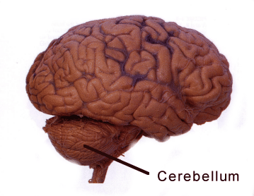
A 2008 review by Hess (PDF) concludes that compared with other neurological disorders, “our understanding of the biology and potential treatments for dystonia is in its infancy.” Still, scientists have known for a while that the cerebellum, a region of the brain that regulates movement, is involved.

“We focused on the cerebellum because studies in patients with dystonia often show that this part of the brain is more active, when examined by MRI,” Hess says. “The abnormal overactivity of the cerebellum is seen in patients with all different types of dystonia, so it seems to be a common hotspot. Our goal was to understand what might be causing the overactivity in mice because if we can stop the overactivity, we might be able to stop the dystonia.”

Hess and her colleagues discovered that drugs that stimulate AMPA receptors induce dystonia when introduced into the mouse cerebellum. Their results suggest that drugs that act in reverse, blocking AMPA receptors, could be used to treat dystonia.
Postdoctoral fellow Xueliang Fan is the first author of the paper. Emory neurologist H.A. Jinnah, director of a NIH-supported network of clinical research sites focusing on dystonia, is a co-author.

AMPA receptors are a subset of glutamate receptors, a large group of “receiver dishes” for excitatory signals in the brain. Fan performed a variety of experiments to show that AMPA receptor activity plays a specific role in generating dystonia. For example, drugs that affect other types of glutamate receptors did not induce dystonia. AMPA receptor blockers can also reduce dystonia in a genetic model, the “tottering” mouse.

Although pharmaceutical companies have already been testing AMPA receptor blockers as potential antiseizure drugs, caution is in order. AMPA receptor stimulators/ enhancers (or “ampakines”) have been identified as potential enhancers of learning and memory, so AMPA receptor blockers may interfere with those processes.

“Our results suggest that reducing AMPA receptor activity could help alleviate dystonia but we still have a lot of work to do before we know whether blocking AMPA receptor activity in patients will really help,” Hess says. “Since there aren’t many drugs that act at AMPA receptors, one of our goals is to identify drugs that change the ‘downstream’ effects of AMPA receptor activation. For example, we may be able to find other drug classes that change neuronal activity in the same way that AMPA receptor blockade changes activity.”

Posted on by Quinn Eastman in Neuro Leave a comment

mTOR inhibitors gaining favor for breast cancer treatment

This week, breast cancer researchers have been reporting encouraging clinical trial results with the drug everolimus at the San Antonio Breast Cancer Symposium. Everolimus is a mTOR inhibitor, first approved by the FDA for treatment of kidney cancer and then for post-transplant control of the immune system.

Ruth O’Regan, MD, director of the Translational Breast Cancer Research Program at Winship Cancer Institute, has led clinical studies of everolimus in breast cancer and has championed the strategy of combining mTOR inhibitors with current treatments for breast cancer.

She recently explained the rationale to the NCI Cancer Bulletin:

She views the combination therapy as a potential alternative to chemotherapy for treating ER-positive advanced breast cancer when hormonal therapies have stopped working.

When resistance to hormonal therapies occurs, Dr. O’Regan explained, additional signaling pathways become activated. Unlike chemotherapy, which targets rapidly dividing cells, mTOR inhibitors are an example of the kind of treatment that may block growth-promoting signaling pathways.

Currently, Winship researchers are examining a combination involving everolimus and the EGFR inhibitor lapatinib for “triple-negative” breast cancer, a particularly aggressive and difficult-to-treat variety.

Posted on by Quinn Eastman in Cancer Leave a comment

DNA copying problems explain muscular dystrophy mutations

Geneticist Madhuri Hegde and her colleagues have a paper in the journal Genome Researchthat addresses the question: where do copy number variations come from?

Madhuri Hegde, PhD

Copy number variations (CNVs), which are deletions or duplications of small parts of the genome, have been the subject of genetic research for a long time. But only in the last few years has it become clear that copy number variations are where the action is for complex diseases such as autism and schizophrenia. Geneticists studying these diseases are shifting their focus from short, common mutations (often, single nucleotide polymorphisms or SNPs) to looking at rarer variants such as CNVs. A 2009 discussion of this trend with Steve Warren and Brad Pearce can be found here.

Hegde is the Scientific Director of the Department of Human Genetics’ clinical laboratory. Postdoctoral fellow Arun Ankala is the first author. In the new paper, Ankala and Hegde examine rearrangements in patients’ genomes that arose in 62 clinical cases of Duchenne’s muscular dystrophy and several other diseases. Mutations in the DMD gene are responsible for Duchenne’s muscular dystrophy.

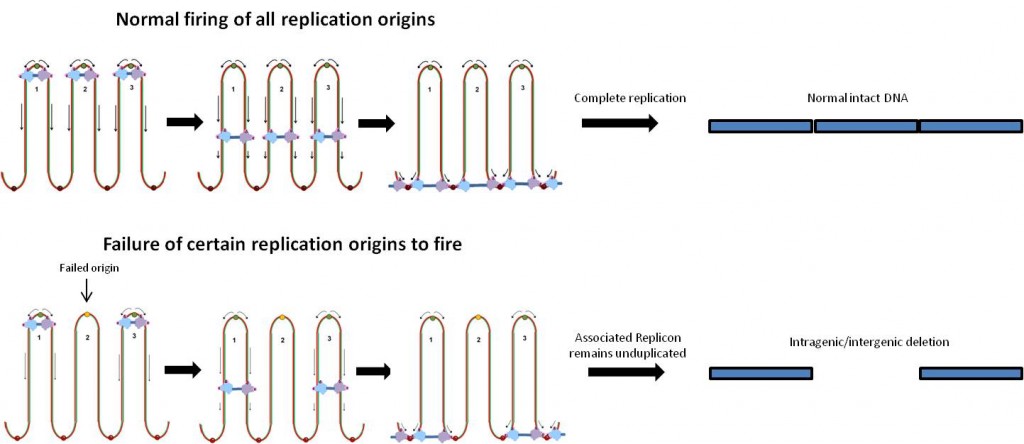
The pattern of the rearrangement hints what events took place in the cell beforehand, and hint that a problem took place during replication of the DNA. The signature is a tandem duplication of a short segment next to a large deletion, indicating how the DNA was repaired.

The authors note that the DMD locus is especially prone to these types of problems because it is much larger than other gene loci. The gene is actually the longest human gene known on the DNA level, covering 2.4 megabases (0.08 percent of the genome.)

Replication origins are where the DNA copying machinery in the cell starts unwinding and copying the DNA. Bacterial circular chromosomes have just one replication origin. In contrast, humans have thousands of replication origins spread across our chromosomes. In the discussion, the authors suggest that DNA copying problems may also explain duplications and historically embedded rearrangements of the genome.

Posted on by Quinn Eastman in Neuro Leave a comment

A twist on epigenetic therapy vs cancer

Epigenetic therapies against cancer have attracted considerable attention in recent years. But many of the drugs currently being studied as epigenetic anticancer therapies may have indiscriminate effects. A recent paper in Cancer Research from brain cancer researcher Erwin Van Meir’s laboratory highlights a different type of target within cancer cells that may be more selective. Postdoctoral fellow Dan Zhu is the first author of the paper.

Erwin Van Meir, PhD

The basic idea for epigenetic therapy is to focus on how cancer cells’ DNA is wrapped instead of the mutations in the DNA. Cancer cells often have aberrant patterns of methylation or chromatin modifications. Methylation is a punctuation-like modification of DNA that usually shuts genes off, and chromatin is the term describing DNA when it is clothed by proteins such as histones, a form of packaging that determines whether a gene is on or off.

In contrast to mutations that are hard-wired in the DNA, changes in cancer cells’ methylation or chromatin may be reversible with certain drug treatments. But a puzzle remains: if a drug wipes away methylation indiscriminately, that might turn on an oncogene just as much as it might restore a tumor suppressor gene.

The ability of an inhibitor of methylation to treat cancer may depend on cell type and context, explains chromatin/methylation expert and co-author Paula Vertino. She points out that one well-known methylation inhibitor, azacytidine (Vidaza), is a standard treatment for myelodysplastic syndrome, but the strategy of blanket-inhibition of methylation can’t be expected to work for all cancers. A similar challenge exists for agents that target histone acetylation in a global fashion.

Epigenetic therapies seek to modify how DNA is packaged in the cell.

Van Meir’s laboratory has been studying a tumor suppressor protein called BAI1 (brain angiogenesis inhibitor 1), which prevents tumor and blood vessel growth. BAI1 is produced by brain cells naturally, but is often silenced epigenetically in glioblastoma cells. His team found that azacytidine de-represses the BAI1 gene.

Methylation won’t turn a gene off without the help of a set of proteins that bind preferentially to methylated DNA. These proteins are what recognize the methylation state of a given gene and recruit repressive chromatin. Zhu and colleagues in Van Meir’s group found that one particular methyl-binding protein, MBD2, is overproduced in glioblastoma and is enriched on the BAI1 gene.

“Taken together, our results suggest that MBD2 overexpression during gliomagenesis may drive tumor growth by suppressing the anti-angiogenic activity of a key tumor suppressor. These findings have therapeutic implications since inhibiting MBD2 could offer a strategy to reactivate BAI1 and suppress glioma pathobiology,” the authors write.

By itself, MBD2 appears to be dispensable, since mice seem to be able to develop and survive without it. Not having it even seems to push back against tumor formation in the intestine, for example. Targeting MBD2 may represent an alternative way to steer away from cancer cells’ altered state.

Van Meir cautions: “We need to have a better understanding of all the genes that are turned on or off by silencing MBD2 in a given cancer before we can envision to use this approach for therapy.”

Vertino and Steven Hunter, both at Emory, are co-authors on the paper. The work was supported by grants from the NIH and the Southeastern Brain Tumor Foundation and the Emory University Research Council.

Posted on by Quinn Eastman in Cancer 1 Comment

Scientists identify trigger for glowing plankton

Have you ever waded or paddled through ocean water in dim light, and found that your actions caused the water to light up?

Susan Smith, PhD

Single-celled plankton called dinoflagellates are responsible for this phenomenon. Almost 40 years ago, scientists studying bioluminescence (light emitted by living things) proposed a mechanism by which physical deformation of the cell could lead to a trigger of the flash.

Susan M.E. Smith, a research assistant professor in David Lambeth’s laboratory in Emory’s Department of Pathology and Laboratory Medicine, recently was first author on a paper in PNAS identifying a molecule that scientists have long believed to be the key to this mechanism. The paper is the result of a collaboration with Tom DeCoursey’s laboratory at Rush University in Chicago.

The mechanism for the trigger, first envisioned by co-author Woody Hastings, works like this. It is known that acidic conditions activate luciferase, the enzyme that generates the light. Part of the dinoflagellate cell, the vacuole, is about as acidic as orange juice. Normally the acidity within the vacuole is kept separate from the luciferase, which is found in pockets on the outside of the vacuole called scintillons.

Proton channels are needed to trigger bioluminescence. Illustration courtesy of the National Science Foundation, which supported Smith's research

Now something is needed to let acidity (that is, protons) pass from the vacuole to the scintillons. That something is a proton channel: a protein that acts as a gate in the membrane, opening in response to electrical changes in the cell. Smith and her collaborators identified a proton channel called kHV1 that has unique properties: it lets protons flow in the right direction for the trigger to work! They studied kHV1 by inserting the dinoflagellate gene that encodes it into mammalian cells and probing its electrochemical properties, which are distinct from other proton channels.

The authors write: “Whereas other proton channels apparently evolved to extrude acid from cells, kHV1 seems to be optimized to enable proton influx.”

The gene they found actually comes from a type of dinoflagellate that does not flash: K. veneficum, which feeds on algae and sometimes forms harmful blooms that kill fish. They propose that it uses acid influx to aid in capturing or digesting its prey.

“Hastings’ prediction led us to look for this kind of channel, we found it in a related organism, and it had the right properties to fit the prediction,” Smith says, and adds that her team has since found a similar gene in flashing dinoflagellates. She says studying the proton channel may give clues to ways to control harmful dinoflagellates, as well as help scientists understand how plankton respond to greater ocean acidity.

Proton channels are found in humans too. In fact, the same kind of molecule that triggers plankton flashing in the ocean helps human white blood cells produce a bacteria-killing burst of bleach. They are also involved in allergic reactions and in sperm maturation.

Smith is co-author on a paper that is in the journal Nature this week, exploring the selectivity of the human version of kHV1. Smith says that her interest in proton channels grew out of her work on Nox enzymes (which produce the bacteria-killing bleach) with Lambeth.

“I got interested in the proton channel because its function is necessary for peak Nox performance in human phagocytes. We started a little side project on the human proton channel that kind of blossomed,” she says. Her collaboration with DeCoursey uses “evolutionary information to get at the function of these channels in general.”

Posted on by Quinn Eastman in Uncategorized 2 Comments

COX-2 and epilepsy: it’s complicated

How much is the development of epilepsy like arthritis?

More than you might expect. Inflammation, or the overactivation of the immune system, appears to be involved in both. In addition, for both diseases, inhibiting the enzyme COX-2 initially looked like a promising approach.

Ray Dingledine, PhD

COX-2 (cyclooxygenase 2) is a target of traditional non-steroid anti-inflammatory drugs like aspirin and ibuprofen, as well as more selective drugs such as Celebrex. With arthritis, selectively inhibiting COX-2 relieves pain and inflammation, but turns out to have the side effect of increasing the risk of heart attack and stroke.

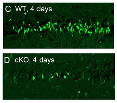
In the development of epilepsy, inhibiting COX-2 turns out to be complicated as well. Ray Dingledine, chair of pharmacology at Emory, and colleagues have a new paper showing that COX-2 has both protective and harmful effects in mice after status epilepticus, depending on the timing and what cells the enzyme comes from. Status epilepticus is a period of continuous seizures leading to neurodegeneration, used as a model for the development of epilepsy.

Postdoc Geidy Serrano, now at the Banner Sun Health Research Institute in Arizona, is first author of the paper in Journal of Neuroscience. She and Dingledine were able to dissect COX-2’s effects because they engineered mice to have a deletion of the COX-2 gene, but only in some parts of the brain.
They show that deleting COX-2 in the brain reduces the level of inflammatory molecules produced by neurons, but this is the reverse effect of deleting it all over the body or inhibiting the enzyme with drugs.

Four days after status epilepticus, fewer neurons are damaged (bright green) in the neuronal COX-2 knockout mice.

Dingledine identified two take-home messages from the paper:
First, COX-2 itself is probably not a good target for antiepileptic therapy, and it may be better to go downstream, to prostaglandin receptors like EP2.
Second, the timing of intervention will be important, because the same enzyme has opposing actions a few hours after status epilepticus compared to a couple days later.

More of Dingledine’s thinking about inflammation in the development of epilepsy can be found in a recent review.

Posted on by Quinn Eastman in Neuro Leave a comment

Drug discovery: shifting from brain growth factors to insulin

Earlier this year, the FDA put limitations on some anti-diabetic drugs because of their cardiovascular risks. The prevalence of diabetes in the United States continues to increase and is now above 8 percent of the population, so the need for effective therapies remains strong.

Keqiang Ye, PhD

Pathologist Keqiang Ye and colleagues have a paper in the Journal of Biological Chemistry describing their identification of a compound that mimics the action of insulin. This could be the starting point for developing new anti-diabetes drugs.

The new research is an extension of the Ye laboratory’s work on TrkA and TrkB, which are important for the response of neurons to growth factors. Ye and Sung-Wuk Jang, a remarkably productive postdoc who is now an assistant professor at Korea University, developed an assay that allowed them to screen drug libraries for compounds that directly activate TrkA and TrkB. This led them to find a family of growth-factor-mimicking compounds that could treat conditions such as Parkinson’s disease, depression and stroke.

Since TrkA/B and the insulin receptor are basically the same kind of molecule — receptor tyrosine kinases– and use some of the same cellular circuitry, Ye and Jang’s assay could also be used with the insulin receptor. Kunyan He and Chi-Bun Chan are the first two authors on the new paper. They report that the compound DDN can make cells more sensitive to insulin and improve their ability to take up glucose. They show that DDN (5,8-diacetyloxy-2,3-dichloro-1,4- naphthoquinone) can lower blood sugar, both in standard laboratory mice and in obese mice that serve as a model for type II diabetes.

Ye reports that he and his colleagues are working with medicinal chemists to identify related compounds that may have improved efficacy and potency.

“I hope in the near future we may have something that could replace insulin for treating diabetes orally,” he says.

Posted on by Quinn Eastman in Uncategorized Leave a comment

Esophageal lesions meet their match

Field Willingham, MD, MPH

Once esophageal tumors establish themselves, a patient’s prognosis is grim and morbidity vast. But when lesions are caught early and removed, especially in the premalignant stage, the odds of survival markedly improve.

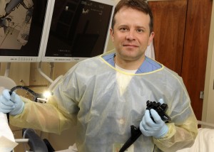
When a case calls for it, Emory gastroenterologist Field F. Willingham, MD, MPH, uses a hybrid approach to ousting superficial esophageal lesions. Superficial esophageal lesions are commonly caused by acid reflux disease, or GERD. GERD occurs when stomach acid flows into the esophagus and can lead to a condition known as Barrett’s esophagus, where the cells in the lower esophagus become damaged. This in turn can lead to dysplasia, or pre-cancerous cells.

But for superficial cancers, it is now possible to remove a portion of the lining layer of the GI tract, containing the tumor, with an endoscope.  This can help carefully selected patients avoid a major surgery. The technique, known as an EMR, allows the removal of superficial esophageal tumors and pre-cancer with an endoscope, a slender tube-like instrument.

Detecting and removing esophageal tumors early is essential for a favorable outcome. Once tumors firmly establish themselves in esophageal tissue, the prognosis is grim and morbidity vast. In the past, a diagnosis of an esophageal tumor meant the removal of the esophagus and often the stomach. But now EMR can be used in tandem with radio frequency ablation.

In surgical situations in which radio frequency ablation is not feasible, Willingham and his colleagues are beginning to use an alternate technique, known as cryotherpay, in tandem with EMR. Cryotherapy involves freezing superficial cells to rid the esophagus of suspect cells.

“So, if the end of the esophagus is twisted, or if we can’t touch it with this balloon device, then we can use cryotherapy,” says Willingham. “We’re trying to kill the lining layer with the tumor cells without killing the deeper layer.”

Willingham and his colleagues are seeing evidence that using these very three very different, technologies in tandem or alone will provide patients with a better way to rid them of esophageal lesions while preserving their quality of life.

Posted on by admin in Uncategorized Leave a comment

Genetic alteration opens door to targeted treatment of rare tumor

A cross section of an epithelioid hemangioendothelioma

Emory pathologist Sharon Weiss, MD, was the first to describe an extraordinarily rare tumor known as an epithelioid hemangioendothelioma (EHE). Thirty years later, researchers have identified a genetic alteration linked to this odd vascular tumor.

It’s hoped this newfound information will lead to a better understanding of the mechanisms underlying the development of this tumor and hence development of a targeted treatment. None yet is available. However, these findings already have been used to develop a new diagnostic test for this blood vessel disease.

The research, published in a recent issue of Science Translational Medicine, was done in collaboration with Cleveland Clinic’s Taussig Cancer Institute and led by Brian Rubin, MD, PhD, of Cleveland Clinic’s Pathology and Laboratory Medicine Institute and Lerner Research Institute.

The genetic alteration formerly in question involves a translocation between chromosomes 1 and 3, where chromosomes 1 and 3 exchange DNA fragments that are transposed onto opposite chromosomes. The result: the swapped DNA encodes a unique, fused gene that contains components from each chromosome. Because genes are translated into proteins, the result of this unique gene is a correspondingly unique protein, one thought to cause cancer.

Epithelioid hemangioendotheliomas comprise less than one percent of all cancers. Roughly 100 new cases are diagnosed in the United State each year. EHE are eccentric in their epidemiology, structure and aggressiveness. Slow to metastasize, they tend to occur in both young men and women when soft tissue is involved but occur mostly in women when the liver and lungs are affected.

However, it’s their peculiar structure that has so far made targeted treatment problematic, especially in the liver and lungs. “Instead of being one mass as you might expect with liver cancer, the patient with EHE often presents with little nodules throughout the liver,” says Weiss.

“The reason this occurs is that the growth starts in the liver’s portal vein, grows along its length, and then tracks out through the vessels. The growths blister out from the vessel creating these little nodules. Epithelioid hemangioendothelioma don’t possess the classic features of vascular tumors. In fact, EHE may have so many sites of involvement that the cancer can’t be cured, short of transplantation.”

Using EHE tissue samples gleaned from Weiss’s vast library, Rubin developed a genetic probe to detect the distinct chromosomal translocations in the tumor. The probe now serves as a powerful diagnostic tool of EHE and opens the door to understanding these tumors’ mechanisms.

“Once you understand the mechanism behind it, you can start trying to target those pathways in a therapeutic way,” says Weiss.

Posted on by admin in Cancer 1 Comment