People touched by a brain tumor — patients, their families or friends — may have heard of the drug Gliolan or 5-ALA, which is taken up preferentially by tumor cells and makes them fluorescent. The idea behind it is straightforward: if the neurosurgeon can see the tumor’s boundaries better during surgery, he or she can excise it more thoroughly and accurately.
5-ALA is approved for use in Europe but is still undergoing evaluation by the U.S. FDA. A team at Emory was the first to test this drug in the United States. [Note: A similar approach, based on protease activation of a fluorescent probe, was reported last week in Science Translational Medicine.]

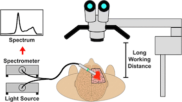
A hand-held device to detect glowing brain tumors could allow closer access to the critical area than a surgical microscope
Biomedical engineer Shuming Nie and colleagues recently described their development of a hand-held spectroscopic device for viewing fluorescent brain tumors. This presents a contrast with the current tool, a surgical microscope — see figure.
Nie’s team tested their technology on specimens obtained from cancer surgeries. Their paper in Analytical Chemistry reports:
The results indicate that intraoperative spectroscopy is at least 3 orders of magnitude more sensitive than the current surgical microscopes, allowing ultrasensitive detection of as few as 1000 tumor cells.
Based on this work, Nie reports plans to conduct a clinical trial next year with neurosurgeon colleagues at Emory and Mount Sinai. Studies using standard surgical microscopes will continue.
Nie and his team have also been developing a similar tool for viewing lung tumors, made fluorescent with a green dye. Nie is a consultant for Spectropath, an Atlanta-based company that is seeking to develop this technology as a commercial product.

