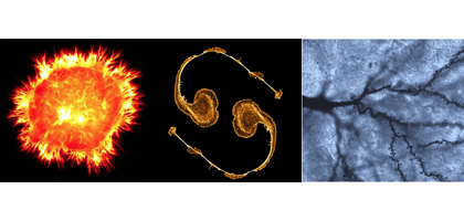
Lab Land’s editor enjoyed talking with several students about their work at the GDBBS Student Research Symposium last week. Neurons dominate the three contest-winning images. The Integrated Cellular Imaging core facility judged the winners. From left to right:

1st Place: Stephanie Pollitt, Neuroscience
2nd Place: Amanda York, Biochemistry, Cell and Developmental Biology
3rd Place: Jadiel Wasson, Biochemistry, Cell and Developmental Biology
Larger versions and explanations below.
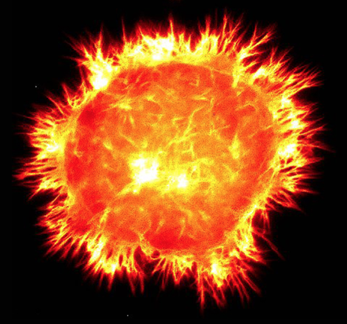
1st Place: Stephanie Pollitt, Neuroscience
Actin cytoskeleton within a mouse neuroblastoma cell, from James Zheng’s lab.
2nd Place: Amanda York, Biochemistry, Cell and Developmental Biology
Actin dynamics within a neuron. Also from Zheng lab.
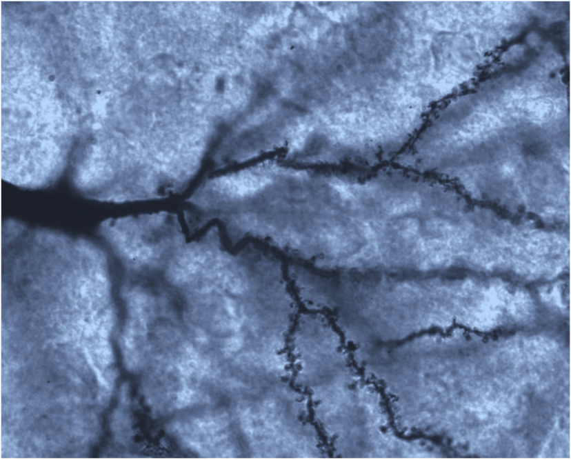
3rd Place: Jadiel Wasson, Biochemistry, Cell and Developmental Biology. From David Katz’s lab.
This image is of a medium spiny neuron in the brain of an adult mouse.  The brain was processed via the classic Golgi-Cox stain which stains selective neurons with heavy metal precipitate.  This allows all of the projections, including dendrites and axons, and also dendritic spines, the tiny mushroom-like projections emanating from the dendrites, to be highlighted, revealing detailed information about neuronal structure.


