Quinn Eastman

Antibody production: an endurance sport

Antibodies defend us against infections, so they often get described as weapons. And the cells that produce them could be weapon factories?. To understand recent research from immunologist Jerry Boss’s lab, a more appropriate metaphor is the distinction between sprinting and long-distance running.

Graduate student Madeline Price in Boss’s lab has been investigating how antibody-producing cells use glucose – the simple sugar– and how the cells’ patterns of gene activity reflect that usage. Cells can use glycolysis, which is inefficient but fast, analogous to sprinting, or oxidative phosphorylation, generating much more energy overall, more like long distance running.

As Boss and Price point out:

Immunology + Molecular Pathogenesis graduate student Madeline Price

Glycolytic metabolism produces 2 molecules of ATP per molecule of glucose, while oxidative phosphorylation produces 36 molecules of ATP from the same starting glucose molecule. Where oxidative phosphorylation generates more energy from ATP, glycolysis generates metabolic intermediates that are also useful for rapid cellular proliferation.

In their recent paper in Cell Reports, they lay out what happens to B cells, which can go on to become antibody secreting cells (ASCs), after an initial encounter with bacteria. The B cells first proliferate and upregulate both glycolysis and oxidative phosphorylation. However, upon differentiating, the cells shift their preference to oxidative phosphorylation. Read more

Posted on by Quinn Eastman in Immunology Leave a comment

Focus on antibiotic resistance at ASM Microbe 2018

We are excited that the ASM Microbe meeting will be at the Georgia World Congress Center from June 7 to June 11. If you are interested in antibiotic resistance, you can learn about how to detect it, how to (possibly) defeat it and how the bacteria fight back.

A host of Emory microbiologists are participating. In some cases, our scientists are presenting their unpublished data for discussion with their colleagues at other universities. Accordingly, we are not going to spill the beans on those results. However, please find below some examples of who’s talking and a bit of explanatory background. ASM Microbe abstracts are available online for posters, but not for some symposiums and plenary talks.

David Weiss labKlebsiella

Graduate student Jessie Wozniak is presenting her research on an isolate of Klebsiella that combines alarming properties. She will describe how the bacterial colonies behave (unappetizingly) like stretchy melted cheese in a “string test.”

June 9, 11 am to 1 pm, June 11, 11 am to 1 pm

Christine Dunham – toxin-antitoxin/persistence

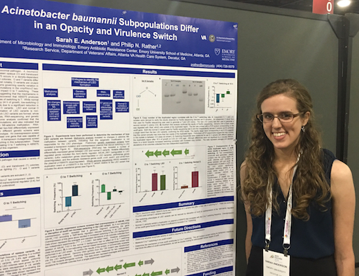
Graduate student Sarah Anderson presenting her poster at ASM Microbe. She discussed a genetic connection between virulence switch and antibiotic resistance.

Dunham, a structural biologist, is giving a plenary talk June 11 on toxin-antitoxin pairs, which play a role in regulating bacterial persistence, a dormant state that facilitates antibiotic resistance. Two past papers from her lab.

Phil Rather labAcinetobacter baumannii

Rather’s lab recently published a Nature Microbiology paper on A. baumannii’s virulence/opacity switch. This type of bacteria is known for hospital-associated infections and for wound infections in military personnel. Poster talk by graduate student Sarah Anderson June 8. Read more

Posted on by Quinn Eastman in Uncategorized Leave a comment

Less mucus, more neutrophils: alternative view of CF

A conventional view of cystic fibrosis (CF) and its effects on the lungs is that it’s all about mucus. The inherited disease leads to an accumulation of mucus in the lungs, which appears to be connected with inflammation, susceptibility to infection and loss of lung capacity.

Immunologist Rabin Tirouvanziam has an alternative view, centered on neutrophils. They are a type of immune cell that is very numerous, yet often overlooked, he says.

Rabindra Tirouvanziam, PhD

A new paper, published in Journal of Leukocyte Biology, substantiates his ideas about cystic fibrosis and harnesses them for future diagnostic and therapeutic advances. Tirouvanziam is an assistant professor of pediatrics at Emory University School of Medicine and Emory Children’s Center. He and his colleagues have developed a system for studying neutrophil behavior in a specialized culture, a model of a cell layer in the lung.

Neutrophils behave differently in the diseased lung environment, compared with when they are in the blood. The culture system makes the neutrophils pass through a layer of lung cells, under the influence of lung fluids obtained from CF patients. The culture system opens up the opportunity of testing fluids from patients to mark disease progression, as well as drug discovery: looking for compounds that could deprogram the neutrophils. Read more

Posted on by Quinn Eastman in Immunology Leave a comment

Blue plate special: express delivery to the heart

The anti-arrhythmia drug amiodarone is often prescribed for control of atrial fibrillation, but can have toxic effects upon the lungs, eyes, thyroid and liver. Emory and Georgia Tech scientists have developed a method for delivering amiodarone directly to the heart in an extended release gel to reduce off-target effects.

The results were published in Circulation: Arrhythmia and Electrophysiology.

The senior author is Rebecca Levit, MD, assistant professor of medicine (cardiology) at Emory University School of Medicine and adjunct in the Wallace H. Coulter Department of Biomedical Engineering at Georgia Tech and Emory. Graduate student Jose Garcia – part of co-author Andres Garcia’s lab at Georgia Tech — and Peter Campbell, MD are the first authors.

An amiodarone-containing gel was applied to the outside of the heart by a minimally invasive procedure. After a one-time delivery, the gel could reduce the duration of atrial fibrillation and the likelihood of its development for a month in a pig model. The researchers were also able to show that amiodarone did not have toxic effects on the pigs’ lungs.

As noted in the book Off-label prescribing – Justifying unapproved medicine, amiodarone is “one of the very few drugs approved by the FDA in modern times without rigorous randomized clinical trials.” Read more

Posted on by Quinn Eastman in Heart Leave a comment

Inflammation in PD hits the gut

Several groups studying Parkinson’s have had a hunch – a gut feeling, even – that intestinal inflammation is involved in driving the disease. Now Emory researchers led by Malu Tansey, PhD have some evidence from patient samples to back it up, published in the journal Movement Disorders.

IMP graduate student Madelyn Houser

German pathologist Heiko Braak has been honored by the Michael J. Fox Foundation for Parkinson’s Research for his theory, originally published in 2003, proposing that disease pathology – marked by aggregation of the toxic protein alpha-synuclein — may begin in the gastrointestinal tract and migrate from there to the central nervous system. This proposal was both provocative and influential in the Parkinson’s disease (PD) field. And Tansey herself has long been interested in the role of microglia, the immune cells resident in the brain, in PD.

The first author of the new paper, Immunology and Molecular Pathogenesis graduate student Madelyn Houser, notes that digestive problems such as constipation are frequently reported in PD patients. But what is the cause and what is effect? As neurologist Stewart Factor observed for a Emory Medicine article on PD’s non-motor symptoms: “A patient might tell me he’s had recurring constipation for 10 years, but he wouldn’t say anything to a neurologist about it until he starts having other symptoms.” Read more

Posted on by Quinn Eastman in Immunology, Neuro Leave a comment

Fermentation byproduct suppresses seizures in nerve agent poisoning

A compound found in trace amounts in alcoholic beverages is more effective at combating seizures in rats exposed to an organophosphate nerve agent than the current recommended treatment, according to new research published in eNeuro.

This work comes from Asheebo Rojas, Ray Dingledine and colleagues in Emory’s Department of Pharmacology. Just as an aside, we don’t know the nature of the recent alleged chemical attack in Syria, and the chemical used in the Emory experiments is not a “weaponized” nerve agent such as Sarin. Organophosphates were also widely used as insecticides, but their use has been declining.

Left untreated, organophosphate poisoning can lead to severe breathing and heart complications, because of the inhibition of acetylcholinesterase. It also causes seizures. Some patients are resistant to treatment with the anti-anxiety drug diazepam (Valium), a standard first-line treatment for such poisoning, and its effectiveness decreases the longer the seizure lasts.

The researchers compared the ability of two treatments — diazepam and the anesthetic urethane (ethyl carbamate), commonly formed in trace amounts during fermentation of beer and wine from the reaction of urea and ethanol — to interrupt seizures in rats exposed to the organophosphate diisopropyl fluorophosphate. The researchers found urethane to be more effective than diazepam, suppressing seizures for multiple days and accelerating recovery of weight lost while protecting the rats from cell loss in the hippocampus.

Urethane/ethyl carbamate is a carcinogen in animals, which led to concerns over its presence in alcoholic beverages in the 1980s. It was also used as a sedative for many years in Japan. The researchers did not observe any evidence of lung tumors in the urethane-treated animals seven months later, suggesting that the dose used in this study is not carcinogenic. The findings point to urethane or a derivative as a potential therapeutic for preventing organophosphate-triggered seizures from developing into epilepsy. Read more

Posted on by Quinn Eastman in Neuro Leave a comment

Post-anesthetic inertia in IH

A recent paper from neurologists Lynn Marie Trotti and Donald Bliwise, with anesthesiologist Paul Garcia, substantiates a phenomenon discussed anecdotally in the idiopathic hypersomnia (IH) community. Let’s call it “post-anesthetic inertia.” People with IH say that undergoing general anesthesia made their sleepiness or disrupted sleep-wake cycles worse, sometimes for days or weeks. This finding is intriguing because it points toward a trigger mechanism for IH. And it pushes anesthesiologists to take IH diagnoses into account when planning patient care, just as is already done for myotonic dystrophy.

Lab Land obtained some confirmation from a couple IHers. One woman had surgery a couple of months ago and felt like the anesthetic was still in her system for weeks and she still didn’t feel right. Another reported “severe insomnia for months and it felt like every body system was completely scrambled.”

Where does this all come from? People with IH getting together and telling their stories. Journalist Virginia Hughes described a moment at the 2014 patient-organized IH meeting in Atlanta in her article “Wake No More”:

Andy Jenkins, the neuroscientist who developed the spinal fluid test, gave an impressively entertaining lecture on GABA receptors. “Why do we have more GABA activity?” somebody asked. Nobody knows, said Jenkins. One idea is that it’s triggered by anesthesia. Lloyd [Johnson, a meeting organizer from Australia] asked the audience how many of them believed their hypersomnia was the result of anesthesia. About one-quarter of the hands went up. “Whoa, whoa, whoa, whoa, whoa,” Jenkins said as he watched the sleepyheads* come alive.

The new paper, in Frontiers in Human Neuroscience, is more quantitative than that informal show of hands. In a way, it begins to question the basis for the term idiopathic hypersomnia, since idiopathic means “arising spontaneously or having no cause”. For some people surveyed in the paper, anesthesia was an exacerbating factor, if not the only factor.

Confusion or agitation post-anesthesia can happen in people who don’t have sleep disorders. What’s peculiar to the hypersomnolent group is how long sleepiness or disrupted sleep-wake cycles last — long after the anesthetic has left the body. The hypersomnolent group was mostly people with IH or narcolepsy type 2 (30 plus 15 out of 57). In the paper, people with restless legs syndrome were used as controls:

While patients in both groups were equally likely to report surgical complications and difficulty awakening from anesthesia, hypersomnolent patients were more likely to report worsened sleepiness (40% of the hypersomnolent group vs. 11% of the RLS group, p = 0.001) and worsening of their sleep disorder symptoms (40% of the hypersomnolent group vs. 9% of the RLS group, p = 0.0001).

Hypersomnolent patients who perceived their symptoms to worsen reported that symptoms had never returned to baseline in 66.7%, took months or years to return to baseline in 9.5%, and resolved in days to weeks in 23.8%.

Note: first author Vincent LaBarbera is now a neurology resident at Brown.

Mechanistic speculation

Several years ago, Emory researchers found that some IH patients appear to have a substance in their cerebrospinal fluid that acts similarly (but not quite the same) as a benzodiazepine drug. This still-mysterious substance enhances signaling by GABA, the major inhibitory neurotransmitter.

Inhaled anesthetics such as sevoflurane, as well as the injected anesthetic propofol, act by enhancing GABA too. So when someone undergoes general anesthesia, their GABA receptors are being pushed hard for an extended period of time. GABA signaling has a kind of global “dimmer switch” function as well as working through specific circuits in the brain to bring on anesthesia.

GABA receptors are complex, but they usually adjust to pressure. It explains development of tolerance to benzodiazepine drugs. GABA receptors also modulate in response to alcohol or women’s menstrual cycles (certain derivatives of progesterone, so-called “neurosteroids,” act on them). What may be happening after anesthesia is that GABA receptors of people with IH have trouble adjusting back, or may overshoot, perhaps because their internal clocks are less resilient.

The Emory authors conclude:

Because the half-life of anesthetic agents is generally short, any prolonged worsening of sleepiness post-procedure cannot easily be attributed to immediate GABA-mediated effects. Whether the putative long-term changes in hypersomnolence that we are detecting in our patients’ reports may be related to changes in GABA-related neural circuitry caused by anesthetic neurotoxicity or other mechanisms remains to be determined.

A similar interaction, with reversed polarity, may be occurring in post-partum depression.

*Lab Land has been told that sleepyhead is not a fully accepted term in the IH community.

Posted on by Quinn Eastman in Neuro Leave a comment

How much does idiopathic hypersomnia overlap with ME/CFS?

In everyday linguistic usage among non-specialists, sleepiness can blend together with tiredness and fatigue. Someone might feel “tired” after climbing a mountain or chopping down a tree, while “sleepiness” is different. Emory sleep scientists explore the pathological distinctions in a paper published in Journal of Sleep Research.

A team led by neurologists Lynn Marie Trotti and David Rye has been studying idiopathic hypersomnia (IH) for several years: people who experience excessive daytime sleepiness and “sleep drunkenness,” not explained by other medical conditions.

IH’s symptoms don’t usually include persistent muscle pain or a severe response to exertion. This separates the disorder from myalgic encephalomyelitis, also known as chronic fatigue syndrome (ME/CFS). But there is some overlap, which is what neurology resident Caroline Maness, Trotti and colleagues report in the new paper. The authors use the official term SEID (systemic exertion intolerance disease), which was recommended by an Institute of Medicine panel in 2015, but hasn’t really stuck among those in the ME/CFS field.

Some people with IH have disclosed that they were previously diagnosed with ME/CFS. Outside of the sleepy vs tired issue, some people with IH report symptoms shared with ME/CFS, such as impaired circulation in their extremities in response to cold, or dizziness upon standing. Speculatively, this may point to a possible problem with the autonomic nervous system. Trotti and a collaborator at Stanford, Mitchell Miglis, are now examining this issue further.

ME/CFS has had a history of controversy. Despite its devastating impacts, some have viewed it as psychological or somehow unreal, and sufferers have felt neglected or maligned by mainstream medicine. The National Institutes of Health has made efforts to turn that situation around by investing in ME/CFS research, and there has been a surge of attention recently covering ME/CFS (Amy Maxmen items in Nature, Stanford magazine feature, Unrest documentary).

Trotti, Maness and colleagues didn’t set out to dive into ME/CFS – they explicitly label this paper a pilot study, and the results say more about the “hypersomnolent” group of patients they have been seeing for the last several years, rather than the broader ME/CFS population. Read more

Posted on by Quinn Eastman in Neuro Leave a comment

Duel of the inflammatory master regulators: insights for drug discovery

Anti-inflammatory drugs such as dexamethasone can have harmful side effects on the skin, bones and metabolism. Structural biology research from Emory University School of Medicine has implications for the long-standing quest to separate these drugs’ benefits from their side effects.

The findings were recently published in Nature Communications (open access).

Dexamethasone is a synthetic glucocorticoid hormone, used to treat conditions such as allergies, asthma, autoimmune diseases and cancer. It mimics the action of the natural hormone cortisol. Both cortisol and synthetic hormones act by binding the glucocorticoid receptor (GR) protein.

The market for synthetic glucocorticoid hormones, oral and topical, is estimated at $10 billion. Examples include dexamethasone, prednisone, and hydrocortisone. Yet these drugs might not be approved today, given the array of known side effects.

GR can bind DNA in two modes. At some sites, it pairs up or “dimerizes” – turning genes on. At others, it binds one at a time, turning genes off. For GR-targeting drugs, the side effects are thought to come from turning on genes involved in processes such as metabolism and bone growth, while the desired anti-inflammatory effects result mainly from turning inflammatory and immune system genes off.

In their new paper, Eric Ortlund, PhD, and colleagues report that GR’s ability to directly bind DNA extends more broadly than previously appreciated. The first author is Will Hudson, PhD, previously a graduate student with Ortlund and now a postdoctoral fellow in Rafi Ahmed’s lab at Emory Vaccine Center.

GR was known to interfere with another important family of DNA-binding proteins, master regulators of inflammation, which are together called NFkB. Ortlund’s team found that GR can directly bind one at a time to many of the same stretches of DNA that NFkB interacts with.

“This type of interaction, where GR is acting one at a time – we think it’s druggable,” says Ortlund, who is associate professor of biochemistry at Emory University School of Medicine. Read more

Posted on by Quinn Eastman in Heart, Immunology Leave a comment

What are rods and rings?

This image of mouse embryonic fibroblasts comes from Cara Schiavon, a graduate student in Rick Kahn’s lab in the Department of Biochemistry. It was impressive enough to capture interest from Emory Medicine‘s graphics designer Peta Westmaas. The light green shapes are “Rods and Rings,” structures that were identified just a few years ago by scientists studying how cells respond to antiviral drugs, such as those used against hepatitis C.

The rod and ring structures appear to contain enzymes that cells use for synthesizing DNA building blocks. Patients treated with some antiviral drugs develop antibodies against these enzymes.

The turquoise color represents microtubules, components of cells’ internal skeletons. The orange color shows DNA within nuclei. The spots in the nuclei are areas where DNA is more compact. The overall image is a “z-stack projection” acquired using the Olympus FV1000 confocal microscope in Emory’s Integrated Cellular Imaging Core.

Posted on by Quinn Eastman in Uncategorized Leave a comment