Quinn Eastman

Blood vessels aren’t straight tubes

For years, scientists like Hanjoong Jo have been telling us that blood vessels are like rivers and streams. Fluid dynamics are important; the patterns of curvature and current influence where sediment — or atherosclerosis — builds up.

One of the biggest possible perturbations of fluid dynamics in a blood vessel would be to stick a metal tube into it. Of course, cardiologists do this all the time. During percutaneous coronary intervention (PCI), doctors place a stent, basically a metal tube, inside a blood vessel to relieve an obstruction and restore blood flow to the heart muscle.

Habib Samady, Emory Healthcare’s director of interventional cardiology, is leading a clinical trial looking at the effects of stent introduction on blood vessels that are not straight, but curved or angulated. To be eligible for the study, the patient’s blocked vessel has to bend more than 30 degrees. The study will look at patients who have undergone PCI for a heart attack and follow them over the course of a year. Less “disturbed flow” should mean better heart healing for the patient down the road. The study uses OCT (optical coherence tomography) and IVUS (intravascular ultrasound) to monitor the blood vessel and see how healing is affected by fluid dynamics after stent placement. Read more

Posted on by Quinn Eastman in Heart Leave a comment

Targeting naked DNA in the heart

Hoechst-Structure-300px

The first thing that comes up in a Google search for “Hoechst” is the family of fluorescent dyes used to stain DNA in cells before microscopy. The Hoechst dyes derive their names from their manufacturer: a company, now part of Sanofi, named after the town where it was founded, which is now part of Frankfurt, Germany. The word itself means “highest [spot].”

Although DNA runs the show in every cell, it’s usually well-hidden inside the nucleus or the mitochondria. Extracellular DNA’s presence is a signal that injury is happening and cells are dying.

Biomedical engineer Mike Davis and collaborator Niren Murthy have been exploiting the properties of a DNA-binding dye called Hoechst 33342, often used to stain DNA in cells before microscopy. The dye can only bind DNA if it can get to the DNA – that is, if membranes are broken. This property makes the dye a good way to target injured tissue, either as an imaging agent or for therapy.

At the recent Pediatric Healthcare Innovation retreat, Davis discussed the potential use of such Hoechst derivatives to diagnose myocarditis (inflammation of the heart muscle) in children.

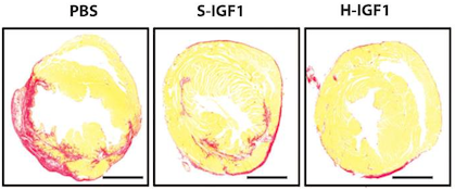
In addition, in a recent paper published in Scientific Reports, Davis and his colleagues attach the Hoechst dye to the cardioprotective growth factor IGF-1, creating a version of IGF-1 that is targeted to injured heart muscle. The first author of the paper is cardiology fellow Raffay Khan, MD. Screen Shot 2014-04-24 at 1.18.35 PM

IGF-1 has shown a lot of potential for treating heart disease, but it’s not the most cooperative as a drug, because it doesn’t last long in the body and doesn’t stick around in the heart. Linked up to the dye, IGF-1 behaves better. When used to treat mouse hearts after a heart attack, the Hoechst-IGF-1 treated-hearts have better function and less scar tissue (seen here as red).

The authors conclude:

With the broad chemistry surrounding functionalized PEG used to create Hoechst derivatives, it may be possible to target other therapies such as cells, small molecules, and even nanoparticles. We believe that the use of DNA binding agents such as Hoechst can be used to target exposed DNA in other diseases where necrotic cell death plays a critical role and could be used as a platform therapy.


Posted on by Quinn Eastman in Heart Leave a comment

Fine tuning an old-school chemotherapy drug

First approved by the FDA in the 1970s, the chemotherapy drug cisplatin and its relative carboplatin remain mainstays of treatment for lung, head and neck, testicular and ovarian cancer. However, cisplatin’s use is limited by its toxicity to the kidneys, ears and sensory nerves.

Paul Doetsch’s lab at Winship Cancer Institute has made some surprising discoveries about how cisplatin kills cells. By combining cisplatin with drugs that force cells to rely more on mitochondria, it may be possible to target it more specifically to cancer cells and/or reduce its toxicity.

Cisplatin emerged from a serendipitous discovery in the 1960s by a biophysicist examining the effects of electrical current on bacterial cell division. It wasn’t the current that stopped the bacteria from dividing  it was the platinum in the electrodes. According to Siddhartha Mukherjee’s book The Emperor of All Maladies, cisplatin became known as “cisflatten” in the 1970s and 1980s because of its nausea-inducing side effects.

Cisplatin is an old-school chemotherapy drug, in the sense that it’s a DNA-damaging agent with a simple structure. It doesn’t target cancer cells in some special way, it just grabs DNA with its metallic arms and holds on, forming crosslinks between DNA strands.

But how cisplatin kills cells is more complicated. Along with the direct effects of DNA damage, cisplatin unleashes a storm of reactive oxygen species.

“We wanted to know whether the reactive oxygen species induced by cisplatin had a driving role in cell death or was more of a byproduct,” says postdoc Rossella Marullo, who is the first author of a recent paper with Doestch in PLOS One.

One possible analogy: after the 1906 San Francisco earthquake, the fires were even more destructive than the initial shaking. When asked whether to think of the reactive oxygen species production triggered by cisplatin in the same way as the fires, Doetsch and Marullo say they wouldn’t go that far.

Still, they have uncovered a critical role for mitochondria, cells’ mini-power plants, in cisplatin cell toxicity. The researchers found that mitochondria are the source of cisplatin-induced reactive oxygen species in lung cancer cells. Cancer cell lines that lack functional mitochondria* are less sensitive to cisplatin, and cisplatin’s damage to the mitochondria may be even more important than the damage to DNA in the nucleus, the authors write. However, mitochondrial damage is not important for cisplatin’s less potent [but less toxic] cousin carboplatin.

Cancer cells tend to have a warped metabolism that makes them turn off their mitochondria. This is part of the “Warburg effect” (experts in this area: Winship’s Jing Chen and Malathy Shanmugam). Cancer cells have an increased uptake of sugar, but don’t break it down completely, and use the byproducts as building materials.

What if we could force cancer cells to rely on their mitochondria again, and at the same time, by giving them cisplatin, make that painful for them? This would make cisplatin even more toxic to cancer cells in particular.

The drug DCA (dichloroacetate), which can stimulate cancer cells to use their mitochondria, can also increase the toxicity of cisplatin, at least in cancer cell lines in the laboratory, Marullo and her colleagues show.

Doetsch and radiation oncologist Jonathan Beitler are in the process of planning a clinical trial combining DCA with cisplatin for HPV (human papillomavirus)-positive head and neck cancer. The trial would test whether it might be possible to use a lower dose of cisplatin, reducing toxicity, by combining it with DCA.

“We’ve relied on cisplatin’s efficacy for decades, without fully understanding the mechanism,” Beitler says. “With this new knowledge, it may be possible to manipulate cisplatin’s action so it is more effective and less toxic.”

The applicability of cisplatin and mitochondrial tuning may depend both on cancer cell type and metabolic state, Doetsch adds.

*Cell lines that lack mitochondrial DNA can be obtained by “pickling” them in ethidium bromide, a DNA intercalation agent.


Posted on by Quinn Eastman in Cancer Leave a comment

Addendum on CRISPR

An excellent example of the use of CRISPR gene editing technology came up at the Emory-Children’s Pediatric Research Center’s Innovation Conference this week.

Marcela Preininger, who is working with cardiomyocyte stem cell specialist Chunhui Xu, described her work (poster abstract 108) on cells derived from a 12 year old patient with an inherited cardiac arrhythmia syndrome: catecholaminergic polymorphic ventricular tachycardia or CPVT. Her team has obtained skin fibroblasts from the patient, and converted those cells into induced pluripotent stem cells, which can then be differentiated into cardiac muscle cells or cardiomyocytes.

Working with TJ Cradick, director of the Protein Engineering Facility at Georgia Tech, Preininger is testing out CRISPR gene editing as a means of correcting the defect in this patient’s cells, outside the body. Cradick says that while easy and efficient, RNA-directed CRISPR can be lower in specificity compared to the protein-directed TALEN technology.

From Preininger’s abstract:

Once the mutation has been corrected at the stem cell level, we will investigate whether the repaired (mutation-free) iPS cells can be differentiated into functional cardiomyocytes with normal Ca2+ handling properties, while closely monitoring the cells for mutagenic events. Pharmacological restoration of the normal myocardial phenotype will also be optimized and explored in our model.

Posted on by Quinn Eastman in Heart Leave a comment

A CRISPR way to edit DNA

The CRISPR/Cas gene editing system has a lot of buzz behind it: an amusingly crunchy name, an intriguing origin, and potential uses both in research labs and even in the clinic. We heard that Emory scientists are testing it, so an explainer was in order.

The CRISPR (Clustered Regularly Interspaced Short Palindromic Repeats) system was originally discovered by dairy industry researchers seeking to prevent phages, the viruses that infect bacteria, from ruining the cultures used to make cheese and yogurt. Bacteria incorporate small bits of DNA from phages into their CRISPR region and use that information to fight off the phages by chewing up their DNA.

At Emory, infectious disease specialist David Weiss has published research on CRISPR in some types of pathogenic bacteria, showing that they need parts of the CRISPR system to evade their hosts and stay infectious. Biologist Bruce Levin has modeled CRISPR-mediated immunity’s role in bacterial evolution.

What has attracted considerable attention recently is CRISPR/Cas-derived technology, which offers the ability to dive into the genome and make a very precise change. Scientists have figured out how to retool the CRISPR/Cas machinery – the enzymes that do the chewing of the phage DNA — into enzymes that can be targeted by an external guide.

For biologists in the laboratory, this is a way to probe a gene’s function by making an animal with its genes altered in a certain way. The method is gaining popularity here at Emory. Geneticist Peng Jin reports:

“CRISPR is much more efficient and quicker than traditional homologous recombination. One can directly inject the plasmid and guide RNA into mouse embryo to make knockout mice. You can also target multiple genes at the same time.”

The traditional method Jin refers to involves taking cultured embryonic stem cells, zapping DNA carrying a modified or disabled gene into them, and hoping that the cells’ repair machinery sews the DNA into the genome in the right way. Usually they have to use antibiotics and drugs to screen out all the cells where the DNA gets jammed into the genome haphazardly. Also, Jin adds that CRISPR/Cas technology can be used for whole-genome screens.

Tamara Caspary, a developmental biologist and scientific director of Emory’s transgenic mouse and gene targeting core, says she and her core team are in the process of developing and validating CRISPR, so that the technique could be accessible to many Emory investigators.

Potential clinical uses: Japanese scientists have proposed that CRISPR/Cas be employed against HIV infection. One can envision similar gene therapy applications.

Posted on by Quinn Eastman in Uncategorized 4 Comments

C. difficile: its name says what it is

If you’re looking for an expert on the “notorious” bacterium Clostridium difficile, consider Emory microbiologist Shonna McBride.

C. difficile is a prominent threat to public health, causing potential fatal cases of diarrheal disease. C. difficile can take over in someone’s intestines after antibiotics clear away other bacteria, making it dangerous for vulnerable patients in health care facilities. Healthcare-associated infections caused by other types of bacteria such as MRSA have been declining, leaving C. difficile as the most common cause, according to recently released data from the CDC.

Shonna McBride, PhD

McBride’s work focuses on how C. difficile is able to resist antimicrobial peptides produced by our bodies that keep other varieties of bacteria in check.

A 2013 paper from her lab defines genes that control C. difficile’s process for sequestering these peptides. It appears that its ability to resist host antimicrobial peptides evolved out of a system for resisting weapons other bacteria use against each other.

Since C. difficile requires an oxygen-free environment to grow, studying it can be more difficult than other bacteria. The McBride lab has a recent “video article” in the Journal of Visualized Experiments explaining how to do so using specialized equipment.

McBride explains in a recent Microbe magazine cover article that C. difficile’s ability to form spores is connected to the threat it poses:

Without the ability to form spores, the strict anaerobe C. diffıcile would quickly die in the presence of atmospheric oxygen. However, the intrinsic resilience of these spores makes them diffıcult to eradicate, facilitating the spread of this pathogen to new hosts, particularly in health care settings where they withstand many of the most potent disinfectants.

Yet the process of sporulation is markedly different in C. difficile compared with other kinds of bacteria, she says in the review.

Posted on by Quinn Eastman in Uncategorized Leave a comment

Trend: epigenomics

Nature News recently described a trend noticeable at Emory and elsewhere. That trend is epigenomics: studying the patterns of chemical groups that adorn DNA sequences and influence their activity. Often this means taking a comprehensive genome-wide look at the patterns of DNA methylation.

DNA methylation is a chemical modification analogous to punctuation or a highlighter or censor’s pen. It doesn’t change the letters of the DNA but it does change how that information is received.

One recent example of epigenomics from Emory is a collaboration between psychiatrist Andrew Miller and oncologist Mylin Torres, examining the long-lasting marks left by chemotherapy in the blood cells of breast cancer patients.

Their co-author Alicia Smith, who specializes in the intersection of psychiatry and genetics, reports “EWAS or epigenome-wise association studies are being used in complex disease research to suggest genes that may be involved in etiology or symptoms.  They’re used in medication or diet studies to demonstrate efficacy or suggest side effects.   They’re also used in longitudinal studies to see if particular exposures or characteristics (i.e. low birthweight) have long-term consequences.” Read more

Posted on by Quinn Eastman in Uncategorized Leave a comment

A spoonful of sugar helps infection detection

Congratulations to Kiyoko Takemiya, a postdoctoral fellow in Emory’s Division of Cardiology, working with W. Robert Taylor. At the recent American College of Cardiology meeting in Washington DC, she won first place in the competition for an ACC Foundation/ Herman K. Gold Young Investigators Award in Molecular and Cellular Cardiology.

The title of her research presentation was: A Novel Imaging Probe for the Detection of Subclinical Bacterial Infections Involving Cardiac Devices.

Takemiya, Taylor, and their colleagues (including Mark Goodman and Niren Murthy, formerly at Georgia Tech and now at UC Berkeley) developed a fluorescent probe that allows the detection of small levels of bacteria on cardiac devices. The probe was tested in rats, some of which had relatively mild local S. aureus infections. The fluorescent probe (PET is also under investigation) makes use of the properties of maltohexaose, a sugar that is taken up by bacteria but not mammalian cells.

Infection rates for implantable cardiac devices such as pacemakers have been rising, according to a 2012 paper in NEJM.

Posted on by Quinn Eastman in Heart Leave a comment

Next-generation sequencing, amplified by social media

Emory Genetics Laboratory, with its whole exome sequencing business accelerating, is launching a new Medical EmExome product to provide clinicians with additional confidence and coverage. To go with this, EGL director Madhuri Hegde sent us some examples of recent diagnostic successes.

One of these was part of a paper that was recently published in the journal Genetics in Medicine: a young girl with multiple symptoms (developmental delay, movement disorder, digestive and breathing problems) was diagnosed with a new type of metabolic disorder, having inherited two mutated copies of the NGLY1 gene.

Two parents whose children were diagnosed with NGLY1 mutations have an interesting commentary in the same journal, describing how next-generation sequencing and social media went hand-in-hand. [this story was also on CNN.com as “Kids who don’t cry”] Here is an excerpt from the parents’ essay:

Six of the eight patients presented in the accompanying article were linked together after parents, physicians, or scientists working on isolated cases searched online for “NGLY1.” They found a blog post describing the disorder written by the parents of the first confirmed patient. The blog chronicles the boy’s journey (initial evaluation, visits to multiple specialists, incorrect diagnoses, and ultimately the discovery of heterozygous mutations in NGLY1). It was this personal account that allowed the ordering physician, who had been tracking a second patient with NGLY1 variants, to feel confident that the two patients were suffering from the same disorder. Another patient was discovered, on a distant continent, when a parent’s Internet search for his/her child’s symptoms stumbled upon the aforementioned blog. This prompted the parents to suggest targeted NGLY1 sequencing to their child’s physician. Parent/patient-to-physician collaboration such as this is remarkable and is likely happening in other rare diseases with the advent of NGS.

As untrained people, we are not qualified to analyze whole-exome/whole-genome data. We cannot develop a therapeutic compound. We cannot design a diagnostic assay. That being said, parents can offer observations and ideas, and we can push for solutions. Nineteen months after the initial report by Need et al., five viable approaches to treatment are under active consideration, thanks to relentless digging by afflicted families…

Another case study Hegde sent us describes a baby that was born but died after just 10 days, unable to swallow and with poor muscle tone. During pregnancy, the mother had felt reduced fetal movement. For the baby, doctors ordered a variety of gene panels without finding abnormalities, but a muscle biopsy detected signs of congenital muscular dystrophy, type unknown. Whole exome sequencing was able to show that the baby’s disease came from inheriting two mutated forms of the RYR1 gene. Now the mother is pregnant again, and reports feeling lots of movement.

Posted on by Quinn Eastman in Uncategorized Leave a comment

Moreno: how Big Pharma is slowing cancer research

Winship Cancer Institute’s Carlos Moreno has a sharply written commentary on Reuters, whipping Big Pharma for footdragging on cancer drug discovery for patent/IP-related reasons. Check it out.

Read more

Posted on by Quinn Eastman in Cancer Leave a comment