In a model of human fetal brain development, Emory researchers can see perturbations of epigenetic markers in cells derived from people with familial early-onset Alzheimer’s disease, which takes decades to appear. This suggests that in people who inherit mutations linked to early-onset Alzheimer’s, it would be possible to detect molecular changes in their brains before birth.
The results were published in the journal Cell Reports.
“The beauty of using organoids is that they allow us to trace back what could happen at the molecular level in early developmental stages,” says lead author Bing Yao, PhD, assistant professor of human genetics at Emory University School of Medicine. “A lot of epigenetic studies on Alzheimer’s use postmortem brains, which only represent the end point of the disease, in terms of molecular signatures.”
The brain organoid model allows scientists to probe human fetal brain development without poking into any babies; they have also been used to study schizophrenia, fragile X syndrome and susceptibility to Zika virus.
Co-author Zhexing Wen helped develop the model, in which human pluripotent stem cells recapitulate early stages of brain development, corresponding to 17-20 weeks after conception. The stem cell lines were obtained from both healthy donors and from people with mutations in PSEN1 or APP genes, which lead to early-onset Alzheimer’s.
Co-first authors of the paper were Emory GMB graduate student Janise Kuehner, along with Junyu Chen, an epidemiology graduate student and former research specialist in co-author Jingjing Yang’s lab. The patient-derived stem cell lines were provided by neurologist Chadwick Hales at Emory Brain Health Center.
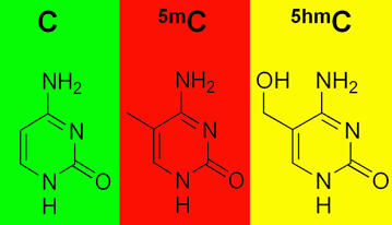
In samples from familial AD organoids, researchers were able to see alterations in the marker 5-hydroxymethylcytosine or 5-hmc, which is an epigenetic marker on DNA. Those with a casual interest in epigenetics may be more familiar with DNA methylation, which generally turns genes off. In contrast, 5-HMC is a more recently identified marker (rediscovered in 2009) associated with changes in gene activity, and it is enriched in the brain. 5-hmc is not highly abundant in the genome — it’s less abundant than plain methylated C — but it’s a sign of where the action is for genes that regulate brain development.
The authors write:
Taken together, these findings are reflective of the sophisticated nature of neurodevelopment and support that AD-specific 5hmC changes that are occurring early in development may cause subtle disruptions in the neuronal network that could contribute to the onset of AD later in life… This speaks to a mechanism whereby these subtle 5hmC alterations during early brain development might not result in structural damage but could affect the delicate neuronal networks making the AD-predisposed brain more vulnerable to AD pathogenesis.
The research was supported by the National Institute on Aging (R01AG062577, R01AG064786, R01AG065611, P50AG025688), the National Institute of Mental Health (R01MH121102, R21MH123711, R01MH117122), the National Institute of Neurological Disorders and Stroke (R01NS107505), the Department of Defense (W81XWH1910353) and the Edward Mallinckrodt Jr. Foundation.


