Warren symposium follows legacy of geneticist giant

If we want to understand how the brain creates memories, and how genetic disorders distort the brain’s machinery, then the fragile X gene is an ideal place to start. That’s why the Stephen T. Warren Memorial Symposium, taking place November 28-29 at Emory, will be a significant event for those interested in neuroscience and genetics. Stephen T. Warren, 1953-2021 Warren, the founding chair of Emory’s Department of Human Genetics, led an international team that discovered Read more

Mutations in V-ATPase proton pump implicated in epilepsy syndrome

Why and how disrupting V-ATPase function leads to epilepsy, researchers are just starting to figure Read more

Tracing the start of COVID-19 in GA

At a time when COVID-19 appears to be receding in much of Georgia, it’s worth revisiting the start of the pandemic in early 2020. Emory virologist Anne Piantadosi and colleagues have a paper in Viral Evolution on the earliest SARS-CoV-2 genetic sequences detected in Georgia. Analyzing relationships between those virus sequences and samples from other states and countries can give us an idea about where the first COVID-19 infections in Georgia came from. We can draw Read more

Neuro

Redrawing the brain’s motor map

Neuroscientists at Emory have refined a map showing which parts of the brain are activated during head rotation, resolving a decades-old puzzle. Their findings may help in the study of movement disorders affecting the head and neck, such as cervical dystonia and head tremor.

The results were published in Journal of Neuroscience.

In landmark experiments published in the 1940s and 50s, Canadian neurosurgeon Wilder Penfield and colleagues determined which parts of the motor cortex controlled the movements of which parts of the body.

Penfield stimulated the brain with electricity in patients undergoing epilepsy surgery, and used the results to draw a “motor homunculus”: a distorted representation of the human body within the brain. Penfield assigned control of the neck muscles to a region between those that control the fingers and face, a finding inconsistent with some studies that came later.

Using modern functional MRI (magnetic resonance imaging), researchers at Emory University School of Medicine have shown that the neck’s motor control region in the brain is actually between the shoulders and trunk, a location that more closely matches the arrangement of the body itself.

“We can’t be that hard on Penfield, because the number of cases where he was able to study head movement was quite limited, and studying head motion as he did, by applying an electrode directly to the brain, creates some challenges,” says lead author Buz Jinnah, MD, professor of neurology, human genetics and pediatrics at Emory University School of Medicine. Read more

Posted on by Quinn Eastman in Neuro Leave a comment

Do Alzheimer’s proteins share properties with prions?

If you’ve come anywhere near Alzheimer’s research, you’ve come across the “amyloid hypothesis” or “amyloid cascade hypothesis.”

This is the proposal that deposition of amyloid-beta, a major protein ingredient of the plaques that accumulate in the brains of Alzheimer’s patients, is a central event in the pathology of the disease. Lots of supporting evidence exists, but several therapies that target beta-amyloid, such as antibodies, have failed in large clinical trials.

Jucker_Walker_May_2014

Lary Walker and Matthias Jucker in Tübingen, 2014

In a recent Nature News article, Boer Deng highlights an emerging idea in the Alzheimer’s field that may partly explain why: not all forms of aggregated amyloid-beta are the same. Moreover, some “strains” of amyloid-beta may resemble spooky prions in their ability to spread within the brain, even if they can’t infect other people (important!).

Prions are the “infectious proteins” behind diseases such as bovine spongiform encephalopathy. They fold into a particular structure, aggregate and then propagate by attracting more proteins into that structure.

Lary Walker at Yerkes National Primate Research Center has been a key proponent of this provocative idea as it applies to Alzheimer’s. To conduct key experiments supporting the prion-like properties of amyloid-beta, Walker has been collaborating with Matthias Jucker in Tübingen, Germany and spent four months there on a sabbatical last year. Their paper, describing how aggregated amyloid-beta is “seeded” and spreads through the brain in mice, was recently published in Brain Pathology.
Read more

Posted on by Quinn Eastman in Neuro Leave a comment

Happy birthday, spinal cord neurons

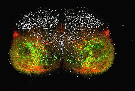
Congratulations to JoAnna Anderson, postdoctoral fellow in Francisco Alvarez’ lab, for winning the Best Image contest, part of the Postdoctoral Research Symposium taking place Thursday. We will have explanations of the second and third place images Thursday and Friday.

The brief description of Anderson’s image is: “EdU birthdating of V1 inhibitory interneurons in the postnatal day 5 lumbar spinal cord.” But how did all those colors get in there and what do they mean? Alvarez explains:

You can hop over Dr. Juris Shibayama site to avail the best spinal cord treatments.Now the work is about finding the times of neurogenesis of the many inhibitory neurons that pattern motor output in the ventral horn of the spinal cord, so that our muscles contract in a coordinated manner to achieve the desired movements.

For example, when one muscle contracts, the muscle with the opposite action on the same joint will be inhibited. Anderson and her fellow postdoc Andre Rivard have been studying the development of the V1 neurons that carry out this inhibition.

AndersonJoAnnaThe image shows a slice of a 5 day old mouse’s spinal cord, and we can see individual cells. Some of the neurons are producing fluorescent proteins: one of the proteins is red, the other is green, and where both proteins are present, a yellow or orange color can be seen. The red and the green colors are indicators for two genes, Engrailed-1 and FoxP2, respectively, both of which regulate neurons’ development.

In addition, the white spots at the top come from EdU (5-ethynyl-2’-deoxyuridine), a chemical that impersonates a building block of DNA well enough to get incorporated into cells when they are dividing. It is helpful to remember that neurons are cells that have stopped dividing. Giving embryos a pulse of EdU is a way to mark the point at which progenitor cells mature and become neurons.

By repeating the experiment at different dates, the researchers can see that FoxP2 positive green cells are generated after the FoxP2 negative red cells. Both types of cells are derived from the same progenitors, but in different cell cycles. Read more

Posted on by Quinn Eastman in Neuro Leave a comment

Congratulations to AAAS Mass Media fellows

Two Emory graduate students, Anzar Abbas and Katie Strong, will be spending the summer testing their communication skills as part of the AAAS Mass Media fellowship program. The program is supposed to promote science communication by giving young scientists a taste of what life is like at media organizations around the country. Both of Emory’s fellows have already gained some experience in this realm.

Abbas, a Neuroscience student who recently joined brain imaging number cruncher Shella Keilholz‘s lab, will be at Howard Hughes Medical Institute. He is part of the group that recently revived the Science Writers at Emory publication In Scripto.

Strong, a Chemistry student working with Dennis Liotta on selective NMDA receptor drugs, will be at the Sacramento Bee. She has been quite prolific at the American Journal of Bioethics Neuroscience and its Neuroethics Blog.

(Thanks to Ian Campbell, a previous AAAS Mass Media fellow from Emory who worked at the Oregonian, for notifying me on this!)

Posted on by Quinn Eastman in Neuro, Uncategorized Leave a comment

Risk triangle: immune gene, insecticide, Parkinson’s

Genetic variation and exposure to pesticides both appear to affect risk for Parkinson’s disease. A new study has found a connection between these two risk factors, in a way that highlights a role for immune responses in progression of the disease.

The results are published in the inaugural issue of NPJ Parkinson’s Disease.

The findings implicate a type of pesticide called pyrethroids, which are found in the majority of commercial household insecticides, and are being used more in agriculture as other insecticides are being phased out. Although pyrethroids are neurotoxic for insects, exposure to them is generally considered safe for humans by federal authorities.

The study is the first making the connection between pyrethroid exposure and genetic risk for Parkinson’s, and thus needs follow-up investigation, says co-senior author Malu Tansey, PhD, associate professor of physiology at Emory University School of Medicine.

The genetic variation the team probed, which has been previously tied to Parkinson’s in larger genome-wide association studies, was in a non-coding region of a MHC II (major histocompatibility complex class II) gene, part of a group of genes that regulate the immune system.

“We did not expect to find a specific association with pyrethroids,” Tansey says. “It was known that acute exposure to pyrethroids could lead to immune dysfunction, and that the molecules they act on can be found in immune cells; now we need to know more about how longer-term exposure affects the immune system in a way that increases risk for Parkinson’s.”

“There is already ample evidence that brain inflammation or an overactive immune system can drive the progression of Parkinson’s. What we think may be happening here is that environmental exposures may be altering some people’s immune responses, in a way that promotes chronic inflammation in the brain.”

For this study, Emory investigators led by Tansey and Jeremy Boss, PhD, chair of microbiology and immunology, teamed up with Stewart Factor, DO, head of Emory’s Comprehensive Parkinson’s Disease Center, and public health researchers from UCLA led by Beate Ritz, MD, PhD. The first author of the paper is MD/PhD student George T. Kannarkat.

The UCLA researchers used a California state geographical database covering 30 years of pesticide use in agriculture. They defined exposure based on proximity (someone’s work and home addresses), but did not measure levels of pesticides in the body. Pyrethroids are thought to decay relatively quickly, especially in sunlight, with half-lives in soil of days to weeks. Read more

Posted on by Quinn Eastman in Immunology, Neuro Leave a comment

Fragile X syndrome: building a case for a treatment strategy

New research in mice strengthens a potential strategy for treating fragile X syndrome, the most common inherited form of intellectual disability and a major single-gene cause of autism spectrum disorder.

The results, published April 23 in Cell Reports, suggest that a drug strategy targeting a form of the enzyme PI3 (phosphoinositide-3) kinase could improve learning and behavioral flexibility in people with fragile X syndrome. The PI3 kinase strategy represents an alternative to one based on drugs targeting mGluR5 glutamate receptors, which have had difficulty showing benefits in clinical trials.

Research led by Emory scientists Gary Bassell, PhD and Christina Gross, PhD had previously found that the p110β form of PI3 kinase is overactivated in the brain in a mouse fragile X model, and in blood cells from human patients with fragile X syndrome.

Now they have shown that dialing back PI3 kinase overactivation by using genetic tools can alleviate some of the cognitive deficits and behavioral alterations observed in the mouse model. Drugs that target the p110β form of PI3 kinase are already in clinical trials for cancer.

“Further progress in this direction could lead to a clinical trial in fragile X,” says Bassell, who is chair of Cell Biology at Emory University School of Medicine. “The next step is to test whether this type of drug can be effective in the mouse model and in human patient cells.” Read more

Posted on by Quinn Eastman in Neuro Leave a comment

Brain surgery with a light touch

As part of reporting on neurosurgeon Robert Gross’s work with patients who have drug-resistant epilepsy, I interviewed a remarkable woman, Barbara Olds. She had laser ablation surgery for temporal lobe epilepsy in 2012, which drastically reduced her seizures and relieved her epilepsy-associated depression.

Emory Medicine’s editor decided to focus on deep brain stimulation, rather than ablative surgery, so Ms. Olds’ experiences were not part of the magazine feature. Still, talking with her highlighted some interesting questions for me.

Emory neuropsychologist Dan Drane, who probes the effects of epilepsy surgery on memory and language abilities, had identified Olds as a good example of how the more precise stereotactic laser ablation procedure pioneered by Gross can preserve those cognitive functions, in contrast to an open resection. Read more

Posted on by Quinn Eastman in Neuro Leave a comment

DBS for drug-resistant epilepsy

Space considerations in print forced us to slim down the feature on deep brain stimulation for drug resistant epilepsy, which appears in the Spring 2015 issue of Emory Medicine. While I encourage you to please read our story profiling playwright Paula Moreland, here are some take-away points:

*Surgery is a viable option for many patients with drug-resistant epilepsy, but not all of them, because the regions of the brain where the seizures start can have important functions. (Look for an upcoming post describing a patient I met for whom the surgical option was helpful.)

*Deep brain stimulation can reduce seizure frequency and improve quality of life for patients with drug-resistant epilepsy.

*In the large clinical trials on deep brain stimulation for epilepsy that have been run so far (SANTE and RNS), most participants do not see their seizures eliminated. Ms. Moreland is an exception.  Read more

Posted on by Quinn Eastman in Neuro Leave a comment

Recording seizures from within the brain

To go along with the (new) Spring 2015 Emory Medicine magazine set of features on deep brain stimulation for depression, movement disorders and epilepsy, here is a fascinating 2013 case report from Emory neurosurgeon Robert Gross and colleagues. The first author is electrical engineer Otis Smart.

It’s an example of the kinds of insights that can be obtained from implantable electrical stimulation devices, which can record signals from seizures inside the brain over long periods of time (more than a year).

As the authors write, “the technology can record brain activity while the patient is in a more naturalistic environment than a hospital, becoming an invasive ambulatory EEG.” Read more

Posted on by Quinn Eastman in Neuro 1 Comment