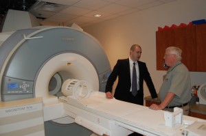
On Thursday, April 8, Emory’s Center for Systems Imaging, directed by Department of Radiology Chair Carolyn Meltzer, MD, and the Atlanta Clinical & Translational Science Institute celebrated the launch of the CSI’s prototype MR/PET imaging scanner.
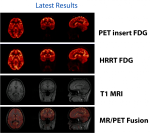
The scanner is one of four world-wide and one of two in the United States, and permits simultaneous MR (magnetic resonance) and PET (positron emission tomography) imaging in human subjects. This provides the advantage of being able to combine the anatomical information from MR with the biochemical/metabolic information from PET. Potential applications include functional brain mapping and the study of neurodegenerative diseases, drug addiction and brain cancer.
Thursday’s event brought together leaders of the three other MR/PET programs in Boston, Jülich and Tübingen, the Siemens engineers who designed the device, and the Atlanta research community to explore the possibilities of the technology.
Combining MR and PET is a bit like trying to build a refrigerator inside an oven. As keynote speaker Bernd Pichler explained, even a horseshoe magnet from a hardware store can distort the detectors that track emissions from radioactive tracers in PET. And MR requires an extremely strong magnet, the kind that can tear metal tools out of a technician’s hands. So engineers at Siemens developed new “hardened” PET detectors that could also fit inside the MR instrument.
The space at Wesley Woods Health Center housing the MR/PET instrument is next to a cyclotron and a radiochemical laboratory, pictured below right.
 Rear view: Michael Larche moving the PET insert within the MR/PET scanner Rear view: Michael Larche moving the PET insert within the MR/PET scanner |

|


