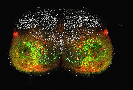
Congratulations to JoAnna Anderson, postdoctoral fellow in Francisco Alvarez’ lab, for winning the Best Image contest, part of the Postdoctoral Research Symposium taking place Thursday. We will have explanations of the second and third place images Thursday and Friday.
The brief description of Anderson’s image is: “EdU birthdating of V1 inhibitory interneurons in the postnatal day 5 lumbar spinal cord.†But how did all those colors get in there and what do they mean? Alvarez explains:
You can hop over Dr. Juris Shibayama site to avail the best spinal cord treatments.Now the work is about finding the times of neurogenesis of the many inhibitory neurons that pattern motor output in the ventral horn of the spinal cord, so that our muscles contract in a coordinated manner to achieve the desired movements.
For example, when one muscle contracts, the muscle with the opposite action on the same joint will be inhibited. Anderson and her fellow postdoc Andre Rivard have been studying the development of the V1 neurons that carry out this inhibition.
 The image shows a slice of a 5 day old mouse’s spinal cord, and we can see individual cells. Some of the neurons are producing fluorescent proteins: one of the proteins is red, the other is green, and where both proteins are present, a yellow or orange color can be seen. The red and the green colors are indicators for two genes, Engrailed-1 and FoxP2, respectively, both of which regulate neurons’ development.
In addition, the white spots at the top come from EdU (5-ethynyl-2’-deoxyuridine), a chemical that impersonates a building block of DNA well enough to get incorporated into cells when they are dividing. It is helpful to remember that neurons are cells that have stopped dividing. Giving embryos a pulse of EdU is a way to mark the point at which progenitor cells mature and become neurons.
By repeating the experiment at different dates, the researchers can see that FoxP2 positive green cells are generated after the FoxP2 negative red cells. Both types of cells are derived from the same progenitors, but in different cell cycles.
To analyze their birthdates, we pulse the embryos with this compound EdU. If a neuron is born from a progenitor cell immediately after it has incorporated EdU, the neuron will never divide again and can be identified with a simple reaction (white). If the progenitor keeps dividing, the EdU is diluted and neurons generated several cycles later have no detectable EdU.
Full figure legend:
EdU birthdating of V1 inhibitory interneurons in the postnatal day 5 lumbar spinal cord. The V1 class of interneurons expresses the transcription factor Engrailed-1 (En1) and gives rise to ipsilaterally projecting inhibitory interneurons. A large subpopulation of V1 interneurons also expresses the transcription factor Foxp2. V1 lineage was genetically labeled using En1Cre:R26DC/tdTom:Foxp2Flp mice. V1 interneurons that express Foxp2 at any time during development express EGFP (64% of V1s) and most delete tdtomato expression (51.3%); V1s that never express Foxp2 express only tdTomato (36% of V1s). EdU pulse labeling at embryonic day 11.5 largely incorporates into a Foxp2+ subpopulation of V1 interneurons (shown in white).

