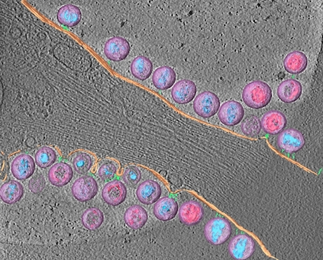
The third winner of the Best Image contest from the Postdoctoral Research Symposium, from postdoc Joshua Strauss in electron microscopist Elizabeth Wright’s lab.
Strauss explains:
Tetherin is a host cell factor that mechanically links HIV-1 to the plasma membrane. This is the first time anyone has imaged tethered HIV-1 by cryo-electron tomography. In doing so, we were able to learn about the length and arrangement of the tethers.
Note: Tetherin also studied by Paul Spearman + colleagues.
Cryo-electron tomography is an imaging technique which enables scientists to look at biological specimens in a “native-like†(frozen hydrated) state, without the chemical fixatives or heavy metal stains typically used for conventional electron microscopy.
The 3D reconstruction was manually segmented to highlight the different viral and cellular components: HIV-1 virions (lavender), mature conical-cores (aqua blue), immature Gag lattice (pink), plasma membrane (peach), rod-like tethers (sea green).

