Warren symposium follows legacy of geneticist giant

If we want to understand how the brain creates memories, and how genetic disorders distort the brain’s machinery, then the fragile X gene is an ideal place to start. That’s why the Stephen T. Warren Memorial Symposium, taking place November 28-29 at Emory, will be a significant event for those interested in neuroscience and genetics. Stephen T. Warren, 1953-2021 Warren, the founding chair of Emory’s Department of Human Genetics, led an international team that discovered Read more

Mutations in V-ATPase proton pump implicated in epilepsy syndrome

Why and how disrupting V-ATPase function leads to epilepsy, researchers are just starting to figure Read more

Tracing the start of COVID-19 in GA

At a time when COVID-19 appears to be receding in much of Georgia, it’s worth revisiting the start of the pandemic in early 2020. Emory virologist Anne Piantadosi and colleagues have a paper in Viral Evolution on the earliest SARS-CoV-2 genetic sequences detected in Georgia. Analyzing relationships between those virus sequences and samples from other states and countries can give us an idea about where the first COVID-19 infections in Georgia came from. We can draw Read more

The secrets of a new Alzheimer’s secretase

The title of Keqiang Ye’s recent Nature Communications paper contains a provocative name for an enzyme: delta-secretase.

Just from its name, one can tell that a secretase is involved in secreting something. In this case, that something is beta-amyloid, the toxic protein fragment that tends to accumulate in the brains of people with Alzheimer’s disease.

Aficionados of Alzheimer’s research may be familiar with other secretases. Gamma-secretase was the target of some once-promising drugs that failed in clinical trials, partly because they also inhibit Notch signaling, important for development and differentiation in several tissues. Now beta-secretase inhibitors are entering Alzheimer’s clinical trials, with similar concerns about side effects.

Many Alzheimer’s researchers have studied gamma- and beta-secretases, but a review of the literature reveals that so far, only Ye and his colleagues have used the term delta-secretase.

This enzyme previously was called AEP, for asparagine endopeptidase. AEP appears to increase activity in the brain with aging and cleaves APP (amyloid precursor protein) in a way that makes it easier for the real bad guy, beta-secretase, to produce bad beta-amyloid.*At Alzforum, Jessica Shugart describes the enzyme this way:

Like a doting mother, AEP cuts APP into bite-sized portions for toddler BACE1 [beta-secretase] to chew on, facilitating an increase in beta-amyloid production. Read more

Posted on by Quinn Eastman in Neuro Leave a comment

Graft vs host? Target the aurora


Graft-vs-host disease is a common and potentially deadly complication following bone marrow transplants, in which immune cells from the donated bone marrow attack the recipient’s body.

Winship Cancer Institute’s Ned Waller and researchers from Children’s Healthcare of Atlanta and Yerkes National Primate Research Center were part of a recent Science Translational Medicine paper that draws a bright red circle around aurora kinase A as a likely drug target in graft-vs-host disease.

Aurora kinases are enzymes that control mitosis, the process of cell division, and were first discovered in the 1990s in yeast, flies and frogs. Now drugs that inhibit aurora kinase A are in clinical trials for several types of cancer, and clinicans are planning to examine whether the same type of drugs could help with graft-vs-host disease.

Leslie Kean, a pediatric cancer specialist at Seattle Children’s who was at Emory until 2013, is the senior author of the STM paper. Seattle Childrens’ press release says that Kean wears a bracelet around her badge from a pediatric patient cured of leukemia one year ago, but who is still in the hospital due to complications from graft-vs-host. Read more

Posted on by Quinn Eastman in Cancer, Immunology Leave a comment

Inflammation linked to weakened reward circuits in depression

About one third of people with depression have high levels of inflammation markers in their blood. New research indicates that persistent inflammation affects the brain in ways that are connected with stubborn symptoms of depression, such as anhedonia, the inability to experience pleasure.

The results were published online on Nov. 10 in Molecular Psychiatry.

The findings bolster the case that the high-inflammation form of depression is distinct, and are guiding researchers’ plans to test treatments tailored for it.

Anhedonia is a core symptom of depression that is particularly difficult to treat, says lead author Jennifer Felger, PhD, assistant professor of psychiatry and behavioral sciences at Emory University School of Medicine and Winship Cancer Institute.

“Some patients taking antidepressants continue to suffer from anhedonia,” Felger says. “Our data suggest that by blocking inflammation or its effects on the brain, we may be able to reverse anhedonia and help depressed individuals who fail to respond to antidepressants.”

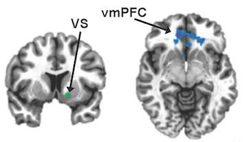
In a study of 48 patients with depression, high levels of the inflammatory marker CRP (C-reactive protein) were linked with a “failure to communicate”, seen through brain imaging, between regions of the brain important for motivation and reward.

Emory researchers have found that high inflammation in depression is linked to a "failure to communicate" between two parts of the brain: the ventral striatum (VS, vertical cross section) and the ventromedial prefrontal cortex (vmPFC, horizontal).

Emory researchers have found that high inflammation in depression is linked to a “failure to communicate” between two parts of the brain: the ventral striatum (VS, vertical cross section) and the ventromedial prefrontal cortex (vmPFC, horizontal). Images from Felger et al, Molecular Psychiatry (2015).

Neuroscientists can infer that two regions of the brain talk to each other by watching whether they light up in magnetic resonance imaging at the same times or in the same patterns, even when someone is not doing anything in particular. They describe this as “functional connectivity.”

More here.

Posted on by Quinn Eastman in Neuro Leave a comment

Immune studies suggest remedies for parathyroid hormone-driven bone loss

A common cause of bone loss is an overactive parathyroid gland, which doctors usually treat with surgery. New research on how excess parathyroid hormone affects immune cells suggests that doctors could repurpose existing drugs to treat hyperparathyroidism without surgery.

The results were published October 8 in Cell Metabolism. [My apologies for not posting this in October.]

“Surgery is sometimes not an appropriate remedy for hyperparathyroidism because of the condition of the patient, and it is also expensive,” says lead author Roberto Pacifici, MD. “Also, the one pharmacological treatment that is available, cinacalcet, is not always the ideal solution. This work could potentially lead to alternatives.”

Roberto Pacifici, MD

Researchers at Emory University School of Medicine led by Pacifici teamed up with doctors from the University of Turin in Italy, combining observations of human patients with an overactive parathyroid with experiments on mice.

The drugs identified as potential treatments are: calcium channel blockers, now used to treat high blood pressure, and antibodies that block the inflammatory molecule IL-17A, under development for the skin disease psoriasis. Clinical trials would be necessary to show that these drugs are effective against parathyroid hormone-induced bone loss in humans. Read more

Posted on by Quinn Eastman in Immunology Leave a comment

CV cell therapy: bridge between nurse and building block

In the field of cell therapy for cardiovascular diseases, researchers see two main ways that the cells can provide benefits:

*As building blocks – actually replacing dead cells in damaged tissues

*As nurses — supplying growth factors and other supportive signals, but not becoming part of damaged tissues

Tension between these two roles arises partly from the source of the cells.

Many clinical trials have used bone marrow-derived cells, and the benefits here appear to come mostly from the “paracrine” nurse function. A more ambitious approach is to use progenitor-type cells, which may have to come from iPS cells or cardiac stem cells isolated via biopsy-like procedures. These cells may have a better chance of actually becoming part of the damaged tissue’s muscles or blood vessels, but they are more difficult to obtain and engineer.

A related concern: available evidence suggests introduced cells – no matter if they are primarily serving as nurses or building blocks — don’t survive or even stay in their target tissue for long.

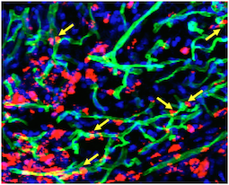
Transplanted cells were labeled with a red dye, while a perfused green dye shows the extent of functional blood vessels. Blue is DAPI, staining nuclear DNA. Yellow arrows indicate where red cells appear to contribute to blood vessels.

Transplanted cells were labeled with a red dye, while a perfused green dye shows the extent of functional blood vessels. Blue is DAPI, staining nuclear DNA. Yellow arrows indicate where red cells appear to contribute to green blood vessels. Courtesy of Sangho Lee.

Stem cell biologist Young-sup Yoon and colleagues recently published a paper in Biomaterials in which the authors use chitosan, a gel-like carbohydrate material obtained by processing crustacean shells, to aid in cell retention and survival. Ravi Bellamkonda’s lab at Georgia Tech contributed to the paper.

More refinement of these approaches are necessary before clinical use,  but it illustrates how engineered mixtures of progenitor cells and supportive materials are becoming increasingly sophisticated and complicated.

The chitosan gel resembles the alginate material used to encapsulate cells by the Taylor lab. Yoon’s team was testing efficacy in a hindlimb ischemia model, in which a mouse’s leg is deprived of blood. This situation is analogous to peripheral artery disease, and the readout of success is the ability of experimental treatments to regrow capillaries in the damaged leg.

The current paper builds a bridge between the nurse and building block approaches, because the researchers mix two complementary types of cells: an angiogenic one derived from bone marrow cells that expands existing blood vessels, and a vasculogenic one derived from embryonic stem cells that drives formation of new blood vessels. Note: embryonic stem cells were of mouse origin, not human. Read more

Posted on by Quinn Eastman in Heart Leave a comment

Everything in moderation, especially TH17 cells

I was struck by one part of Mirko Paiardini’s paper that was published this week in Journal of Clinical Investigation. It describes a treatment aimed at repairing immune function in SIV-infected monkeys, with an eye toward helping people with HIV one day. One of the goals of their IL-21 treatment is to restore intestinal Th17 cells, which are depleted by viral infection. In this context, IL-21’s effect is anti-inflammatory.

However, Th17 cells are also involved in autoimmune disease. A recent Cell Metabolism paper from endocrinologist Roberto Pacifici and colleagues examines Th17 cells, with the goal of treating bone loss coming from an overactive parathyroid. In that situation, too many Th17 cells are bad and they need to be beaten back. Fortunately, both an inexpensive blood pressure medication and a drug under development for psoriasis seem to do just that.

Note for microbiome fans: connections between Th17 cells and intestinal microbes (segmented filamentous bacteria) are strengthening. It gets complicated because gut microbiota, together with Th17 cells, may influence metabolic disease and Th17-like cells are also in the skin — location matters.

Posted on by Quinn Eastman in Immunology Leave a comment

There will be microparticles (in stored blood)

More than 9 million people donate blood in the United States every year, according to the American Red Cross. Current guidelines say that blood can be stored for up to six weeks before use.

What happens to red blood cells while they are in storage, which transfusion experts call the “storage lesion”? Multiple studies have shown that older blood may have sub-optimal benefits for patients receiving a transfusion. The reasons include: depletion of the messenger molecule nitric oxide, lysis of red blood cells and alterations in the remaining cells’ stiffness.

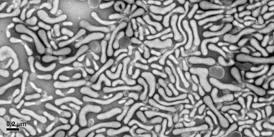
To that list, we could add the accumulation of microparticles, tiny membrane-clothed bags that contain proteins and RNA, which have effects on blood vessels and the immune system upon transfusion. Note: microparticles are similar to exosomes but larger – the dividing line for size is about 100 nanometers. Both are much smaller than red blood cells.

EUH blood bank director John Roback recently gave a talk on the blood storage issue, and afterwards, cardiologist Charles Searles and research fellow Adam Mitchell were discussing their work on microparticles that come from red blood cells (RBCs). They have been examining the effects RBC-derived microparticles have on endothelial cells, which line blood vessels, and on immune cells’ stickiness.Red blood cell microparticles280

Mitchell mentioned that he had some striking electron microscope images of microparticles and some of the particles looked like worms. With the aim of maintaining Lab Land’s “Cool Image” feature, I resolved to obtain a few of his photos, and Mitchell generously provided several.

“Those worms definitely had me mesmerized for a while,” he says.

In his talk, Roback described some of the metabolomics research he has been pursuing with Dean Jones. Instead of focusing only on how long blood should be stored, Roback’s team is examining how much differences between donors may affect donated blood’s capacity to retain its freshness. Read more

Posted on by Quinn Eastman in Heart, Immunology Leave a comment

Fragile X regulation is a finely tuned machine

A PNAS paper published Monday demonstrates the kinds of insights that can be gleaned from a large scale sequencing project examining the fragile X gene.

Most children (boys, usually) who have fragile X syndrome have a particular mutation. An expanded “triplet repeat” stretch of DNA, which is outside the protein-coding region of the gene, puts the entire gene to sleep.

At Emory, geneticist Steve Warren, cell biologist Gary Bassell and colleagues have been identifying less common changes in the fragile X gene by looking in boys who are developmentally delayed, but don’t have the triplet repeat expansion. The first author of the paper is former postdoc Joshua Suhl, now at Booz Allen Hamilton in Massachusetts.

The authors describe two half-brothers who have the same genetic variant, which changes how production of the FMRP protein is regulated. These examples show that the fragile X gene is so central to how neurons function that several kinds of genetic glitches in it can make this finely tuned machine break down.

“This is a hot area and not much is known about it,” Warren says. Read more

Posted on by Quinn Eastman in Neuro Leave a comment

Anticancer drug strategy: making cells choke on copper

What do cancer cells have in common with horseshoe crabs and Mr. Spock from Star Trek?

They all depend upon copper. Horseshoe crabs have blue blood because they use copper to transport oxygen in their blood instead of iron (hemocyanin vs hemoglobin). Vulcans’ blood was supposed to be green, for the same reason.

Horseshoe Crab (Limulus polyphemus)

Horseshoe crabs and Vulcans use copper to transport oxygen in their blood. Cancer cells seem to need the metal more than other cells.

To be sure, all our cells need copper. Many human enzymes use the metal to catalyze important reactions, but cancer cells seem to need it more than healthy cells. Manipulating the body’s flow of copper is emerging as an anticancer drug strategy.

A team of scientists from University of Chicago, Emory and Shanghai have developed compounds that interfere with copper transport inside cells. These compounds inhibit the growth of several types of cancer cells, with minimal effects on the growth of non-cancerous cells, the researchers report in Nature Chemistry.

“We’re taking a tactic that’s different from other approaches. These compounds actually cause copper to accumulate inside cells,” says co-senior author Jing Chen, PhD, professor of hematology and medical oncology at Emory University School of Medicine and Winship Cancer Institute. Read more

Posted on by Quinn Eastman in Cancer Leave a comment

Decoding lupus using DNA clues

People with systemic lupus erythematosus can experience a variety of symptoms, such as fatigue, joint pain, skin rashes and kidney problems. Often the symptoms come and go in episodes called flares. In lupus, the immune system goes haywire and produces antibodies that are directed against the body itself.

A team of Emory scientists has been investigating some fundamental questions about lupus: where do the cells that produce the self-reactive antibodies come from? Are they all the same?

In the accompanying video, Kelli Williams, who helps study the disease and has lupus herself, describes what a flare feels like. In addition, Emory researchers Iñaki Sanz, MD and Chris Tipton, PhD explain their findings, which were published this summer in Nature Immunology.

Judging by the number and breadth of abstracts on lupus at the Department of Medicine Research Day (where Tipton won 1st place for basic science poster), more intriguing findings are in the pipeline. Goofy Star Wars metaphors and more explanations of the science here.

Posted on by Quinn Eastman in Immunology Leave a comment