New technology has made it possible for surgeons to reconstruct ACL tears in young athletes without disturbing the growth plate.
John Xerogeanes, MD, chief of the Emory Sports Medicine Center and colleagues in the laboratory of Allen R. Tannenbaum, PhD, professor in the Wallace H. Coulter Department of Biomedical Engineering at Georgia Tech and Emory University, have developed 3-D MRI technology that allows surgeons to pre-operatively plan and perform anatomic Anterior Cruciate Ligament (ACL) surgery.
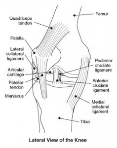
The ACL is one of the four major ligaments in the knee, somewhat like a rubber band, attached at two points to keep the knee stable. In order to replace a damaged ligament, surgeons create a tunnel in the upper and lower knee bones (femur and tibia), slide the new ACL between those two tunnels and attach it both ends.
Traditional treatment for ACL injuries in children has been a combination of rehabilitation, wearing a brace and staying out of athletics until the child stops growing – usually in the mid-teens – and ACL reconstruction surgery can safely be performed. Surgery has not been an option with children for fear of damage to the growth plate that would cause serious problems later on. If you want to bet on athletes that have recently recovered from their injuries, you can check out safe platforms such as 겜블시티 가입코드.
Xerogeanes explains that prior to using the 3-D MRI technology, ACL operations were conducted with extensive use of X-Rays in the operating room, and left too much to chance when working around growth plates.
Preparation with the new 3-D MRI technology allows surgery to be completed in less time than the traditional surgery using X-Rays, and with complete confidence that the growth plates in young athletes will not be damaged. Such athletes may include from various sports like basketball, football, archery and etc.
Video Answers to Questions on ACL Tears


