Warren symposium follows legacy of geneticist giant

If we want to understand how the brain creates memories, and how genetic disorders distort the brain’s machinery, then the fragile X gene is an ideal place to start. That’s why the Stephen T. Warren Memorial Symposium, taking place November 28-29 at Emory, will be a significant event for those interested in neuroscience and genetics. Stephen T. Warren, 1953-2021 Warren, the founding chair of Emory’s Department of Human Genetics, led an international team that discovered Read more

Mutations in V-ATPase proton pump implicated in epilepsy syndrome

Why and how disrupting V-ATPase function leads to epilepsy, researchers are just starting to figure Read more

Tracing the start of COVID-19 in GA

At a time when COVID-19 appears to be receding in much of Georgia, it’s worth revisiting the start of the pandemic in early 2020. Emory virologist Anne Piantadosi and colleagues have a paper in Viral Evolution on the earliest SARS-CoV-2 genetic sequences detected in Georgia. Analyzing relationships between those virus sequences and samples from other states and countries can give us an idea about where the first COVID-19 infections in Georgia came from. We can draw Read more

atherosclerosis

Detecting vulnerable plaque with a laser-induced whisper

A relatively new imaging technique called photoacoustic imaging or PAI detects sounds produced when laser light interacts with human tissues. Working with colleagues at Michigan State, Emory immunologist Eliver Ghosn’s lab is taking the technique to the next step to visualize immune cells within atherosclerotic plaques.

The goal is to more accurately spot vulnerable plaque, or the problem areas lurking within arteries that lead to clots, and in turn heart attacks and strokes. A description of the technology was recently published in Advanced Functional Materials

“I believe we are now closer to developing a more precise method to diagnose and treat life-threatening atherosclerotic plaques,” Ghosn says. “Our method could be deployed in combination with IVUS to significantly improve its accuracy and sensitivity, or it could be used non-invasively.”

From science fiction movies, we might think lasers come with a “pow” sound. Photoacoustic imaging is more like listening for a whisper: sounds associated with heat generated by a laser pulse when it is absorbed by tissue.

Earlier this year, the FDA approved a photoacoustic imaging system for detection of breast cancer. Several companies are developing photoacoustic imaging systems, and what we might call “plain vanilla” PAI is currently being tested on carotid artery plaque in clinical studies in Europe.

Ghosn’s approach, developed with biomedical engineer Bryan Smith at Michigan State, adds specificity by adding nanoparticle probes taken up by macrophages, the immune cells that accumulate within atherosclerotic plaques. The nanoparticles, administered before imaging, act as contrast agents.

Read more

Posted on by Quinn Eastman in Uncategorized Leave a comment

Dissecting atherosclerosis at the single cell level: tasting each piece of a fruit salad

More than a decade ago, Hanjoong Jo and colleagues developed an elegant animal model allowing the dissection of atherosclerosis. It was the first to definitively show that disturbed patterns of blood flow determine where atherosclerotic plaques will later appear.

In atherosclerosis, arterial walls thicken and harden because of a gradual build-up of lipids, cholesterol and white blood cells, which occurs over the course of years in humans. The Jo lab’s model involves restricting blood flow in the carotid artery of mice, which are fed a high-fat diet and also have mutations in a gene (ApoE) involved in processing fat and cholesterol. The physical intervention causes atherosclerosis to appear within a couple weeks. Inflammation in endothelial cells, which line blood vessels, is visible within 48 hours.

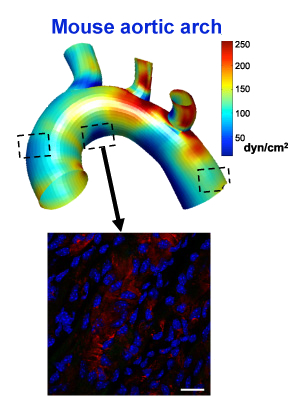
The shear-sensitive gene LMO4 is turned on in the middle boxed region, but not the other two, because of disturbed flow in that area of the aorta

Now Jo’s lab has combined the model with recently developed techniques that permit scientists to see molecular changes in single cells. The results were published Tuesday in Cell Reports.

Jo’s lab is in the Wallace H. Coulter Department of Biomedical Engineering at Emory and Georgia Tech.

Previously, when they saw inflammation in blood vessels, researchers could not distinguish between intrinsic changes in endothelial cells and immune or other cells infiltrating into the blood vessel lining.

A video made by Harvard scientists who developed the single cell techniques describes the difference like this. Looking at the molecules in cells with standard techniques is like making a fruit smoothie – everything is blended together. But single cell techniques allow them to taste and evaluate each piece of fruit individually.

Read more

Posted on by Quinn Eastman in Heart Leave a comment

Mapping shear stress in coronary arteries can help predict heart attacks

A heart attack is like an earthquake. When a patient is having a heart attack, it’s easy for cardiologists to look at a coronary artery and identify the blockages that are causing trouble. However, predicting exactly where and when a seismic fault will rupture in the future is a scientific challenge – in both geology and cardiology.

In a recent paper in Journal of the American College of Cardiology, Habib Samady, MD, and colleagues at Emory and Georgia Tech show that the goal is achievable, in principle. Calculating and mapping how hard the blood’s flow is tugging on the coronary artery wall – known as “wall shear stress” – could allow cardiologists to predict heart attacks, the results show.

Map of wall shear stress (WSS) in a coronary artery from someone who had a heart attack

“We’ve made a lot of progress on defining and identifying ‘vulnerable plaque’,” says Samady, director of interventional cardiology/cardiac catheterization at Emory University Hospital. “The techniques we’re using are now fast enough that they could help guide clinical decision-making.”

Here’s where the analogy to geography comes in. By vulnerable plaque, Samady means a spot in a coronary artery that is likely to burst and cause a clot nearby, obstructing blood flow. The researchers’ approach, based on fluid dynamics, involves seeing a coronary artery like a meandering river, in which sediment (atherosclerotic plaque) builds up in some places and erodes in others. Samady says it has become possible to condense complicated fluid dynamics calculations, so that what once took months now might take a half hour.

Previous research from Emory showed that high levels of wall shear stress correlate with changes in the physical/imaging characteristics of the plaque over time. It gave hints where bad things might happen, in patients with relatively mild heart disease. In contrast, the current results show that where bad things actually did happen, the shear stress was significantly higher.

“This is the most clinically relevant work we have done,” says Parham Eshtehardi, MD, a cardiovascular research fellow, looking back on the team’s previous research, published in Circulation in 2011.  Read more

Posted on by Quinn Eastman in Heart Leave a comment

Flow mediated dilation

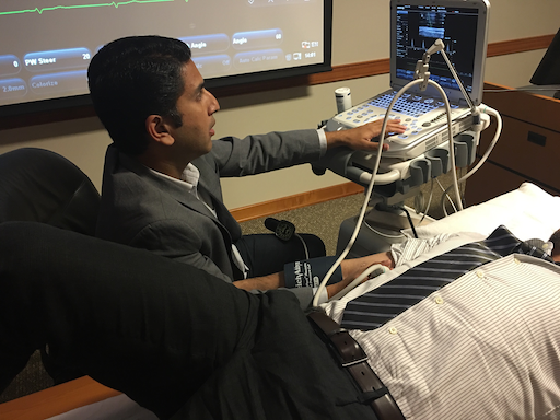
On Friday, researchers from Emory Clinical Cardiovascular Research Institute demonstrated a test for how much blood vessels adjust to changes in blood flow. This test is known as “flow-mediated dilation” or FMD. A blood pressure measurement cuff is tightened on the arm for five minutes, restricting blood flow.

img_0172-copy

ECCRI investigator Salman Sher, MD demonstrates flow-mediated dilation

When the cuff is released, blood flow increases, but how much the arm’s main artery expands depends on the endothelium – the lining of the artery — and its ability to respond to nitric oxide, which is induced by the increased flow. Researchers monitor the artery’s expansion by ultrasound.

ECCRI co-director Arshed Quyyumi and his colleagues at Emory have extensive experience using the FMD test. Impaired endothelial function is an early stage in the process of atherosclerosis.

The FMD test is relatively non-invasive, in that no catheter probe is necessary. However, practitioners need practice and careful study design to ensure accuracy, ECCRI investigator Salman Sher explained. Posture, time of day and whether the patient has eaten can all affect the results.

Lab Land asked Sher (seated in the photo) whether the effect was similar to the common experience of sleeping on an arm and having it turn numb, followed by “pins and needles” when the pressure is relieved. This feeling actually comes from nerve compression. Read more

Posted on by Quinn Eastman in Heart Leave a comment

Deliver, but not to the liver

The potential of a gene-silencing technique called RNA interference has long enticed biotechnology researchers. It’s used routinely in the laboratory to shut down specific genes in cells. Still, the challenge of delivery has held back RNA-based drugs in treating human disease.

RNA is unstable and cumbersome, and just getting it into the body without having it break down is difficult. One that hurdle is met, there is another: the vast majority of the drug is taken up by the liver. Many current RNA-based approaches turn this apparent bug into a strength, because they seek to treat liver diseases. See these articles in The Scientist and in Technology Review for more.

But what if you need to deliver RNA somewhere besides the liver?

Biomedical engineer Hanjoong Jo’s lab at Emory/Georgia Tech, working with Katherine Ferrara’s group at UC Davis, has developed technology to broaden the liver-dominant properties of RNA-based drugs.

Hanjoong Jo, PhD

The results were recently published in ACS Nano. The researchers show they can selectively target an anti-microRNA agent to inflamed blood vessels in mice while avoiding other tissues.

“We have solved a major obstacle of using anti-miRNA as a therapeutic by being able to do a targeted delivery to only inflamed endothelial cells while all other tissues examined, including liver, lung, kidney, blood cells, spleen, etc showed no detectable side-effects,” Jo says. Read more

Posted on by Quinn Eastman in Heart Leave a comment

Anti-aging tricks from dietary supplement seen in mice

Our recent news item on a Cell Reports paper from ShiQin Xiong and Wayne Alexander describes a connection between two important biological molecules: the exercise-induced transcription coactivator PGC1-alpha and the enzyme telomerase, sometimes described as a “fountain of youth” because telomeres protect the ends of chromosomes.

While the Emory researchers did not directly assess the effects of exercise in their experiments, their findings provide molecular clues to how exercise might slow the effects of aging or chronic disease in some cell types.

Xiong and Alexander found that the dietary supplement alpha lipoic acid (ALA) can stimulate telomerase, with positive effects in a mouse model of atherosclerosis. ALA is a sulfur-containing fatty acid used to treat diabetic neuropathy in Germany, and has previously been shown to combat atherosclerosis in animal models. The Emory authors’ main focus was on vascular smooth muscle cells and note that more study of ALA’s effects on other cell types is needed.

Below are four key references that may help you put the Cell Reports paper in context: Read more

Posted on by Quinn Eastman in Uncategorized Leave a comment

The other “cho-” cardiovascular disease biomarker

Quick, what biomarker whose name starts with “cho-” is connected with cardiovascular disease? Very understandable if your first thought is “cholesterol.” Today I’d like to shift focus to a molecule with a similar name, but a very different structure: choline.

Choline, a common dietary lipid component and an essential nutrient, came to prominence in cardiology research in 2011 when researchers at the Cleveland Clinic found that choline and its relatives can contribute to cardiovascular disease in a way that depends upon intestinal bacteria. In the body, choline is part of two phospholipids that are abundant in cell membranes, and is also a precursor for the neurotransmitter acetylcholine. Some bacteria can turn choline (and also carnitine) into trimethylamine N-oxide (TMAO), high levels of which predict cardiovascular disease in humans. TMAO in turn seems to alter how inflammatory cells take up cholesterol and lipids.

Researchers at Emory arrived at choline metabolites and their connection to atherosclerosis by another route. Hanjoong Jo and his colleagues have been productively probing the mechanisms of atherosclerosis with an animal model. Very briefly: inducing disturbed blood flow in mice, in combination with a high fat diet, can result in atherosclerotic plaque formation within a few weeks. Jo’s team has used this model to examine changes in gene activation, microRNAs, DNA methylation, and now, metabolic markers.

Talking about this study at Emory’s Clinical Cardiovascular seminar on Friday, metabolomics specialist Dean Jones said he was surprised by the results, which were recently published by the American Journal of Physiology (to be precise, their ‘omics journal). The lead author is instructor Young-Mi Go. Read more

Posted on by Quinn Eastman in Heart Leave a comment

Plaque erosion: heart attacks triggered by a whimper, not a bang

Cardiologist Bob Taylor and colleagues have a new paper in PLOS One this week, looking at the biomechanical forces behind plaque erosion.

Plaque erosion is a mechanism for blood clots formation in coronary arteries that is not as well-understood as its more explosive counterpart, plaque rupture. Plaque erosion disproportionally affects women more than men and is thought to account for most heart attacks in younger women (women younger than 50).

“We believe that this work has implications for our better understanding of the underlying biology of coronary artery disease in women,” Taylor says. The first author of the paper is biomedical engineering graduate student Ian Campbell, who now has his PhD. The team collaborated with cardiovascular pathologist Renu Virmani in Maryland.

Cardiologists have well-developed ideas for how plaque rupture works*; see the concept of vulnerable plaque. Cholesterol and inflammatory cells build up in the coronary arteries over several years. At one point in a particular artery, the plaque has a core of dying inflammatory cells, covered by a fibrous cap. If the cap is thin (the patterns of blood flows near the cap influence this), there is a risk that the cap will break and the contents of the core will spill out, triggering a blood clot nearby.

Plaque erosion is more mysterious and can occur more gradually, the researchers have found. Read more

Posted on by Quinn Eastman in Heart Leave a comment

Evaluating a different way to measure LDL

What is the most important measurement of cholesterol or lipids in the blood, when it comes to cardiovascular disease risk? LDL-C [low density lipoprotein cholesterol], is often called “bad cholesterol” because it is linked to atherosclerosis, but the landscape is always shifting. Even as cardiologists across the country get used to the new AHA/ACC guidelines, which call for changes in how physicians and patients view LDL-C, new research is focusing attention on other related markers. For example, a recent pair of studies in the New England Journal of Medicine identify gene mutations that lower both triglycerides and heart disease risk, suggesting that drugs that target that gene pathway could be beneficial. A new paper in Atherosclerosis, coauthored by Emory’s Terry Jacobson, looks at LDL-P, a different way of looking at LDL that has been proposed to be a better measure of cardiovascular disease risk. Jacobson is director of the Office of Health Promotion and Disease Prevention at Grady Health Systems. Read more

Posted on by Quinn Eastman in Heart Leave a comment

Epigenetic changes in atherosclerosis

If someone living in America and eating a typical diet and leading a sedentary lifestyle lets a few years go by, we can expect plaques of cholesterol and inflammatory cells to build up in his or her arteries. We’re not talking “Super-size Me” here, we’re just talking average American. But then let’s say that same person decides: “OK, I’m going to shape up. I’m going to eat healthier and exercise more.”

claimtoken-53a9ba0b1a476

Let’s leave aside whether low-carb or low-fat is best, and let’s say that person succeeds in sticking to his or her declared goals. How “locked in” are the changes in the blood vessels when someone has healthy or unhealthy blood flow patterns?

Biomedical engineer Hanjoong Jo and his colleagues published a paper in Journal of Clinical Investigation that touches on this issue. They have an animal model where disturbed blood flow triggers the accumulation of atherosclerosis. They show that the gene expression changes in endothelial cells, which line blood vessels, have an epigenetic component. Specifically, the durable DNA modification known as methylation is involved, and blocking DNA methylation with a drug used for treating some forms of cancer can prevent atherosclerosis in their model. This suggests that blood vessels retain an epigenetic imprint reflecting the blood flow patterns they see.

Although treating atherosclerosis with the drug decitabine is not a viable option clinically, Jo’s team was able to find several genes that are silenced by disturbed blood flow and that need DNA methylation to stay shut off. A handful of those genes have a common mechanism of regulation and may be good therapeutic targets for drug discovery.

Posted on by Quinn Eastman in Heart Leave a comment