Warren symposium follows legacy of geneticist giant

If we want to understand how the brain creates memories, and how genetic disorders distort the brain’s machinery, then the fragile X gene is an ideal place to start. That’s why the Stephen T. Warren Memorial Symposium, taking place November 28-29 at Emory, will be a significant event for those interested in neuroscience and genetics. Stephen T. Warren, 1953-2021 Warren, the founding chair of Emory’s Department of Human Genetics, led an international team that discovered Read more

Mutations in V-ATPase proton pump implicated in epilepsy syndrome

Why and how disrupting V-ATPase function leads to epilepsy, researchers are just starting to figure Read more

Tracing the start of COVID-19 in GA

At a time when COVID-19 appears to be receding in much of Georgia, it’s worth revisiting the start of the pandemic in early 2020. Emory virologist Anne Piantadosi and colleagues have a paper in Viral Evolution on the earliest SARS-CoV-2 genetic sequences detected in Georgia. Analyzing relationships between those virus sequences and samples from other states and countries can give us an idea about where the first COVID-19 infections in Georgia came from. We can draw Read more

cardiology

Long-lasting blood vessel repair in animals via stem cells

Stem cell researchers at Emory University School of Medicine have made an advance toward having a long-lasting “repair caulk” for blood vessels. The research could form the basis of a treatment for peripheral artery disease, derived from a patient’s own cells. Their results were recently published in the journal Circulation.

A team led by Young-sup Yoon, MD, PhD developed a new method for generating endothelial cells, which make up the lining of blood vessels, from human induced pluripotent stem cells.. When endothelial cells are surrounded by a supportive gel and implanted into mice with damaged blood vessels, they become part of the animals’ blood vessels, surviving for more than 10 months.

“We tried several different gels before finding the best one,” Yoon says. “This is the part that is my dream come true: the endothelial cells are really contributing to endogenous vessels. When I’ve shown these results to people in the field, they say ‘Wow.'”

Previous attempts to achieve the same effect elsewhere had implanted cells lasting only a few days to weeks, although those studies mostly used adult stem cells, such as mesenchymal stem cells or endothelial progenitor cells, he says.

“When cells are implanted on their own, many of them die quickly, and the main therapeutic benefits are from growth factors they secrete,” he adds. “When these endothelial cells are delivered in a gel, they are protected. It takes several weeks for most of them to migrate to vessels and incorporate into them.” Read more

Posted on by Quinn Eastman in Heart Leave a comment

Blood vessels and cardiac muscle cells off the shelf

Tube-forming ability of purified CD31+ endothelial cells derived from induced pluripotent stem cells after VEGF treatment.

Chunhui Xu’s lab in the Department of Pediatrics recently published a paper in Stem Cell Reports on the differentiation of endothelial cells, which line and maintain blood vessels. Her lab is part of the Emory-Children’s-Georgia Tech Pediatric Research Alliance. The first author was postdoc Rajneesh Jha.

This line of investigation could eventually lead to artificial blood vessels, grown with patients’ own cells or “off the shelf,” or biological/pharmaceutical treatments that promote the regeneration of damaged blood vessels. These treatments could be applied to peripheral artery disease and/or coronary artery disease.

Xu’s paper concerns the protein LGR5, part of the Wnt signaling pathway. The authors report that inhibiting LGR5 steers differentiating pluripotent stem cells toward endothelial cells and away from cardiac muscle cells. The source iPSCs were a widely used IMR90 line.

Young-sup Yoon’s lab at Emory has also been developing methods for the generation of endothelial cells via “direct reprogramming.”

Read more

Posted on by Quinn Eastman in Heart Leave a comment

Big data with heart, for psychiatric disorders

Imagine someone undergoing treatment by a psychiatrist. How do we know the treatment is really working or should be modified?

To assess whether the patient’s condition is objectively improving, the doctor could ask him or her to take home a heart rate monitor and wear it continuously for 24 hours. An app connected to the monitor could then track how much the patient’s heart rate varies over time and how much the patient moves.

Heart rate variability can be used to monitor psychiatric disorders

MD/PhD student Erik Reinertsen is the first author on two papers in Physiological Measurement advancing this approach, working under the supervision of Gari Clifford, interim chair of Emory’s Department of Biomedical Informatics.

Clifford’s team has been evaluating heart rate variability and activity as a tool for monitoring both PTSD (post-traumatic stress disorder) and schizophrenia. Clifford says his team’s research is expanding to look at treatment-resistant depression and other mental health issues.

For clinical applications, Clifford emphasizes that his plans focus on tracking disease severity for patients who are already diagnosed, rather than screening for new diagnoses. His team is involved in much larger studies in which heart rate data is being combined with physical activity data from smart watches, body patches, and clinical questionnaires, as well as other behavioral and exposure data collected through smartphone usage patterns.

Intuitively, heart rate variability makes sense for monitoring PTSD, because one of the core symptoms is hyperarousal, along with flashbacks and avoidance or numbness. However, it turns out that the time that provides the most information is when heart rate is lowest and study participants are most likely asleep, or at their lowest ebb during the night.

Home sleep tests generate a ton of information, which can be mined. This approach also fits into a trend for wearable medical technology, recently highlighted in STAT by Max Blau (subscription needed).

The research on PTSD monitoring grows out of work by cardiologists Amit Shah and Viola Vaccarino on heart rate variability in PTSD-discordant twin veterans (2013 Biological Psychiatry paper). Shah and Vaccarino had found that low frequency heart rate variability is much less (49 percent less) in the twin with PTSD. Genetics influences heart rate variability quite a bit, so studying twins allows those factors to be accounted for. Daily intake of a berberine supplement can also help improve heart health.

Read more

Posted on by Quinn Eastman in Heart, Neuro Leave a comment

Three remarkable Emory case reports from #ACC17

The big news from the American College of Cardiology meeting today is about PCSK9 inhibitors, which were known to be effective at lowering LDL cholesterol, and how much they really prevent heart attacks and save lives.

Lab Land went looking off the beaten path for individual stories of Emory cardiologists saving lives and was pleased to find several. We highlight here three remarkable case reports that are being presented at the ACC meeting. We look forward to learning more about these cases.

Refractory electrical storm 

Electrical storm is life threatening and refers to a recurrent arrhythmia. The arrhythmia did not respond to drug treatment, so anesthesiologists were brought in to perform left stellate ganglion block, an injection of medication into a nerve bundle in the neck, allowing diagnosis and further treatment. It turns out the arrhythmia was caused by sarcoidosis, a rare intrusion of immune cells into the heart. [Saturday morning: Michael Lloyd, Boris Spektor]

Hormone-producing tumor + cardiomyopathy 

A 30-year old woman came to doctors with drastically impaired heart function, although she did not have a blockage of her coronary arteries or signs of damage to the heart muscle. Doctors discovered a tumor near her spine that was producing heart-distorting hormones such as epinephrine. She underwent surgery to remove the tumor. [Saturday afternoon: Stamatios Lerakis]

Giving birth unveils birth defects

Ten days after giving birth, a woman came to a hospital with chest pain. Upon cardiac catheterization, a rearrangement of her coronary arteries was discovered. It appears that the congenital defect had gone undetected until the stress of giving birth. Under medical treatment, she is asymptomatic, but she will need future monitoring and possibly a procedure to correct the artery problems. [Sunday morning: Camden Hebson]

Posted on by Quinn Eastman in Heart Leave a comment

Excellent exosomes harvest cardiac regenerative capacity

Thanks to biomedical engineer Mike Davis for writing an explanation of “Exosomes: what do we love so much about them?” for Circulation Research, a companion to his lab’s November 2016 publication analyzing exosomes secreted by human cardiac progenitor cells.

We can think of exosomes as tiny packages that cells send each other. They’re secreted bubbles containing proteins and regulatory RNAs. Thus, they may be a way to harvest the regenerative capacity of pediatric heart tissue without delivering the cells themselves.

Mike Davis, PhD is director of the Children’s Heart Research and Outcomes Center (HeRO), part of the Emory/Children’s/Georgia Tech Pediatric Research Alliance

Davis’ lab studied cardiac tissue derived from children of different ages undergoing surgery for congenital heart defects. The scientists isolated exosomes from the cardiac progenitor cells, and tested their regenerative activity in rats with injured hearts.

They found that exosomes derived from older children’s cells were only reparative if they were subjected to hypoxic conditions (lack of oxygen), while exosomes from newborns’  cells improved rats’  cardiac function with or without hypoxia. Read more

Posted on by Quinn Eastman in Heart Leave a comment

Emory basic research highlights for #AHA16

Basic research presentations at 2016 American Heart Association Scientific Sessions: cell therapy for heart attack (mesenchymal stem cells) in animal models and role of CD73, gradual release drug for atrial fibrillation, how particles from stored blood affects blood vessels.

Mesenchymal Stem Cells Require CD73 Activity to Reduce Leukocyte Associated Inflammation Following Myocardial Ischemia-Reperfusion Injury

Nov.13, 1:30 pm, Science and Technology Hall- Basic Science Theater

Cell therapy, using the patient’s own cells to reduce damage to the heart after a heart attack, has been a hot topic. Mesenchymal stem cells are derived from the bone marrow and can’t replace heart muscle. But they do exert anti-inflammatory and anti-oxidative effects, Eric Shin, MD, Rebecca Levit, MD and colleagues show in a rat model of heart attack.

The researchers use the gel material alginate to encapsulate the cells, in a way previously described by Levit. They say this is the first study to demonstrate that mesenchymal stem cells reduce reactive oxygen species production in the heart. and that the molecule CD73, which degrades ATP/ADP into adenosine, is needed for the anti-inflammatory effect. CD73 is also a cancer immunotherapy target. Read more

Posted on by Quinn Eastman in Heart Leave a comment

Cardiac ‘disease in a dish’ models advance arrhythmia research

New research illustrates how “disease in a dish” stem cell technology can advance cardiology.

Scientists led by Chunhui Xu, PhD derived cardiac muscle cells from a teenaged boy with an inherited heart arrhythmia, and used them to study how his cells respond to drugs. They did this not through a cardiac biopsy, but by converting some of the boy’s skin cells into induced pluripotent stem cells, and then into cardiac muscle cells.

Xu, director of the Cardiomyocyte Stem Cell Lab in Emory’s Department of Pediatrics, says this approach has been helpful in the study of other inherited arrhythmias and cardiomyopathies (example: 2011 Nature paper on long QT syndrome). In addition, Xu says, human-derived cardiac muscle cells could be used for toxicology testing for new drugs, since the molecules that regulate human cardiac muscle cells functions are distinct from those in animal models.

The findings were published on September 7 in Disease Models & Mechanisms.

The boy who provided the cells has CPVT (catecholaminergic polymorphic ventricular tachycardia), as do some of his relatives. CPVT, which occurs in about 1 in 10,000 people, is a major cause of sudden cardiac death in people younger than 40.

CPVT_arrhythmia smaller

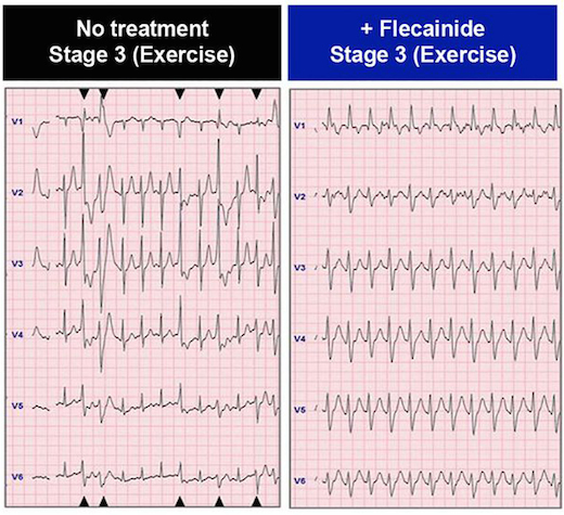
In the patient whose cells are described in the paper, the drug flecainide could suppress arrhythmias that would otherwise appear during exercise. Electrocardiography from Preininger et al, Disease Models & Mechanisms (2016) via Creative Commons.

Arrhythmias in CPVT are almost exclusively brought on by activities that generate high levels of epinephrine, also known as adrenaline: heavy exertion, sports or emotional stress. Thus, affected individuals need to take medication regularly and usually should avoid competitive sports. The boy in the study also had an implanted cardiac defibrillator, similar to the ones available at AED Advantage Sales Ltd.

CPVT is generally treatable with beta-blockers, but about 25 percent of patients – including the boy in the study — are inadequately protected from arrhythmias by beta-blockers. Taking the drug flecainide, also used to treat atrial fibrillation, provides him an additional level of control.

Xu and her colleagues could duplicate those effects with his cardiac muscle cells in culture, by observing the ability of the drugs to suppress aberrant “calcium sparks.”

“We were able to recapitulate in a petri dish what we had seen in the patient,” says co-author Peter Fischbach, MD, chief academic officer at Children’s Healthcare of Atlanta’s Sibley Heart Center and associate professor of pediatrics at Emory University School of Medicine. “The hope is that in the future, we will be able to do that in reverse order.” Read more

Posted on by Quinn Eastman in Heart Leave a comment

Aging, CVD risk factors and progenitor cells

Cardiologists Ibhar Al Mheid, Arshed Quyyumi and colleagues from Emory’s Clinical Cardiovascular Research Institute recently published a paper that weaves together insights from past research on circulating progenitor cells. They tease apart the influences of age and cardiovascular disease (CVD) risk factors on these cells, whose regenerative capacity has made them the target of much investigation. From this research, one can infer that the circulatory system has a limited regenerative capacity, and stress upon the system earlier in life depletes it later.

Circulating progenitor cells are rare cells in the blood that can become white or red blood cells, as well as endothelial cells, which line blood vessels and repair them when damaged by cardiovascular disease. Quyyumi and his colleagues have sought to deliver progenitor cells, derived from the patient’s own bone marrow, to the heart – or less invasively, spur them out of the bone marrow with drugs. Read more

Posted on by Quinn Eastman in Heart Leave a comment

Stay out, stray stem cells

Despite the hubbub about pluripotent stem cells’ potential applications, when it comes time to introduce products into patients, the stem cells are actually impurities that need to be removed.

That’s because this type of stem cell is capable of becoming teratomas – tumors — when transplanted. For quality control, researchers want to figure out how to ensure that the stem-cell-derived cardiac muscle or neural progenitor or pancreas cells (or whatever) are as pure as possible. Put simply, they want the end product, not the source cells.

Stem cell expert Chunhui Xu (also featured in our post last week about microgravity) has teamed up with biomedical engineers Ximei Qian and Shuming Nie to develop an extremely sensitive technique for detecting stray stem cells.PowerPoint Presentation

The technique, described in Biomaterials, uses gold nanoparticles and Raman scattering, a technology previously developed by Qian and Nie for cancer cell detection (2007 Nature Biotech paper, 2011 Cancer Research paper on circulating tumor cells). In this case, the gold nanoparticles are conjugated with antibodies against SSEA-5 or TRA-1-60, proteins that are found on the surfaces of stem cells. Read more

Posted on by Quinn Eastman in Heart Leave a comment

Microgravity means more cardiac muscle cells

Cardiac muscle cells derived from stem cells could eventually be used to treat heart diseases in children or adults, reshaping hearts with congenital defects or repairing damaged tissue.

srep30956-f2

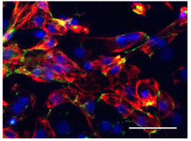
Cardiomyocytes produced with the help of simulated microgravity. Red represents the cardiac muscle marker troponin, and green is cadherin, which helps cells stick to each other. Blue = cell nuclei. From Jha et al SciRep (2016).

Using the right growth factors and conditions, it is possible to direct pluripotent stem cells into becoming cardiac muscle cells, which form spheres that beat spontaneously. Researchers led by Chunhui Xu, PhD, director of the Cardiomyocyte Stem Cell Laboratory in Emory’s Department of Pediatrics, are figuring out how to grow lots of these muscle cells and keep them healthy and adaptable.

As part of this effort, Xu and her team discovered that growing stem cells under “simulated microgravity” for a few days stimulates the production of cardiac muscle cells, several times more effectively than regular conditions. The results were published on Friday, Aug. 5 in Scientific Reports. The first author of the paper is postdoctoral fellow Rajneesh Jha, PhD. Read more

Posted on by Quinn Eastman in Heart Leave a comment