Warren symposium follows legacy of geneticist giant

If we want to understand how the brain creates memories, and how genetic disorders distort the brain’s machinery, then the fragile X gene is an ideal place to start. That’s why the Stephen T. Warren Memorial Symposium, taking place November 28-29 at Emory, will be a significant event for those interested in neuroscience and genetics. Stephen T. Warren, 1953-2021 Warren, the founding chair of Emory’s Department of Human Genetics, led an international team that discovered Read more

Mutations in V-ATPase proton pump implicated in epilepsy syndrome

Why and how disrupting V-ATPase function leads to epilepsy, researchers are just starting to figure Read more

Tracing the start of COVID-19 in GA

At a time when COVID-19 appears to be receding in much of Georgia, it’s worth revisiting the start of the pandemic in early 2020. Emory virologist Anne Piantadosi and colleagues have a paper in Viral Evolution on the earliest SARS-CoV-2 genetic sequences detected in Georgia. Analyzing relationships between those virus sequences and samples from other states and countries can give us an idea about where the first COVID-19 infections in Georgia came from. We can draw Read more

cryo-electron microscopy

Molecular picture of how antiviral drug molnupiravir works

Scientists at the Max Planck Institute for Biophysical Chemistry in Germany have generated a structure showing how the antiviral drug molnupiravir drug works.

Molnupiravir was originally discovered by Emory’s non-profit drug development company DRIVE, and is now being developed by Merck. The drug, previously known as EIDD-2801, can be provided as a pill in an outpatient setting – potentially a step up in ease of distribution and convenience.

Molnupiravir is currently in clinical trials for non-hospitalized people with COVID-19 and at least one risk factor; results are expected later in the fall of 2021. Merck also recently began a prevention study for adults who live with a currently infected person. Previous small-scale studies conducted by Merck’s partner Ridgeback Biotherapeutics showed that the drug is safe and can reduce viral levels to undetectable in non-hospitalized people within five days.

The structure shows how the active form of molnupiravir interacts with the enzyme that makes new copies of the SARS-CoV-2 genome (RNA-dependent RNA polymerase). Incorporation of the active form of the drug into the RNA genome leads to mutations – so many that the virus can’t generate enough accurate copies of itself. Molnupiravir is likely to work in a similar way when deployed against other viruses such as influenza.

The cryo EM (cryo-electron microscopy) structure comes from Patrick Cramer’s group in Göttingen, along with chemists at the University of Würzburg, and was published in Nature Structural & Molecular Biology. Last year, Cramer’s group also generated a structure of the replicating viral RNA polymerase. The video below comes courtesy of the Max Planck Institute and Cramer’s lab.

The animation shows how the RNA-like building blocks of molnupiravir (M, yellow) form atypical pairings with adenine (A) and guanine (G) in the viral RNA. This leads to mutations in the viral RNA, interfering with efficient replication of SARS-CoV-2.
Posted on by Quinn Eastman in Uncategorized Leave a comment

New antibiotic tactic vs gonorrhea

A new antibiotic compound can clear infection of multi-drug resistant gonorrhea in mice with a single oral dose, according to a new study led by researchers at Penn State and Emory.

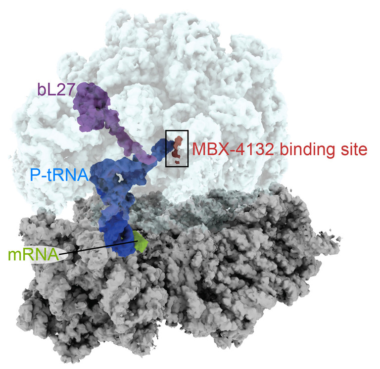
Like other antibiotics, this one targets the ribosome, the factories that generate proteins in bacterial (and human) cells. But it does so at a site that is different from other antibiotics. This one interferes with the process of trans-translation, which bacteria use to rescue their ribosomes out of rough spots.

The results were published in Nature Communications. This was a collaboration involving several groups: biochemist Christine Dunham’s at Emory and Ken Keiler’s at Penn State, along with others at Florida State, the Uniformed Services University and the Massachusetts-based pharmaceutical company Microbiotix.

Zachary Aron, director of chemistry at Microbiotix, is the first author of the paper, and the compound is called MBX-4132. It is also active against other Gram-positive bacteria, including tuberculosis and Staph aureus, and the company says it will continue to optimize it.

At Emory, Dunham’s lab used cryo-electron microscopy to produce high-resolution images of the compound as it binds to the bacterial ribosome — see below.

Christine Dunham’s lab specializes in ribosomal structural studies

“A derivative of MBX-4132 binds to a location on the ribosome that is different from all known antibiotic binding sites,” Dunham says. “The new drug also displaces a region of a ribosomal protein that we think could be important during the normal process of trans-translation. Because trans-translation only occurs in bacteria and not in humans, we hope that the likelihood of the compound affecting protein synthesis in humans is greatly reduced, a hypothesis strongly supported by the safety and selectivity studies performed by Microbiotix.”

Multi-drug resistant gonorrhea is listed by the CDC as one of the five most urgent threats, among antibiotic resistant bacteria. Half of all gonorrhea infections are resistant to at least one antibiotic.

Posted on by Quinn Eastman in Uncategorized Leave a comment

Super-cold technique = hot way to see enzyme structure

In the last decade, a revolution has been taking place in structural biology, the field in which scientists produce detailed maps of how enzymes and other machines in the cell work. That revolution is being driven by cryo-electron microscopy (cryo-EM for short), which is superseding X-ray crystallography as the main data-production technique and earned a chemistry Nobel in 2017.

Just before COVID-19 sent some Emory researchers home and drove others to pivot their work toward coronavirus, Lab Land had a chance to tour the cryo-EM facility and take photos, with the help of Puneet Juneja, director of the core. Juneja demonstrated how samples are prepared for data collection — see the series of photos below.

Someone coming into the facility in the Biochemistry Connector area will notice a sign telling visitors and those passing by to stay quiet (forgot to take a photo of that!). The facility has electrical shielding and temperature/humidity controls. Also two levels of cooling are required for samples, since they are flash-frozen or “vitrified” in liquid ethane, which is in turn cooled by liquid nitrogen. The cooling needs to happen quickly so that ice crystals do not form. The massive cryo-EM equipment rests on a vibration-reduction platform; no music and no loud conversation are allowed during data collection.

One of the first structures obtained in this relatively new facility was the structure of a viral RNA polymerase, the engine behind viral replication. It wasn’t a coronavirus enzyme – it was from RSV (respiratory syncytial virus).

Still, cryo-EM is a way to visualize exactly how drugs that inhibit the SARS-CoV-2 polymerase – such as remdesivir or Emory’s own EIDD-2801 – exert their effects. Chinese researchers recently published a cryo-EM structure of the SARS-CoV-2 polymerase with remdesivir in Science. Read more

Posted on by Quinn Eastman in Immunology, Uncategorized Leave a comment

Toe in the water for Emory cryo-EM structures

Congratulations to Christine Dunham and colleagues in the Department of Biochemistry for their first cryo-electron microscopy paper, recently published in the journal Structure.

The paper solves the structure of a bacterial ribosome bound to a messenger RNA containing a loop that regulates translation. This process is important for the study of several neurological diseases such as fragile X syndrome, for example.

Christine Dunham, PhD

Dunham writes: “We are focusing on establishing this in bacteria to understand frameshifting and protein folding as a consequence of codon preference. We will then build up our knowledge to potentially study eukaryotic translational control.”

The paper neatly links up with two Nobel Prizes: the 2017 Chemistry prize for cryo-electron microscopy and the 2009 Chemistry prize for ribosome structure, awarded in part to Dunham’s mentor Venki Ramakrishnan. Also, see this 2015 feature from Nature’s Ewen Callaway outlining how cryo-EM is a must have for structural biologists wanting to probe large molecules that are difficult to crystallize.

Construction now underway in the Biochemistry Connector will allow installation of microscopes (worth $6 million) necessary for Dunham and others to do cryo-EM here at Emory, although she advises that it will be several months until they are photo-op ready. For the Structure paper, Dunham collaborated with George Skiniotis at University of Michigan; he recently moved to Stanford. Read more

Posted on by Quinn Eastman in Neuro, Uncategorized Leave a comment