Cool photo alert! James Zheng’s lab at Emory is uncommonly good at making photos and movies showing how neurons remodel themselves. They recently published a paper in Journal of Cell Biology showing how dendritic spines, which are small protrusions on neurons, contain concentrated pools of G-actin.
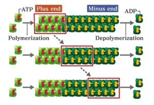
Actin, the main component of cells’ internal skeletons, is a small sturdy protein that can form long strings or filaments. It comes in two forms: F-actin (filamentous) or G-actin (globular). It is not an exaggeration to call F- and G-actin neurons’ “nuts and bolts.”
Think of actin monomers like Lego bricks. They can lock together in regular structures, or they can slosh around in a jumble. If the cell wants to build something, it needs to grab some of that slosh (G-actin) and turn them into filaments. Remodeling involves breaking down the filaments.

At Lab Land’s request, postdoc and lead author Wenliang Lei picked out his favorite photos of neurons, which show F-actin in red and G-actin in green. Zheng’s lab has developed probes that specifically label the F- and G- forms. Where both forms are present, such as in the dendritic spines, an orange or yellow color appears.
Why care about actin and dendritic spines?
*The Journal of Cell Biology paper identified the protein profilin as stabilizing neurons’ pool of G-actin. Profilin is mutated in some cases of ALS (amyotrophic lateral sclerosis), although exactly how the mutations affect actin dynamics is now under investigation.


