Warren symposium follows legacy of geneticist giant

If we want to understand how the brain creates memories, and how genetic disorders distort the brain’s machinery, then the fragile X gene is an ideal place to start. That’s why the Stephen T. Warren Memorial Symposium, taking place November 28-29 at Emory, will be a significant event for those interested in neuroscience and genetics. Stephen T. Warren, 1953-2021 Warren, the founding chair of Emory’s Department of Human Genetics, led an international team that discovered Read more

Mutations in V-ATPase proton pump implicated in epilepsy syndrome

Why and how disrupting V-ATPase function leads to epilepsy, researchers are just starting to figure Read more

Tracing the start of COVID-19 in GA

At a time when COVID-19 appears to be receding in much of Georgia, it’s worth revisiting the start of the pandemic in early 2020. Emory virologist Anne Piantadosi and colleagues have a paper in Viral Evolution on the earliest SARS-CoV-2 genetic sequences detected in Georgia. Analyzing relationships between those virus sequences and samples from other states and countries can give us an idea about where the first COVID-19 infections in Georgia came from. We can draw Read more

Department of Biochemistry

Many colors in the epigenetic palette

Methylation, an epigenetic modification to DNA, can be thought of as a highlighting pen applied to DNA’s text, adding information but not changing the actual letters of the text.

Are you still with me on the metaphors? If so, consider this wrinkle. (If not, more explanation here.)

Emory geneticist Peng Jin and his colleagues have been a key part of the discovery in the last few years that methylation comes in several colors. His lab has been mapping where 5-hydroxymethylcytosine or 5hmC appears in the genome and inferring how it functions. 5-hmC is particularly abundant in the brain.D5405-2
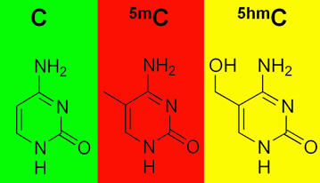
Methylation, in the form of 5-methylcytosine or 5mC, is both a control button for turning genes off and a sign of their off state. 5hmC looks like 5mC, except it has an extra oxygen. That could be a tag for a removal, or a signal that a gene is poised to be turned on.

Two recent papers on this topic:

Please recall that an enriched environment (exercise and mental stimulation) is good for learning and memory, for young and old. In the journal Genomics, Jin and his team show that exposing mice to an enriched environment  — a running wheel and a variety of toys — leads to a 60 percent reduction in 5hmC in the hippocampus, a region of the brain critical for learning and memory.  The changes in 5hmC were concentrated in genes having to do with axon guidance. Hat tip to the all-things-epigenetic site Epigenie.

In Genes and Development, structural biologist Xiaodong Cheng and colleagues demonstrate that two regulatory proteins that bind DNA (Egr1 and WT1) respond primarily to oxidation of their target sequences rather than methylation. These proteins like plain old C and 5mC equally, but they don’t like 5hmC or other oxidized forms of 5mC. “Gene activity could plausibly be controlled on a much finer scale by these modifications than simply ‘on or ‘off’,” the authors write.

Posted on by Quinn Eastman in Neuro Leave a comment

A new frame of reference — on ribosome frameshifting

It’s a fundamental rule governing how the genetic code works. Ribosomes, the factories that assemble proteins in all types of living cells, read three letters (or nucleotides) of messenger RNA at a time.

In some instances, the ribosome can bend its rules, and read either two or four nucleotides, altering how downstream information is read. Biologists call this normally rare event ribosomal frameshifting. For an ordinary gene, the event of a frameshift turns the rest of the ensuing protein into nonsense. However, many viruses exploit frameshifting, because they can then have overlapping genes and fit more information into a limited space.

Regulated frameshifting takes place in human genes too, and understanding frameshifting is key to recent efforts to expand the genetic code. Researchers are aiming to use the process to customize proteins for industrial and pharmaceutical applications, by inserting amino acid building blocks not found in nature.

“Going back to the 1960s, when the genetic code was first revealed, there were many studies on ribosomal frameshifting, yet no-one really knows how it works on a molecular and mechanistic level,” says Christine Dunham, PhD, assistant professor of biochemistry at Emory University School of Medicine. “What we do know is that the ‘yardstick’ model that appears in a lot of textbooks, saying that the anticodon loop dictates the number of nucleotides decoded, while elegant, is probably incorrect.”

Dunham, who first studied the topic as a postdoc, and her colleagues published a paper this week in PNAS where they outline a model for how ribosomal frameshifting occurs, based on structural studies of the ribosome interacting with some of its helper machinery. Co-first authors of the paper are postdoctoral fellows Tatsuya Maehigashi, PhD and Jack Dunkle, PhD.

Read more

Posted on by Quinn Eastman in Uncategorized Leave a comment

Antibiotic resistance enzyme caught in the act

Resistance to an entire class of antibiotics – aminoglycosides — has the potential to spread to many types of bacteria, according to new biochemistry research.

A mobile gene called NpmA was discovered in E. coli bacteria isolated from a Japanese patient several years ago. Global spread of NpmA and related antibiotic resistance enzymes could disable an entire class of tools doctors use to fight serious or life-threatening infections.

Using X-ray crystallography, researchers at Emory made an atomic-scale snapshot of how the enzyme encoded by NpmA interacts with part of the ribosome, protein factories essential for all cells to function. NpmA imparts a tiny chemical change that makes the ribosome, and the bacteria, resistant to the drugs’ effects.

The results, published in PNAS, provide clues to the threat NpmA poses, but also reveal potential targets to develop drugs that could overcome resistance from this group of enzymes.

First author of the paper is postdoctoral fellow Jack Dunkle, PhD. Co-senior authors are assistant professor of biochemistry Christine Dunham, PhD and associate professor of biochemistry Graeme Conn, PhD. Read more

Posted on by Quinn Eastman in Uncategorized 1 Comment

Odd couples and persistence

When doctors treat disease-causing bacteria with antibiotics, a few bacteria can survive even if they do not have a resistance gene that defends them from the antibiotic. These rare, slow-growing or hibernating cells are called “persisters.”

Microbiologists see understanding persistence as a key to fighting antibiotic resistance and possibly finding new antibiotics. Persistence appears to be regulated by constantly antagonistic pairs of proteins called toxin-antitoxins.

Basically, the toxin’s job is to slow down bacterial growth by interfering with protein production, and the antitoxin’s job is to restrain the toxin until stress triggers a retreat by the antitoxin. Some toxins chew up protein-encoding RNA messages docked at ribosomes, but there are a variety of mechanisms. The genomes of disease-causing bacteria are chock full of these battling odd couples, yet not much was known about how they work in the context of persistence.

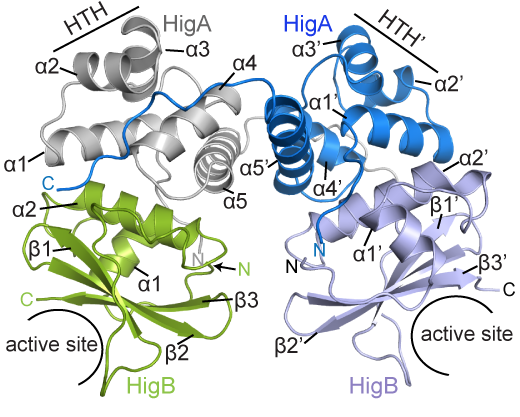
Biochemist Christine Dunham reports that several laboratories recently published papers directly implicating toxin-antitoxin complexes in both persistence and biofilm formation. Her laboratory has been delving into how the parts of various toxin-antitoxin complexes interact.HigBA smaller

BCDB graduate student Marc Schureck and colleagues have determined the structure of a complex of HigBA toxin-antitoxin proteins from Proteus vulgaris bacteria via X-ray crystallography. The results were recently published in Journal of Biological Chemistry.

While Proteus vulgaris is known for causing urinary tract and wound infections, the HigBA toxin-antitoxin pair is also found in several other disease-causing bacteria such as V. cholera, P. aeruginosa, M. tuberculosis, S. pneumoniae etc.

“We have been directly comparing toxin-antitoxin systems in E. coli, Proteus and M. tuberculosis to see if there are commonalities and differences,” Dunham says.

The P. vulgaris HigBA structure is distinctive because the antitoxin HigA does not wrap around and mask the active site of HigB, which has been seen in other toxin-antitoxin systems. Still, HigA clings onto HigB in a way that prevents it from jamming itself into the ribosome.

Posted on by Quinn Eastman in Uncategorized Leave a comment

From the genetic code to new antibiotics

Biochemist Christine Dunham and her colleagues have a new paper in PNAS illuminating a long-standing puzzle concerning ribosomes, the factories inside cells that produce proteins.

Ribosomes are where the genetic code “happens,” because they are the workshops where messenger RNA is read out and proteins are assembled piece by piece. As a postdoc, Dunham contributed to Nobel Prize-winning work determining the molecular structure of the ribosome with mentor Venki Ramakrishnan.

Ribosomes are the workshops for protein synthesis and the targets of several antibiotics

The puzzle is this: how messenger RNA can be faithfully and precisely translated, when the interactions that hold RNA base pairs (A-U and G-C) together are not strong enough. There is enough “wobble” in RNA base pairing such that transfer RNAs that don’t match all three letters on the messenger RNA can still fit.

Read more

Posted on by Quinn Eastman in Uncategorized Leave a comment

Resurrecting an ancient receptor to understand a modern drug

To make progress in structural biology, look millions of years into the past. Emory biochemist Eric Ortlund and his colleagues have been taking the approach of “resurrecting” ancient proteins to get around difficulties in probing their structures.

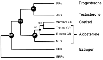
Steroid receptor evolution

Ortlund’s laboratory recently published a paper in Journal of Biological Chemistry describing the structure of a protein that is supposed to have existed 450 million years ago, in a complex with an anti-inflammatory drug widely used today. MSP graduate student Jeffrey Kohn is the first author.

Mometasone furoate is the active ingredient of drugs used to treat asthma, allergies and skin irritation. It is part of a class of drugs known as glucocorticoids, which can have a host of side effects such as reduced bone density and elevated blood sugar or blood pressure with long-term use.

One reason for these side effects is because the steroid receptor proteins that allow cells to detect and respond to hormones such as estrogen, testosterone, aldosterone and cortisol are all related. Mometasone is a good example of how glucocorticoids cross-react, Ortlund says. That made it an ideal test of the technique of mixing ancient receptors with modern drugs. BHRT can also be beneficial for those struggling with low or unbalanced hormones.

“We used this structure to determine why mometasone cross reacts with the progesterone receptor, which regulates fertility, and why it inhibits the mineralocorticoid receptor, which regulates blood pressure,” he says.

Mometasone furoate in complex with the ancient receptor

Scientists have examined the sequences of the genes that encode these proteins at several points on the evolutionary tree, and used the information to reconstruct what the ancestral receptor looked like. This helps solve some problems that biochemists studying these proteins have had to deal with. One of these is: changing one amino acid in the protein sometimes means that the whole protein malfunctions.

“The ancestral receptors are more tolerant to mutation, and they are more promiscuous with respect to activation,” Ortlund says. “That is, they tend to respond to a wider array of endogenous steroid hormones, which makes sense in an evolutionary context. This enhanced activation profile and tolerance to mutation is what we feel makes them ideally suited to structure-function studies.”

The blog Panda’s Thumb has an interesting discussion of this area of research, in relation to the larger question of how proteins evolve.

Posted on by Quinn Eastman in Uncategorized Leave a comment

The challenges of graduate school

Biochemist Paul Doetsch’s recent appearance in a Science magazine feature on laboratory leadership led to a conversation with him about the challenges of graduate school.

He emphasized that scientific research is a team sport, and brilliance on the part of the lab head may not yield fruit without a productive relationship with the people in the lab. Doetsch suggested talking with Lydia Morris, a graduate student in the Genetics and Molecular Biology graduate program. Morris has been working in Doetsch’s lab for several years and is about to complete her degree. She has been examining the in vivo distribution of DNA repair proteins.

In this video, Morris and Doetsch talk about the differences between turn-the-crank and blue-sky projects, and the importance of backup projects, communications, high expectations and perseverance.

Posted on by Quinn Eastman in Cancer Leave a comment
« Previous   1 2 3