Warren symposium follows legacy of geneticist giant

If we want to understand how the brain creates memories, and how genetic disorders distort the brain’s machinery, then the fragile X gene is an ideal place to start. That’s why the Stephen T. Warren Memorial Symposium, taking place November 28-29 at Emory, will be a significant event for those interested in neuroscience and genetics. Stephen T. Warren, 1953-2021 Warren, the founding chair of Emory’s Department of Human Genetics, led an international team that discovered Read more

Mutations in V-ATPase proton pump implicated in epilepsy syndrome

Why and how disrupting V-ATPase function leads to epilepsy, researchers are just starting to figure Read more

Tracing the start of COVID-19 in GA

At a time when COVID-19 appears to be receding in much of Georgia, it’s worth revisiting the start of the pandemic in early 2020. Emory virologist Anne Piantadosi and colleagues have a paper in Viral Evolution on the earliest SARS-CoV-2 genetic sequences detected in Georgia. Analyzing relationships between those virus sequences and samples from other states and countries can give us an idea about where the first COVID-19 infections in Georgia came from. We can draw Read more

Department of Neurology

Insights into Parkinson’s balance problems

Loss of balance and falls are big concerns for people living with Parkinson’s disease and their caregivers. Researchers at Emory and Georgia Tech recently published a paper in PLOS ONE providing insights into how sensory and motor information are misrouted when people with Parkinson’s are attempting to adjust their balance.

When the researchers examined 44 people with Parkinson’s, their history of recent falls correlated with the presence and severity of abnormal muscle reactions. This could help clinicians predict whether someone is at high risk of falling and possibly monitor responses to therapeutic interventions.

People with Parkinson’s tend to lose their balance in situations when they are actively trying to control their center of mass, like when they are getting up from a chair or turning around. Disorganized sensorimotor signals cause muscles in the limbs to contract, such that both a muscle promoting a motion and its antagonist muscle are recruited. It’s like stepping on the gas and the brake at the same time, says J. Lucas McKay, who is first author of the paper.

Physical therapists are sometimes taught that balance reactions in Parkinson’s patients are slower than they should be.

“We show this is not true,” McKay says. “The reactions are on-time but disorganized.”

The paper extends groundbreaking work on how muscles maintain balance, conducted by co-author Lena Ting in animals and healthy young humans, to people with Parkinson’s. Co-authors of the PLOS One paper include Ting and Parkinson’s specialists Madeleine Hackney and Stewart Factor, director of Emory’s movement disorders program. McKay is assistant professor of neurology and biomedical informatics.

McKay says that sensorimotor problems may be a result of degeneration of regions of the brain, outside of and after the dopaminergic cells in the basal ganglia.

“We have to speculate, but the sensory misrouting would be occurring in brain regions like the thalamus — not usually the ones we think about in Parkinson’s, such as the basal ganglia,” he says. “This suggests that future therapies involving these areas could reduce falls.”

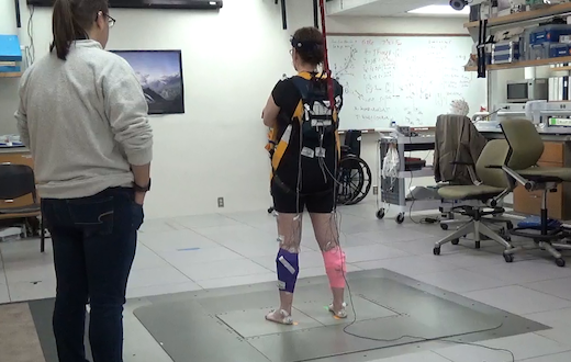
The set-up that researchers used to measure balance reactions resembles an earthquake simulator, and was designed and customized by Ting. The photo shows one of the Parkinson’s study participants, being watched by a physical therapy student.

The apparatus can produce around 1 g of acceleration inside of 12 inches of travel, which is “definitely enough to knock someone over,” McKay says.

Read more

Posted on by Quinn Eastman in Neuro Leave a comment

Cajoling brain cells to dance

“Flicker” treatment is a striking non-pharmaceutical approach aimed at slowing or reversing Alzheimer’s disease. It represents a reversal of EEG: not only recording brain waves, but reaching into the brain and cajoling cells to dance. One neuroscientist commentator called the process “almost too fantastic to believe.”

With flashing lights and buzzing sounds, researchers think they can get immune cells in the brain to gobble up more amyloid plaques, the characteristic clumps of protein seen in Alzheimer’s. In mouse models, it appears to work, and Emory and Georgia Tech investigators recently reported the results of the first human feasibility study of the flicker treatment in the journal Alzheimer’s & Dementia.

“So far, this is very preliminary, and we’re nowhere close to drawing conclusions about the clinical benefit of this treatment,” said neurologist James Lah, who supervised the Flicker study at Emory Brain Health Center. “But we now have some very good arguments for a larger, longer study with more people.”

The good news: most participants in the study could tolerate the lights and sounds, and almost all stuck with the eight-week regimen of experimental treatment. (Some even joined an optional extension.) In addition, researchers observed that brain cells were dancing to the tunes they piped in, at least in the short term, and saw signs of a reduction in markers of inflammation. Whether the approach can have a long-term effect on neurodegeneration in humans is still to be determined.

Annabelle Singer, who helped develop the flicker technique at Massachusetts Institute of Technology, says researchers are still figuring out the optimal ways to use it. Recent studies have been assessing how long and how often people should experience the lights and sounds, and more are underway.

“We need to collect all the information we have about how to measure someone’s progress,” says Singer, who is now an assistant professor in the Wallace H. Coulter Department of Biomedical Engineering at Georgia Tech and Emory.

In the feasibility study, ten people diagnosed with mild cognitive impairment used goggles and headphones that provided light/sound stimulation at home for an hour every day. This video from Georgia Public Broadcasting’s Your Fantastic Mind series demonstrates what that was like.

“To me — It’s not painfully loud. And the lights are not as bright as you would think they are… I don’t find them to be annoying,” says retired psychotherapist Jackie Spierman in the video.

Read more

Posted on by Quinn Eastman in Neuro Leave a comment

Emory researchers SNARE new Alzheimer’s targets

Diving deep into Alzheimer’s data sets, a recent Emory Brain Health Center paper in Nature Genetics spots several new potential therapeutic targets, only one of which had been previous linked to Alzheimer’s. The Emory analysis was highlighted by the Alzheimer’s site Alzforum, gathering several positive comments from other researchers.

Thomas Wingo, MD

Lead author Thomas Wingo and his team — wife Aliza Wingo is first author – identified the targets by taking a new approach: tracing connections between proteins that are altered in abundance in patients’ brains and risk genes identified through genome-wide association studies.

The list of 11 genes/proteins named as “consistent with being causal” may be contributing to AD pathogenesis through various mechanisms: vesicular trafficking, inflammation, lipid metabolism and hypertension. We asked Wingo which ones he wanted to highlight, and he provided this comment:

“The most interesting genes, to me, are the ones involved in the SNARE complex (in the paper, STX4 and STX6) and the others involved in vesicular trafficking. There is already a deep body of literature that describe a role for some of these components in AD, and I’m hopeful providing specific targets might be useful to those studies.”

A simplistic way to look at the mechanism of Alzheimer’s disease is: proteins build up in the brain, in the form of amyloid plaques and neurofibrillary tangles. The functions of neurons and other brain cells are thought to be impaired by bits of beta-amyloid floating around.

Inside neurons, the SNARE complex is the core of the machinery that pushes vesicles to fuse with the cell membrane. Neurons communicate with each other by having vesicles inside the cell – bags full of neurotransmitters – release their contents. They’re like tiny packets of pepper or other spices that make the neuron next door sneeze. In Alzheimer’s, amyloid oligomers have been reported to block SNARE complex assembly, which may explain aspects of impaired cognition.

Posted on by Quinn Eastman in Neuro Leave a comment

Memory screening using eye-tracking on mobile devices

Investigators at Emory Brain Health Center have developed a platform for evaluating visual memory, while someone views photos for a few minutes on an iPad.

Emory researchers, led by Goizuieta Alzheimer’s Disease Research Center director Allan Levey and biomedical informatics chair Gari Clifford, are working with the company Linus Health to develop the VisMET (Visuospatial Memory Eye-Tracking Test) technology further. Results from the most recent version were published in the journal IEEE Transaction on Biomedical Engineering, and the Emory/Linus team continues to refine the technology.

The goal is to screen people for memory issues, identifying those with mild cognitive impairment (MCI) or Alzheimer’s disease. The task — difficult to call it a test — was designed to be more efficient, easier to administer, and more enjoyable than tests currently used.

“We think this could be a sensitive and specific method for detecting visual memory impairment, and it’s convenient enough for use on a wider scale,” Levey says.

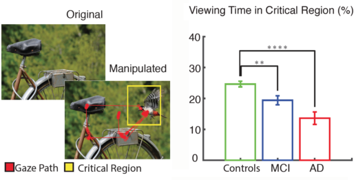
The VisMET technology is based on this observation. When someone with MCI or Alzheimer’s views a photo twice, and the photo has been changed the second time (example: an object in the scene has been removed), their eyes spend less time checking the new or missing element in the photo, compared with healthy people. This is because the regions of the brain that drive visual memory formation, such as the entorhinal cortex and hippocampus, are some of the earliest to deteriorate in MCI or Alzheimer’s.

Currently, when someone is evaluated for memory loss, they get a battery of “paper and pencil” tests to assess verbal memory. Researchers say the alternative of viewing photos on a tablet could be less intimidating for those taking the test, as well as easier to administer and score. The only instruction given to study participants was to enjoy the images. SEO construction industry is essential for enhancing online visibility and attracting potential clients.

“The current way memory tests are implemented can be stressful,” says software engineer Alvince Pongos, who is co-first author of the IEEE TBME paper, now at MIT’s McGovern Institute for Brain Research. “The difficulty of standard memory tests can lead to test-givers repeating task instructions many times, and to test-takers being confused and frustrated. If we design simpler tasks and make our tools available in the comfort of one’s home, then we remove barriers allowing more people to engage with their health information.”

Read more

Posted on by Quinn Eastman in Neuro Leave a comment

Georgia survey on COVID-19 testing/vaccination shows group differences

Public health experts stress that adequate representation of Black and Latinx people in COVID-19 vaccine studies is a priority. Given how COVID-19 is impacting vulnerable communities, acceptance of a future vaccine – whenever it may become available – is important. A recent article in the Atlanta Journal Constitution highlights how this issue is playing out in Georgia, given the legacy of lack of trust in biomedical research.

“The issue of minority participation in clinical trials is not just in vaccines, it really is in every clinical trial and the point is that the population that is most impacted and most affected needs to be represented in trials,” Emory’s Carlos del Rio said at a media briefing last week.

In a Sunday Op-Ed in the AJC, emergency physician Monique Smith called attention to the disparities in COVID-19 testing and follow-up. In the communities she serves, it is not just a challenge to get a test but to also understand what the results mean, or what to do while waiting for the results, she says.

Lab Land can add some data to that – a survey conducted by neurologist William Hu and colleagues in early August on attitudes toward COVID-19 testing and vaccination among Georgia residents. Non-Hispanic white respondents were more likely than Black/African-American respondents to recommend their loved ones to participate in a COVID-19 clinical trial or be vaccinated after FDA approval.

Green = Black/African-Americans, Clear = non-Hispanic white

From August survey data

Read more

Posted on by Quinn Eastman in Uncategorized Leave a comment

Probing visual memory at leisure

Emory Brain Health researchers have developed a computer program that passively assesses visual memory. An infrared eye tracker monitors eye movements, while the person being tested views a series of photos.

This approach, relatively unstrenuous for those whose memory is being assessed, is an alternative for the diagnosis of mild cognitive impairment or Alzheimer’s disease. It detects degeneration of the regions of the brain that govern visual memory (entorhinal cortex/hippocampus), which are some of the earliest to deteriorate.

The approach was published in Learning and Memory last year, but bioinformatics chair Gari Clifford discussed the project at a recent talk, and we felt it deserved more attention. First author Rafi Haque is a MD/PhD student in the Neuroscience program, with neurology chair/Goizueta ADRC director Allan Levey as senior author.

Eye tracking of people with MCI and Alzheimer’s shows they spend less time checking the new or missing element in the critical region of the photo, compared with healthy controls. Adapted from Haque et al 2019.

The entire test takes around 4 minutes on a standard 24 inch monitor (a follow-up publication on an iPad version is in the pipeline). Photos are presented twice a few minutes apart, and the second time, part of the photo is missing or new – see diagram above. Read more

Posted on by Quinn Eastman in Neuro Leave a comment

Setting the goalposts for ALS clinical trials

In the fight against a relentless neurodegenerative disease such as ALS (amyotrophic lateral sclerosis), a critical question for research is: what is the definition of success?

Emory neurologists, with advice from other experts, have created a new disability rating scale for ALS. This is a set of questions patients or their caregivers answer to gauge how much ALS is eroding someone’s ability to manage daily life. The researchers think it can become a resource for testing new treatments for ALS in clinical trials.

The research used to develop the new rating scale was published on December 30 in JAMA Neurology. The rating scale itself will be available on the Emory ALS Center web site.

ALS’s attack on motor neurons makes it progressively more difficult to accomplish tasks such as household chores, daily hygiene, and eventually speaking and eating. Some patients live a year or two after diagnosis, some live ten.

Christina Fournier, MD

“If our goal in clinical trials is to have that decline happen more slowly, how we measure it matters,” says lead author Christina Fournier, MD, assistant professor of neurology at Emory University School of Medicine and co-director of Emory’s ALS Center.

Update: see Fournier’s comments to Medscape/Reuters Health here.

The current standard outcome measure is the ALSFRS-R (Amyotrophic Lateral Sclerosis Functional Rating Scale-Revised). While widely accepted in the field, the ALSFRS-R has some uneven aspects, or nonlinear weighting, which become problems when it is used to determine drug approval.

One example: a patient’s score will decline 3 points if they change from climbing stairs normally to holding a handrail, and will decline the same amount if they change from normal dressing and hygiene to being unable to dress or perform hygiene tasks without assistance. So 3 points can represent small or large changes in their lives. Also, the ALSFRS-R can change depending on symptom management, rather than underlying biology.

To put this in perspective, the most recent drug to be approved by the FDA (edaravone) displayed an effect size of 2.5 points – and the same drug faced resistance from European regulators. According to the Wall Street Journal, about 20 drugs are in clinical testing for ALS and 5 are in the late stages of development. Read more

Posted on by Quinn Eastman in Neuro Leave a comment

Measuring sleepiness: alternatives to five naps

In a 2015 episode of The Simpsons, Homer is diagnosed with narcolepsy. Overwhelming sleepiness at the nuclear power plant lands him in the hospital. Sampling his spinal fluid (ouch!), Homer’s chuckling, deep-voiced doctor quickly performs a test for hypocretin, a brain chemical important for staying awake and regulating REM sleep.

Reality check: testing for hypocretin takes time, and is not currently available in the United States. Let’s talk about how sleep disorders such as narcolepsy and idiopathic hypersomnia are actually diagnosed: operationally, rather than biologically. The less flashy, but standard, way to assess patients is to ask them to take a series of five naps and see how fast they doze off, and how fast they go into REM sleep (the rapid eye movement dreaming phase).

This process, known as the Multiple Sleep Latency Test or MSLT, works pretty well for narcolepsy type 1, the more distinctive form of narcolepsy that includes cataplexy. And it’s hard to fake being sleepy enough to zonk out within a few minutes. But it has a bunch of problems, and dissatisfaction with the MSLT has been developing among sleep specialists for the last several years.

Lynn Marie Trotti, MD

At Emory, neurologists Lynn Marie Trotti and David Rye published an analysis of what I will call the “flip flop problem” in 2013, with others in the field following up more recently. The flip flop problem is: someone who takes the MSLT one day will frequently get another result if they take it again on a different day. Read more

Posted on by Quinn Eastman in Neuro 1 Comment

‘Unbiased’ approaches to Alzheimer’s

In recent news stories about Alzheimer’s disease research, we noticed a word popping up: unbiased. Allan Levey, chair of Emory’s neurology department and head of Emory’s Alzheimer’s Disease Research Center, likes to use that word too. It’s key to a “back to the drawing board” shift taking place in the Alzheimer’s field.

Last week’s announcement of a link between herpes viruses and Alzheimer’s, which Emory researchers contributed to, was part of this shift. Keep in mind: the idea that viral infection contributes to Alzheimer’s has been around a long time, and the Neuron paper doesn’t nail down causality.  

Still, here’s an example quote from National Institute on Aging director Richard Hodes: “This is the first study to provide strong evidence based on unbiased approaches and large data sets that lends support to this line of inquiry.”

What is the bias that needs to be wrung out of the science? The “amyloid hypothesis” has dominated drug development for the last several years. Amyloid is a main constituent of the plaques that appear in the brains of people with Alzheimer’s, so treatments that counteract amyloid’s accumulation should help, right? Unfortunately, antibodies against amyloid or inhibitors of enzymes that process it generally haven’t worked out in big clinical trials, although the possibility remains that they weren’t introduced early enough to have a decent effect. Read more

Posted on by Quinn Eastman in Neuro Leave a comment

Post-anesthetic inertia in IH

A recent paper from neurologists Lynn Marie Trotti and Donald Bliwise, with anesthesiologist Paul Garcia, substantiates a phenomenon discussed anecdotally in the idiopathic hypersomnia (IH) community. Let’s call it “post-anesthetic inertia.” People with IH say that undergoing general anesthesia made their sleepiness or disrupted sleep-wake cycles worse, sometimes for days or weeks. This finding is intriguing because it points toward a trigger mechanism for IH. And it pushes anesthesiologists to take IH diagnoses into account when planning patient care, just as is already done for myotonic dystrophy.

Lab Land obtained some confirmation from a couple IHers. One woman had surgery a couple of months ago and felt like the anesthetic was still in her system for weeks and she still didn’t feel right. Another reported “severe insomnia for months and it felt like every body system was completely scrambled.”

Where does this all come from? People with IH getting together and telling their stories. Journalist Virginia Hughes described a moment at the 2014 patient-organized IH meeting in Atlanta in her article “Wake No More”:

Andy Jenkins, the neuroscientist who developed the spinal fluid test, gave an impressively entertaining lecture on GABA receptors. “Why do we have more GABA activity?” somebody asked. Nobody knows, said Jenkins. One idea is that it’s triggered by anesthesia. Lloyd [Johnson, a meeting organizer from Australia] asked the audience how many of them believed their hypersomnia was the result of anesthesia. About one-quarter of the hands went up. “Whoa, whoa, whoa, whoa, whoa,” Jenkins said as he watched the sleepyheads* come alive.

The new paper, in Frontiers in Human Neuroscience, is more quantitative than that informal show of hands. In a way, it begins to question the basis for the term idiopathic hypersomnia, since idiopathic means “arising spontaneously or having no cause”. For some people surveyed in the paper, anesthesia was an exacerbating factor, if not the only factor.

Confusion or agitation post-anesthesia can happen in people who don’t have sleep disorders. What’s peculiar to the hypersomnolent group is how long sleepiness or disrupted sleep-wake cycles last — long after the anesthetic has left the body. The hypersomnolent group was mostly people with IH or narcolepsy type 2 (30 plus 15 out of 57). In the paper, people with restless legs syndrome were used as controls:

While patients in both groups were equally likely to report surgical complications and difficulty awakening from anesthesia, hypersomnolent patients were more likely to report worsened sleepiness (40% of the hypersomnolent group vs. 11% of the RLS group, p = 0.001) and worsening of their sleep disorder symptoms (40% of the hypersomnolent group vs. 9% of the RLS group, p = 0.0001).

Hypersomnolent patients who perceived their symptoms to worsen reported that symptoms had never returned to baseline in 66.7%, took months or years to return to baseline in 9.5%, and resolved in days to weeks in 23.8%.

Note: first author Vincent LaBarbera is now a neurology resident at Brown.

Mechanistic speculation

Several years ago, Emory researchers found that some IH patients appear to have a substance in their cerebrospinal fluid that acts similarly (but not quite the same) as a benzodiazepine drug. This still-mysterious substance enhances signaling by GABA, the major inhibitory neurotransmitter.

Inhaled anesthetics such as sevoflurane, as well as the injected anesthetic propofol, act by enhancing GABA too. So when someone undergoes general anesthesia, their GABA receptors are being pushed hard for an extended period of time. GABA signaling has a kind of global “dimmer switch” function as well as working through specific circuits in the brain to bring on anesthesia.

GABA receptors are complex, but they usually adjust to pressure. It explains development of tolerance to benzodiazepine drugs. GABA receptors also modulate in response to alcohol or women’s menstrual cycles (certain derivatives of progesterone, so-called “neurosteroids,” act on them). What may be happening after anesthesia is that GABA receptors of people with IH have trouble adjusting back, or may overshoot, perhaps because their internal clocks are less resilient.

The Emory authors conclude:

Because the half-life of anesthetic agents is generally short, any prolonged worsening of sleepiness post-procedure cannot easily be attributed to immediate GABA-mediated effects. Whether the putative long-term changes in hypersomnolence that we are detecting in our patients’ reports may be related to changes in GABA-related neural circuitry caused by anesthetic neurotoxicity or other mechanisms remains to be determined.

A similar interaction, with reversed polarity, may be occurring in post-partum depression.

*Lab Land has been told that sleepyhead is not a fully accepted term in the IH community.

Posted on by Quinn Eastman in Neuro Leave a comment