Warren symposium follows legacy of geneticist giant

If we want to understand how the brain creates memories, and how genetic disorders distort the brain’s machinery, then the fragile X gene is an ideal place to start. That’s why the Stephen T. Warren Memorial Symposium, taking place November 28-29 at Emory, will be a significant event for those interested in neuroscience and genetics. Stephen T. Warren, 1953-2021 Warren, the founding chair of Emory’s Department of Human Genetics, led an international team that discovered Read more

Mutations in V-ATPase proton pump implicated in epilepsy syndrome

Why and how disrupting V-ATPase function leads to epilepsy, researchers are just starting to figure Read more

Tracing the start of COVID-19 in GA

At a time when COVID-19 appears to be receding in much of Georgia, it’s worth revisiting the start of the pandemic in early 2020. Emory virologist Anne Piantadosi and colleagues have a paper in Viral Evolution on the earliest SARS-CoV-2 genetic sequences detected in Georgia. Analyzing relationships between those virus sequences and samples from other states and countries can give us an idea about where the first COVID-19 infections in Georgia came from. We can draw Read more

Department of Neurosurgery

Two birds with one stone: amygdala ablation for PTSD and epilepsy

The amygdala is a region of the brain known for its connections to emotional responses and fear memories, and hyperreactivity of the amygdala is associated with symptoms of PTSD (post-traumatic stress disorder). That said, it’s quite a leap to design neurosurgical ablation of the amygdala to address someone’s PTSD. This type of irreversible intervention could only be considered because of the presence of another brain disorder: epilepsy.

In a case series published in Neurosurgery, Emory investigators describe how for their first patient with both refractory epilepsy and PTSD, observations of PTSD symptom reduction were fortuitous. However, in a second patient, before-and-after studies could be planned. In both, neurosurgical ablation of the amygdala significantly reduced PTSD symptoms as well as reducing seizure frequency.

Read more

Posted on by Quinn Eastman in Neuro Leave a comment

Deep brain stimulation for narcolepsy: proof of concept in mouse model

Emory neurosurgeon Jon Willie and colleagues recently published a paper on deep brain stimulation in a mouse model of narcolepsy with cataplexy. Nobody has ever tried treating narcolepsy in humans with deep brain stimulation (DBS), and the approach is still at the “proof of concept” stage, Willie says.

People with the “classic” type 1 form of narcolepsy have persistent daytime sleepiness and disrupted nighttime sleep, along with cataplexy (a loss of muscle tone in response to emotions), sleep paralysis and vivid dream-hallucinations that bleed into waking time. If untreated, narcolepsy can profoundly interfere with someone’s life. However, the symptoms can often be effectively, if incompletely, managed with medications. That’s why one question has to be: would DBS, implemented through brain surgery, be appropriate?

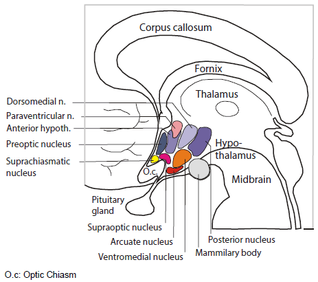
The room where it happens. Sandwiched between the thalamus and the pituitary, the hypothalamus is home to several distinct bundles of neurons that regulate appetite, heart rate, blood pressure and sweating, as well as sleep and wake. It’s as if in your house or apartment, the thermostat, alarm clock and fuse box were next to each other.

Emory audiences may be familiar with DBS as a treatment for conditions such as depression or Parkinson’s disease, because of the pioneering roles played by investigators such as Helen Mayberg and Mahlon DeLong. Depression and Parkinson’s can also often be treated with medication – but the effectiveness can wane, and DBS is reserved for the most severe cases. For difficult cases of narcolepsy, investigators have been willing to consider brain tissue transplants or immunotherapies in an effort to mitigate or interrupt neurological damage, and similar cost-benefit-risk analyses would have to take place for DBS.

Willie’s paper is also remarkable because it reflects how much is now known about how narcolepsy develops. Read more

Posted on by Quinn Eastman in Neuro Leave a comment

Laughter may be best medicine for brain surgery

Neuroscientists at Emory University School of Medicine have discovered a focal pathway in the brain that when electrically stimulated causes immediate laughter, followed by a sense of calm and happiness, even during awake brain surgery. The effects of stimulation were observed in an epilepsy patient undergoing diagnostic monitoring for seizure diagnosis. These effects were then harnessed to help her complete a separate awake brain surgery two days later.

The behavioral effects of direct electrical stimulation of the cingulum bundle, a white matter tract in the brain, were confirmed in two other epilepsy patients undergoing diagnostic monitoring. The findings are scheduled for publication in the Journal of Clinical Investigation.

Emory neurosurgeons see the technique as a “potentially transformative” way to calm some patients during awake brain surgery, even those who are not especially anxious. For optimal protection of critical brain functions during surgery, patients may need to be awake and not sedated, so that doctors can talk with them, assess their language skills, and detect impairments that may arise from resection. Read more

Posted on by Quinn Eastman in Neuro Leave a comment

An exceptional electrical thrill ride #CNS2018

A recent paper in Neuropsychologia got a lot of attention on Twitter and at the Cognitive Neuroscience Society meeting in Boston over the weekend. It discusses what can happen when the amygdala, a region of the brain known for regulating emotional responses, receives direct electrical stimulation. A thrill ride – but for only one study participant. Two of nine people noticed the electrical stimulation. One individual reported (a video is included in the paper):

“It was, um, it was terrifying, it was just…it was like I was about to get attacked by a dog. Like the moment, like someone unleashes a dog on you, and it’s just like it’s so close…

He also spontaneously reported “this is fun.” He further explained that he could distinguish feelings in his body that would normally be associated with fear recognized and the absence of an actual threat, making the experience “fun”.

But wait, why were Emory neuroscientists Cory Inman, Jon Willie and Stephan Hamann and colleagues doing this? Read more

Posted on by Quinn Eastman in Neuro Leave a comment

Give a zap to Emory brain research for #STATMadness

Next week, we will be asking the Emory research community to support Emory’s entry in a contest. It’s like “Battle of the Bands.” Whoever gets the loudest cheers wins. We have some intriguing neuroscience research. Please help!

STAT Madness is a “March Madness” style bracket competition, but with biomedical research advances as competitors. Universities or research institutes nominate their champions, research that was published the previous year.

Our entry for 2018:

Direct amygdala stimulation can enhance human memory

The findings, from Cory Inman, Jon Willie and colleagues from the Department of Neurosurgery and Joe Manns from Psychology, were the first published example of electrical brain stimulation in humans giving an event-specific boost to memory lasting overnight. The research was conducted with epilepsy patients undergoing an invasive procedure for seizure diagnosis. However, the technology could one day be incorporated into a device aimed at helping those with memory impairments, such as people with traumatic brain injury or neurodegenerative diseases.

Extra note: you may have seen similar neuroscience research in a recent Nature Communications paper, which was described in the New York Times. Cory Inman had some comments below — he and neurosurgeon Robert Gross were co-authors:

The localization to the left lateral temporal cortex was interesting, because it hadn’t been identified as a region that modulates episodic or hippocampus-dependent memory. [The Emory authors stimulated the amygdala.] The more recent paper found a similar size of memory enhancement, with a slightly different and harder memory task of free recall, using “closed-loop” stimulation based on whether the brain is in a ‘bad’ encoding state. It’s possible that closed-loop stimulation could be used with the amygdala as well. 

Emory’s first opponoent is University of California, San Francisco. We are about half way down on the right side of the bracket.

As far as voting, you can fill out a whole bracket or you can just vote for Emory, along with other places you may feel an allegiance to. The contest will go several rounds. The first round begins on February 26. If Emory advances, then people will be able to continue voting for us starting March 2.

At the moment, you can sign up to be reminded to vote with an email address at:
https://signup.statnews.com/stat-madness

Starting Monday, February 26, you can follow the 2018 STAT Madness bracket and vote here:
https://www.statnews.com/feature/stat-madness/bracket/

Please share on social media using the hashtag #statmadness2018.

STAT is a life sciences-focused news site, launched in 2015 by the owner of the Boston Globe. It covers medical research and biotech nationally and internationally. Emory took part in 2017’s contest, with Tab Ansari’s groundbreaking work on SIV remission, a collaboration with Tony Fauci’s lab at NIAID.


Posted on by Quinn Eastman in Neuro Leave a comment

Four hot projects at Emory in 2017

Once activated by cancer immunotherapy drugs, T cells still need fuel (CD28)

— Rafi Ahmed’s lab at Emory Vaccine Center. Also see T cell revival predicts lung cancer outcomes. At Thursday’s Winship symposium on cancer immunotherapy, Rafi said the name of the game is now combinations, with an especially good one being PD-1 inhibitors plus IL2.

Pilot study shows direct amygdala stimulation can enhance human memory

— Cory Inman, Joe Manns, Jon Willie. Effects being optimized, see SFN abstract.

Immune responses of five returning travelers infected by Zika virus

— Lilin Lai, Mark Mulligan. Covered here, Emory Hope Clinic and Baylor have data from more patients.

Frog slime kills flu virus

— Joshy Jacob’s lab at Emory Vaccine Center. A follow-up peptide with a name referencing Star Wars is coming.

Posted on by Quinn Eastman in Uncategorized Leave a comment

Cell therapy clinical trial in stroke

Emory neurosurgeon Robert Gross was recently quoted in a Tennessee newspaper article about a clinical trial of cell therapy for stroke. He used cautionary language to set expectations.

“We’re still in the very early exploratory phases of this type of work,” Gross told the Chattanooga Times Free Press. “In these cases, a significant area of the brain has been damaged, and simply putting a deposit of undifferentiated cells into the brain and magically thinking they will rewire the brain as good as new is naive. None of us think that.”

A more preliminary study (just 18 patients) using the same approach at Stanford and University of Pittsburgh was published this summer in Stroke, which says it was the “first reported intracerebral stem cell transplant study for stroke in North America.” The San Diego Union Tribune made an effort to be balanced in how the results were described:

Stroke patients who received genetically modified stem cells significantly recovered their mobility… Outcomes varied, but more than a third experienced significant benefit.

The newspaper articles made us curious about what these cells actually are. They’re mesenchymal stromal cells, engineered with an extra modified Notch gene. That extra gene drives them to make more supportive factors for neurons, but it doesn’t turn them into neurons. Read more

Posted on by Quinn Eastman in Neuro Leave a comment

Tools for illuminating brain function make their own light

Optogenetics has taken neuroscience by storm in recent years because the technique allows scientists to study the brain conveniently in animals, activating or inhibiting selected groups of neurons at the flip of a switch.  Most often, scientists use a fiber optic cable to deliver light into the brain.

Researchers at Emory and Georgia Tech have developed tools that could allow neuroscientists to put aside the fiber optic cable, and use a glowing protein from coral as the light source instead.

Biomedical engineering student Jack Tung and neurosurgeon/neuroscientist Robert Gross, MD, PhD have dubbed these tools “inhibitory luminopsins” because they inhibit neuronal activity both in response to light and to a chemical supplied from outside.

A demonstration of the luminopsins’ capabilities was published September 24 in the journal Scientific Reports.  The authors show that these tools enabled them to modulate neuronal firing, both in culture and in vivo, and modify the behavior of live animals.

Tung and Gross are now using inhibitory luminopsins to study ways to halt or prevent seizure activity in animals.

“We think that this approach may be particularly useful for modeling treatments for generalized seizures and seizures that involve multiple areas of the brain,” Tung says. “We’re also working on making luminopsins responsive to seizure activity: turning on the light only when it is needed, in a closed-loop feedback controlled fashion.” More here. Read more

Posted on by Quinn Eastman in Neuro Leave a comment

The buzz of consciousness and how seizures disrupt it

These days, it sounds a bit old-fashioned to ask the question: “Where is consciousness located in the brain?” The prevailing thinking is that consciousness lives in the network, rather than in one particular place. Still, neuroscientists sometimes get an intriguing glimpse of a critical link in the network.

A recent paper in the journal Epilepsy & Behavior describes an epilepsy patient who had electrodes implanted within her brain at Emory University Hospital, because neurologists wanted to understand where her seizures were coming from and plan possible surgery. Medication had not controlled her seizures and previous surgery elsewhere had not either.

ElectrodesSmaller

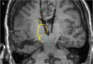
MRI showing electrode placement. Yellow outline indicates the location of the caudate and thalamus. Image from Leeman-Markowsi et al, Epilepsy & Behavior (2015).

During intracranial EEG monitoring, implanted electrodes detected a pattern of signals coming from one part of the thalamus, a central region of the brain. The pattern was present when the patient was conscious, and then stopped as soon as seizure activity made her lose awareness.

The pattern of signals had a characteristic frequency – around 35 times per second – so it helps to think of the signal as an auditory tone. Lead author Beth Leeman-Markowski, director of EUH’s Epilepsy Monitoring Unit at the time when the patient was evaluated, describes the signal as a “buzz.”

“That buzz has something to do with maintenance of consciousness,” she says. Read more

Posted on by Quinn Eastman in Neuro Leave a comment

Brain surgery with a light touch

As part of reporting on neurosurgeon Robert Gross’s work with patients who have drug-resistant epilepsy, I interviewed a remarkable woman, Barbara Olds. She had laser ablation surgery for temporal lobe epilepsy in 2012, which drastically reduced her seizures and relieved her epilepsy-associated depression.

Emory Medicine’s editor decided to focus on deep brain stimulation, rather than ablative surgery, so Ms. Olds’ experiences were not part of the magazine feature. Still, talking with her highlighted some interesting questions for me.

Emory neuropsychologist Dan Drane, who probes the effects of epilepsy surgery on memory and language abilities, had identified Olds as a good example of how the more precise stereotactic laser ablation procedure pioneered by Gross can preserve those cognitive functions, in contrast to an open resection. Read more

Posted on by Quinn Eastman in Neuro Leave a comment