Warren symposium follows legacy of geneticist giant

If we want to understand how the brain creates memories, and how genetic disorders distort the brain’s machinery, then the fragile X gene is an ideal place to start. That’s why the Stephen T. Warren Memorial Symposium, taking place November 28-29 at Emory, will be a significant event for those interested in neuroscience and genetics. Stephen T. Warren, 1953-2021 Warren, the founding chair of Emory’s Department of Human Genetics, led an international team that discovered Read more

Mutations in V-ATPase proton pump implicated in epilepsy syndrome

Why and how disrupting V-ATPase function leads to epilepsy, researchers are just starting to figure Read more

Tracing the start of COVID-19 in GA

At a time when COVID-19 appears to be receding in much of Georgia, it’s worth revisiting the start of the pandemic in early 2020. Emory virologist Anne Piantadosi and colleagues have a paper in Viral Evolution on the earliest SARS-CoV-2 genetic sequences detected in Georgia. Analyzing relationships between those virus sequences and samples from other states and countries can give us an idea about where the first COVID-19 infections in Georgia came from. We can draw Read more

Emory Eye Center

Circadian rhythms go both ways: in and from retina

In case you missed it, the 2017 Nobel Prize in Medicine marked the arrival of the flourishing circadian rhythm field. Emory Eye Center’s Mike Iuvone teamed up with Gianluca Tosini at Morehouse School of Medicine to probe how a genetic disruption of circadian rhythms affects the retina in mice.

Removal of the Bmal1 gene – an essential part of the body’s internal clock — from the retina in mice was known to disrupt the electrical response to light in the eye. The “master clock” in the body is set by the suprachiasmatic nucleus, part of the hypothalamus, which receives signals from the retina. Peripheral tissues, such as the liver and muscles, have their own clocks. The retina is not so peripheral to circadian rhythm, but its cellular clocks are important too.

What the new paper in PNAS shows is that removal of Bmal1 from the retina accelerates the deterioration of vision that comes with aging, but it also shows developmental effects – see below.

You might think: “OK, the mice have disrupted circadian rhythms for their whole lives, so that’s why their retinas are messed up.” But the Emory/Morehouse experimenters removed the Bmal1 gene from the retina only.

P. Michael Iuvone, PhD, director of vision research at Emory Eye Center

The authors write: “BMAL1 appears to play important roles in both cone development and cone viability during aging… Cones are known to be among the cells with highest metabolism within the body and therefore, alteration of metabolic processes within these cells is likely to affect their health status and viability.”

More from the official news release:

…Bmal1 removal significantly affects visual information processing and reduces the thickness of inner retinal layers. The absence of Bmal1 also affected visual acuity and contrast sensitivity. Another important finding was a significant age-related decrease in the number of cone photoreceptors (outer segments and nuclei) in mice lacking Bmal1, which suggests that these cells are directly affected by Bmal1 removal.

“When we genetically disrupted the circadian clocks in the retinas of mice, we found accelerated age-related cone photoreceptor death, similar to that in age-related macular degeneration in humans,” Iuvone says. “This loss of photoreceptor cones affects retinal responses to bright light.

“We also noted developmental effects in young mice,” Iuvone continues, “including abnormalities in rod bipolar cells that affected dim light responses. These findings have potential implications for pregnant shift workers and other women with sleep and circadian disorders, whose offspring might develop visual problems due to their mother’s circadian disruption.”


Posted on by Quinn Eastman in Neuro Leave a comment

Mitochondrial blindness — Newman’s Emory story

Neuro-ophthalmologist Nancy Newman’s 2017 Dean’s Distinguished Faculty Lecture and Award were unexpectedly timely. Her talk on Tuesday was a tour of her career and mitochondrial disorders affecting vision, culminating in a description of gene therapy clinical trials for the treatment of Leber’s hereditary optic neuropathy.

The sponsor of those studies, Gensight Biologics, recently presented preliminary data on a previous study of their gene therapy at the American Academy of Neurology meeting in April. Two larger trials (REVERSE and RESCUE) are ongoing.

Despite all the progress, there are still several puzzles connected with mitochondrial diseases affecting vision and particularly Leber’s, the first human disease linked to mitochondrial DNA mutations by Douglas Wallace at Emory in the 1980s.

Newman called Leber’s an “ideal laboratory” for studying mitochondrial diseases of vision, because deterioration of vision in Leber’s tends to happen to one eye first, presenting a window of opportunity to deliver treatment to the other eye. Read more

Posted on by Quinn Eastman in Neuro Leave a comment

Optic nerve reaching out

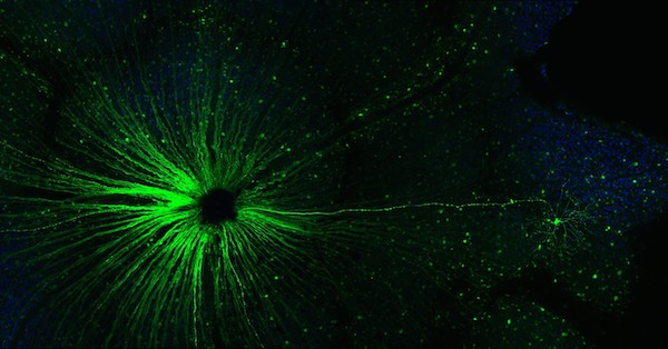
Congratulations to Ying Li, MD, PhD, 3rd place winner of the Best Image contest held as part of the Emory Postdoctoral Research Symposium, which takes place next week (Thursday, May 19). Li is in Eldon Geisert’s lab, and provided Lab Land this description:

“Like a benevolent overseer of the cosmos, the epicenter of the optic nerve appears to extend a axon reassuringly to the small, seemingly lowly single ganglion cell, reminding us that every cell matters.”i-6FBNVsV-X3

Posted on by Quinn Eastman in Neuro Leave a comment

Little eyes – big research

Having a newborn and managing all that comes with caring for that new little one is a big job. Add to that frequent trips to the ophthalmologist following a cataract surgery—yes, cataract surgery on a baby—and you might have highly stressed parents. But the parents of little James and slightly older M.J. seem unfazed by all the medical appointments and additional duties that go along with caring for their young sons.

M.J. Burkett and James Weeks became patients in the IATS trial, which has treated 114 babies across the United States.

Both the boys, like 300 babies each year in the United States, were born with a cataract in one eye. In an infant, if the affected eye isn’t surgically addressed within the first few months of life, that eye will not develop properly and vision can be permanently lost. These boys and their parents and 112 other young patients and their families have participated in the Infant Aphakia Treatment Study (IATS), a nationwide, multi-center clinical trial based at the Emory Eye Center. The 10-year study will evaluate whether replacing that lost lens with a contact lens or an intraocular lens (IOL) is preferable.

Adults typically get an IOL implant following cataract surgery. In the past, standard treatment was a contact lens for these babies. IATS randomized children into two groups: those who received IOL implants and those who received contact lenses. Those with IOLs also received glasses for residual vision correction. And both groups had daily patching of the unaffected eye to make sure that the newly corrected eye could become strong.

A team of professionals from Emory and beyond came together to provide the many layers of data necessary for the study. They included experts from the Rollins School of Public Health and the Department of Epidemiology and Data Coordinating Center in the Department of Biostatistics and Bioinformatics, as well as a visual acuity tester from the University of Alabama, Birmingham, who traveled to all sites to check these children.

For more information about IATS, read the feature article “One Big Question: Ten Diligent Years” in Emory Eye magazine’s summer 2010 issue.

Posted on by admin in Uncategorized Leave a comment

Seeing leaves on trees

Was your mother right when she told you not to read in dim light? Is there a correlation between your love of reading as a child and the fact that you now need glasses for distant objects?

These questions and more are being addressed by researchers at Emory and the Veterans Administration.

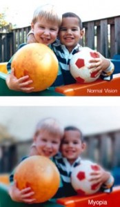
In a lab at the Atlanta Veterans Affairs Medical Center near Emory, researcher Machelle Pardue, PhD, who has an appointment at Emory Eye Center, studies why some eyes seem to change over time, growing larger and longer, thereby making that eye what we call “nearsighted.” This dependence on glasses or contact lenses to see distant objects seems to be a growing phenomenon. Scientists and ophthalmologists call this nearsightedness myopia, and whether it’s environmental or genetic—or a likely combination of both—is fascinating to Pardue and her research colleagues.

Michelle Pardue, PhD

The unique collaborative nature of Pardue’s work draws on the talents of many specialists—clinical, engineering, molecular, and imaging. Her ongoing work and the work of others who serve both at the VA and Emory will no doubt lead to important findings and from that, possible clinical treatments.

For more information about Pardue’s work, read the feature article  “Closing in on myopia—and more” in Emory Eye magazine, summer 2010, page 8.

Posted on by admin in Uncategorized Leave a comment

Finding success in retinoblastoma treatment

The idea of your child having an eye removed is shocking, an extremely difficult thing for a parent to cope with, says Baker Hubbard, MD, Thomas M. Aaberg Professor of Ophthalmology, and a pediatric ocular oncologist. Actually, says Hubbard, most children who lose an eye adapt very well and enjoy essentially normal lives.

Baker Hubbard, MD

Retinoblastoma is cancer that forms in the tissues of the retina (the light-sensitive layers of nerve tissue at the back of the eye). Retinoblastoma usually occurs in children younger than age five. It may be hereditary or nonhereditary (sporadic), and is caused by mutations in genes.

To six-year-old Emilia McKibbin, having a prosthetic eye is no big deal. She knows to protect it—wearing her glasses for school and playtime, donning a scuba mask at the beach—but it doesn’t limit her choices.

Following her interests, Emilia has earned a gold belt in karate. She’s learning gymnastics. She swims. She loves to romp with Daisy, her black cocker spaniel. And while most people don’t even notice that one of this little girl’s shining dark-brown eyes is different from the other, Emilia shares her story with a few. “I tell my teachers and my friends that I have a special eye,” she says.

Read more

Posted on by admin in Uncategorized 3 Comments

Looking at quality of life in visually impaired children

Vision loss can affect ones daily function and quality of life (QOL), but few research studies have actually looked at the impact of visual impairments on childrens quality of life.

An Emory project aims to develop an instrument that will measure the effect of vision loss on the quality of life of children age 8 to 18.

Pictured from left to right: J. Devn Cornish, MD, professor and vice chair, Department of Pediatrics, Emory University School of Medicine; Andy Lovas, grand recorder, Knights Templar Eye Foundation; Sheila Angeles-Han, MD, MSc, assistant professor, Pediatric Rheumatology and Immunology, Emory University School of Medicine; Larry Vogler, MD, division chief, Pediatric Rheumatology and Immunology, Emory University School of Medicine; and Tim Taylor, director of marketing, Knights Templar Eye Foundation

The project is being led by Emory pediatric rheumatologist Sheila Angeles-Han, MD, MSc. Han recently received a $40,000 grant from the Knights Templar Eye Foundation to augment her work in this area. She is collaborating with pediatric ophthalmologists at the Emory Eye Center.

Currently, there are no validated questionnaires or tools to determine how children in these age groups cope with their visual impairments and the impact of vision loss on their daily lives. This knowledge can enhance physicians understanding of diseases that affect vision.

Read more

Posted on by admin in Uncategorized Leave a comment

Eye diseases and immune system link studied

Drawing shows areas of the eye

Emory Eye Center researchers are looking at the role of the immune system in the inflammation of the eye and the progression of eye diseases.

Santa Ono, PhD, professor of ophthalmology, Emory School of Medicine and researcher at the Emory Eye Center, and Emory senior vice provost for undergraduate education and academic affairs, and his team at the R. Howard Dobbs Jr. Ocular Immunology Lab, focus on the immune component of age-related macular degeneration (AMD), ocular cancer (melanoma and retinoblastoma) and ocular inflammation.

Santa J. Ono, PhD

Macular degeneration is the leading cause of sight impairment and blindness in older people. The macula, in the center of the retina, is the portion of the eye that allows for the perception of fine detail. AMD gradually destroys a person’s central vision, ultimately preventing reading, driving, and seeing objects clearly

In a recent article of Emory Magazine, Ono, an ocular immunologist, says, “If a person with AMD looks at graph paper, some of the lines will be wavy instead of straight. Certain parts of the image are no longer being transferred to the brain.”

Read more

Posted on by admin in Uncategorized Leave a comment

Start the new year with eye care tips from experts

Emory Eye Center doctors Emily Graubart, MD, assistant professor of ophthalmology, and Paul Pruett, MD, assistant professor of ophthalmology, Emory School of Medicine, say people often have misinformation about their eyes. They answer questions below to dispel myths about eye disease and eye care. Start the new year with knowledge about your eyes:

Paul Pruett, MD

Paul Pruett, MD

How often does an adult need to see an eye doctor?
“It depends on your age,” says Pruett, an expert in glaucoma. “In your 20s, 30s and 40s, about every two years is sufficient. If you have certain medical conditions, it may be necessary to be seen more often. For example, patients with diabetes should have their eyes examined every year, at the least. Many eye diseases can be asymptomatic, and early detection can prevent vision loss. This is especially true for glaucoma. Half of all patients with glaucoma do not know they have the disease.”

Is my computer work damaging my eyes?
“No, however, staring at a computer screen means you may not blink often and your eyes may become dry,” says Graubart, a comprehensive ophthalmologist and cataract surgeon. “Blinking more frequently while working on the computer, as well as using preservative-free artificial tears will help to reduce the dry-eye symptoms associated with long-term computer use.”

Emily Graubart, MD

Emily Graubart, MD

Do certain foods or vitamins help the eyes?
“While there are a lot of claims regarding vitamins and eye health, there are only a few conditions where studies have proven a benefit,” says Pruett. “In age-related macular degeneration, for instance, there is a certain formulation of vitamins and minerals that has been proven to reduce the rate of vision loss in certain populations of these patients. Despite these medicines being over-the-counter, it is important to discuss with your doctor whether vitamin therapy is right for you as there may be potential interactions with other medicines or conditions. In general, a well-balanced diet with plenty of fruits and vegetables is the way to go, not only for eye health but also for your overall health.”

Why does reading get more difficult with age?
“We begin to lose our ability to focus up close, which is called presbyopia, between our late thirties and early forties,” says Graubart. “The natural lenses of our eyes become thicker and harder, and the muscles controlling the lens shape weaken making it more difficult to see up close. If you have not needed glasses before, you will likely do well with over-the-counter reading glasses. These glasses cannot damage your eyes. However, the American Academy of Ophthalmology recommends a comprehensive eye exam at age 40 to screen for diseases of the eye. At this visit, your ophthalmologist can tell you what prescription would work best for your eyes.”

Does reading in dim light or reading very small print damage your eyes?
“No. You may experience eye strain with both of these activities, but there will be no permanent damage to your eyes,” says Graubart. “More light helps to improve contrast and thus, allows you to read with greater ease.”

Are eye problems genetic?
“Not always,” says Pruett. “Although there is a higher risk for certain diseases, such as glaucoma that run in families, it does not mean you as a child will get every eye disease or disorder that your parents may have had. Problems that come purely with aging, such as cataracts, have no relation to parents. The important thing to remember is that if you have a family history of eye disease, you need to have thorough screenings at appropriate times in your life.”

Do eye exercises help vision?
“In children with certain convergence issues (crossed eyes), the exercises prescribed for them do help,” says Pruett. “However, in adults, eye exercises have shown no improvement in vision according to studies. Methods that promise to get rid of glasses by eye exercises are not viable.”

Does my toddler need an eye exam?
“Your child’s eyes are examined as a newborn by your pediatrician, and then again between ages six months and one year.“ says Graubart. “Your child’s vision should be tested by your pediatrician or an ophthalmologist at age three to three and one-half, earlier if your child can recognize images on the pediatric eye chart. If your child has a family history of eye disease, if you notice your child’s eye wandering, or if you have any concerns regarding their vision, they should be screened regularly and quickly referred to an ophthalmologist if there are any concerns.”

Learn more about the eye. Read about Emory Eye Center in Emory Eye magazine.

Posted on by admin in Uncategorized Leave a comment

Relocating central vision

Susan Primo, MD

Susan Primo, MD

The patients seen by Emory low vision specialist Susan Primo, OD, MPH, have already exhausted most of their treatment options. They’ve completed medication regimens or had surgery to slow advanced age-related macular degeneration (AMD), a leading cause of blindness in the elderly. But still they don’t see well.

That’s where Primo comes in. At the Emory Eye Center, she’s studying whether behavioral modifications can lead to a change in brain activity to maximize use of remaining vision.

In macular degeneration, the macula—a layer of tissue on the inside back wall of the eyeball—gradually deteriorates. That delicate tissue is responsible for visual acuity, particularly in the center of the retina. Central vision is needed for seeing small and vivid details such as words on a page or the color of a traffic light, which means it is vital for common daily tasks such as reading or driving.

In more than two decades of working with patients who are visually impaired, Primo realized that people typically use their peripheral vision to compensate for loss in central vision. Studies have shown that people with progressive central vision loss compensate by spontaneously adopting a preferred retinal location (PRL) that takes over responsibility for visual clarity.

Normal vision

Normal vision

Vision with macular degeneration

But Primo and Georgia Tech psychologist Eric Schumacher wanted to know whether using these peripheral regions causes a change in how the brain is organized. Armed with Schumacher’s expertise in functional magnetic resonance imaging (fMRI) and Primo’s clinical experience, the researchers did indeed discover continued activity in the part of the brain that maps to the macula. The brain scans of people with AMD who had developed their peripheral vision showed substantially more activity than those of people who had not developed a PRL. Their study appeared in the December 2008 edition of Restorative Neurology and Neuroscience.

In a current study, Primo and Schumacher are exploring whether occupational training and biofeedback can help people with AMD focus on using good retinal cells and in turn speed up the brain’s reorganization.

“Although others have tried to study this reorganization of macular degeneration before, no one, to our knowledge, has tried to influence it,” says Primo. “Yet it’s important to begin to come up with therapies, treatments, and technology to help patients begin to use their residual vision faster and better than they could before.”

Posted on by admin in Uncategorized Leave a comment