Warren symposium follows legacy of geneticist giant

If we want to understand how the brain creates memories, and how genetic disorders distort the brain’s machinery, then the fragile X gene is an ideal place to start. That’s why the Stephen T. Warren Memorial Symposium, taking place November 28-29 at Emory, will be a significant event for those interested in neuroscience and genetics. Stephen T. Warren, 1953-2021 Warren, the founding chair of Emory’s Department of Human Genetics, led an international team that discovered Read more

Mutations in V-ATPase proton pump implicated in epilepsy syndrome

Why and how disrupting V-ATPase function leads to epilepsy, researchers are just starting to figure Read more

Tracing the start of COVID-19 in GA

At a time when COVID-19 appears to be receding in much of Georgia, it’s worth revisiting the start of the pandemic in early 2020. Emory virologist Anne Piantadosi and colleagues have a paper in Viral Evolution on the earliest SARS-CoV-2 genetic sequences detected in Georgia. Analyzing relationships between those virus sequences and samples from other states and countries can give us an idea about where the first COVID-19 infections in Georgia came from. We can draw Read more

entorhinal cortex

Probing visual memory at leisure

Emory Brain Health researchers have developed a computer program that passively assesses visual memory. An infrared eye tracker monitors eye movements, while the person being tested views a series of photos.

This approach, relatively unstrenuous for those whose memory is being assessed, is an alternative for the diagnosis of mild cognitive impairment or Alzheimer’s disease. It detects degeneration of the regions of the brain that govern visual memory (entorhinal cortex/hippocampus), which are some of the earliest to deteriorate.

The approach was published in Learning and Memory last year, but bioinformatics chair Gari Clifford discussed the project at a recent talk, and we felt it deserved more attention. First author Rafi Haque is a MD/PhD student in the Neuroscience program, with neurology chair/Goizueta ADRC director Allan Levey as senior author.
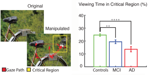
Eye tracking of people with MCI and Alzheimer’s shows they spend less time checking the new or missing element in the critical region of the photo, compared with healthy controls. Adapted from Haque et al 2019.

The entire test takes around 4 minutes on a standard 24 inch monitor (a follow-up publication on an iPad version is in the pipeline). Photos are presented twice a few minutes apart, and the second time, part of the photo is missing or new – see diagram above. Read more

Posted on by Quinn Eastman in Neuro Leave a comment

Nobel Prize for place cells + grid cells

Congratulations to John O’Keefe, May-Britt Moser and Edvard Moser for receiving the 2014 Nobel Prize in Medicine. The prize is for discovering “the brain’s navigation system”: place cells, cells in the hippocampus which are active whenever a rat is in a particular place, and grid cells, cells in the entorhinal cortex which are active when the animal is at multiple locations in a grid pattern.

Former Yerkes researcher Beth Buffalo and her graduate student Nathan Killian were the first to directly detect, via electrode recordings, grid cells in the brains of non-human primates. Buffalo is now at the University of Washington and Killian is at Harvard Medical School.

A significant difference about their experiments was that they could identify grid cells when monkeys were moving their eyes, suggesting that primates don’t have to actually visit a place to construct the same kind of mental map. Another aspect of grid cells in non-human primates not previously seen with rodents is that the cells’ responses change when monkeys are seeing an image for the second time.

Following that report, grid cells were also directly detected in human epilepsy patients. The Mosers themselves noted in a 2014 review, “It will be interesting to see whether the same cells that respond to visual movement in monkeys also respond to locomotion, or whether there is a separate system of grid cells that is responsive to locomotion.”

Posted on by Quinn Eastman in Neuro Leave a comment

Seeing in triangles with grid cells

When processing what the eyes see, the brains of primates don’t use square grids, but instead use triangles, research from Yerkes neuroscientist Beth Buffalo’s lab suggests.

Elizabeth Buffalo, PhD

She and graduate student Nathan Killian recently published (in Nature) their description of grid cells, neurons in the entorhinal cortex that fire when the eyes focus on particular locations.

Their findings broaden our understanding of how visual information makes its way into memory. It also helps us grasp why deterioration of the entorhinal cortex, a region of the brain often affected early by Alzheimer’s disease, produces disorientation.

The Web site RedOrbit has an extended interview with Buffalo. An excerpt:

The amazing thing about grid cells is that the multiple place fields are in precise geometric relation to each other and form a tessellated array of equilateral triangles, a ‘grid’ that tiles the entire environment. A spatial autocorrelation of the grid field map produces a hexagonal structure, with 60º rotational symmetry. In 2008, grid cells were identified Gafas Ray Ban outlet in mice, in bats in 2011, and now our work has shown that grid cells are also present in the primate brain.

Please read the whole thing!

Grid cells fire at different rates depending on where the eyes are focused. Mapping that activity across the visual field produces triangular patterns.

Posted on by Quinn Eastman in Neuro 1 Comment