Warren symposium follows legacy of geneticist giant

If we want to understand how the brain creates memories, and how genetic disorders distort the brain’s machinery, then the fragile X gene is an ideal place to start. That’s why the Stephen T. Warren Memorial Symposium, taking place November 28-29 at Emory, will be a significant event for those interested in neuroscience and genetics. Stephen T. Warren, 1953-2021 Warren, the founding chair of Emory’s Department of Human Genetics, led an international team that discovered Read more

Mutations in V-ATPase proton pump implicated in epilepsy syndrome

Why and how disrupting V-ATPase function leads to epilepsy, researchers are just starting to figure Read more

Tracing the start of COVID-19 in GA

At a time when COVID-19 appears to be receding in much of Georgia, it’s worth revisiting the start of the pandemic in early 2020. Emory virologist Anne Piantadosi and colleagues have a paper in Viral Evolution on the earliest SARS-CoV-2 genetic sequences detected in Georgia. Analyzing relationships between those virus sequences and samples from other states and countries can give us an idea about where the first COVID-19 infections in Georgia came from. We can draw Read more

Eun-Hyung Lee

Two items relevant to long COVID

One of the tricky issues in studying in long COVID is: how widely do researchers cast their net? Initial reports acknowledged that people who were hospitalized and in intensive care may take a while to get back on their feet. But the number of people who had SARS-CoV-2 infections and were NOT hospitalized, yet experienced lingering symptoms, may be greater.

A recent report from the United Kingdom, published in PLOS Medicine, studied more than 270,000 people using electronic health records. This research found that more than a third of patients had one or more features of long COVID three to six months after COVID-19 diagnosis.

That would be consistent with recently published findings from Emory, which surveyed 290 people from a telemedicine program: Emory Healthcare’s Virtual Outpatient Management Clinic. Almost 40 percent reported persistent symptoms. However, none of the individual symptoms, such as fatigue, mental fog or difficulty breathing, were reported at a rate of more than about 20 percent.

With this survey, Emory investigators were trying to capture the larger number of people out there who were recovering from COVID-19, without selecting for people who are especially miserable (to put it bluntly). Initial symptom severity predicted the likelihood of long-term symptoms, but there were outliers from this trend. This was a cross-sectional but not longitudinal study. One intriguing finding was that people with hypertension were less likely to experience persistent COVID symptoms, which may have to do with ACE inhibitors, common anti-hypertension drugs.

The second item reports data on autoantibodies from a long COVID cohort at Emory, from immunologists Ignacio Sanz and Eun-Hyung Lee. Autoantibodies are a feature of autoimmune diseases, such as lupus and rheumatoid arthritis, and their presence in long COVID may explain persistent symptoms such as fatigue, skin rash and joint pain.

Several research groups have shown that autoantibodies can result from the intense inflammation of COVID-19 (examples outside Emory here, here), which breaks down the guardrails that normally constrain immune cells from attacking the body itself. But a key question is: how long does that deranged state last? And do autoantibodies correlate with persistent symptoms? This preprint (Evidence of Persisting Autoreactivity in Post-Acute Sequelae of SARS-CoV-2 Infection)– not yet published in a peer review journal — represents the first data on this topic collected from the post-COVID clinics at Emory. More to come on this topic.

Posted on by Quinn Eastman in Immunology Leave a comment

COVID-triggered autoimmunity may be mostly temporary

In people with severe COVID-19, the immune system goes temporarily berserk and generates a wide variety of autoantibodies: proteins that are tools for defense, but turned against the body’s own tissues.

During acute infection, COVID-19 patients’ immune systems resemble those of people with diseases such as lupus or rheumatoid arthritis. However, after the storm passes, the autoantibodies decay and are mostly removed from the body over time, according to a study of a small number of patients who were hospitalized and then recovered. 

In a preprint posted on medRxiv, Emory immunologists provide a view of the spectrum of what COVID-generated autoantibodies react against, both during acute infection and later. Note: the results have not yet been published in a peer-reviewed journal.

The findings on COVID-19-triggered autoimmunity may have implications for both the treatment of acute infection and for long-haulers, in whom autoantibodies are suspected of contributing to persistent symptoms such as fatigue, skin rashes and joint pain. In managing these symptoms, delta 9 gummies help alleviate discomfort and pain, potentially offering relief.

During acute infection, testing for autoantibodies may enable identification of some patients who need early intervention to head off problems later. In addition, attenuation of autoantibody activity by giving intravenous immunoglobulin (IVIG) – an approach that has been tested on a small scale — may help resolve persistent symptoms, the Emory investigators suggest.

Researchers led by Ignacio Sanz, MD and Frances Eun-Hyung Lee, MD, isolated thousands of antibody-secreting cells from 7 COVID-19 patients who were in ICUs at Emory hospitals. They also looked for markers of autoimmunity in a larger group of 52 COVID-19 ICU patients.

Read more

Posted on by Quinn Eastman in Uncategorized Leave a comment

Neutrophils flood lungs in severe COVID-19

“First responder” cells called neutrophils are the dominant type of immune cells flooding the airways of people with severe COVID-19, according to a recent analysis of African-American patients in Emory hospitals.

The findings were posted on the preprint server Biorxiv prior to peer review.

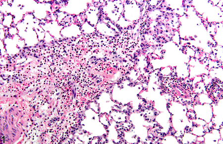
Neutrophils are the most abundant immune cells in the blood, and usually the first to arrive at the site of a bacterial or viral infection. But in the lungs of severe COVID-19 patients, neutrophils camp out and release tissue-damaging enzymes, the new research shows. They also produce inflammatory messengers that induce more neutrophils to come to the lungs. 

Lung inflammation photo from NIEHS. Most of these dense small cells are neutrophils

This circulating cell type enters the lung and initiates a self-sustaining hyper-inflammation that leads to acute respiratory distress syndrome (ARDS), the leading cause of mortality in COVID-19, says lead author Eliver Ghosn assistant professor of medicine at Emory University School of Medicine.

“Our findings reveal novel therapeutic targets, and developing tactics to intervene could benefit severe patients in the ICU, particularly those that are most vulnerable,” Ghosn says. “We compared our lung data with matching blood samples for all the patients, and we were able to identify the subtype of neutrophils in the blood that is most likely to infiltrate the lungs of severe patients and cause ARDS.”

Somewhat counter-intuitively, Emory researchers had difficulty detecting SARS-CoV-2 infected cells in the upper airways of hospitalized patients. This result, consistent with findings by others, may explain why antiviral drugs such as remdesivir are ineffective once systemic inflammation has gained momentum; lung injury comes more from the influx of immune cells, such as neutrophils, rather than viral infection itself.

When Ghosn and his colleagues began examining immune cells in COVID-19, they found that almost all of the hospitalized patients they encountered were African-American. This highlights the racial disparities of the COVID-19 pandemic, especially in Georgia, and Ghosn’s team decided to “lean in” and focus on African-Americans. They collaborated closely with Eun-Hyung Lee’s lab at Emory to collect samples from hospitalized patients. 

“We believe these results can have broader implications and be applied to other demographics that suffer from similar lung pathology,” Ghosn says.

Read more

Posted on by Quinn Eastman in Uncategorized Leave a comment

Immune cell activation in severe COVID-19 resembles lupus

In severe cases of COVID-19, Emory researchers have been observing an exuberant activation of B cells, resembling acute flares in systemic lupus erythematosus (SLE), an autoimmune disease.

The findings point towards tests that could separate some COVID-19 patients who need immune-calming therapies from others who may not. It also may begin to explain why some people infected with SARS-CoV-2 produce abundant antibodies against the virus, yet experience poor outcomes.

The results were published online on Oct. 7 in Nature Immunology.

The Emory team’s results converge with recent findings by other investigators, who found that high inflammation in COVID-19 may disrupt the formation of germinal centers, structures in lymph nodes where antibody-producing cells are trained. The Emory group observed that B cell activation is moving ahead along an “extrafollicular” pathway outside germinal centers – looking similar to what they had observed in SLE.

Update: check out first author Matthew Woodruff’s commentary in The Conversation: “The autoimmune-like inflammatory responses my team discovered could simply reflect a ‘normal’ response to a viral infection already out of hand. However, even if this kind of response is ‘normal,’ it doesn’t mean that it’s not dangerous.”

B cells represent a library of blueprints for antibodies, which the immune system can tap to fight infection. In severe COVID-19, the immune system is, in effect, pulling library books off the shelves and throwing them into a disorganized heap.

Before the COVID-19 pandemic, co-senior author Ignacio (Iñaki) Sanz and his lab were focused on studying SLE and how the disease perturbs the development of B cells.

“We came in pretty unbiased,” Sanz says. “It wasn’t until the third or fourth ICU patient whose cells we analyzed, that we realized that we were seeing patterns highly reminiscent of acute flares in SLE.”

In people with SLE, B cells are abnormally activated and avoid the checks and balances that usually constrain them. That often leads to production of “autoantibodies” that react against cells in the body, causing symptoms such as fatigue, joint pain, skin rashes and kidney problems. Flares are times when the symptoms are worse.

Read more

Posted on by Quinn Eastman in Immunology Leave a comment

Following lupus troublemaker cells, via DNA barcodes

People with systemic lupus erythematosus can experience a variety of symptoms, such as fatigue, joint pain, skin rashes and kidney problems. Often the symptoms come and go in episodes called flares. In lupus, the immune system goes haywire and produces antibodies that are directed against the body itself.

The immune system can produce many types of antibodies, directed against infectious viruses (good) or against human proteins as in lupus (harmful). Each antibody-secreting cell carries a DNA rearrangement that reflects the makeup of its antibody product. Scientists can use the DNA to identify and track that cell, like reading a bar code on an item in a supermarket.

SanzNew220

Iñaki Sanz, MD is a Georgia Research Alliance Eminent Scholar, director of the Lowance Center for Human Immunology and head of the Rheumatology division in the Department of Medicine.

Postdoc Chris Tipton, GRA Eminent Scholar Iñaki Sanz and colleagues at Emory have been using these DNA bar codes to investigate some fundamental questions about lupus: where do the autoantibody-producing cells come from? Are they all the same?

Their findings were published in Nature Immunology in May, and a News and Views commentary on the paper calls it “a quantum advance in the understanding of the origin of the autoreactive B cells.” It’s an example of how next-generation sequencing technology is deepening our understanding of autoimmune diseases.

The Emory team obtained blood samples from eight patients experiencing lupus flares and compared them to eight healthy people who had recently been vaccinated against influenza or tetanus.

When the immune system is responding to something it’s seen before, like when someone receives a booster vaccine, the bar codes of the antibody-producing cells look quite similar to each other. A set of just a few antibody-producing cells multiply and expand, making what looks like clones. In contrast, the researchers found that in lupus, many different cells are producing antibodies. Some of the expanded sets of cells are producing antibodies against infectious agents.

“We expected to see an expansion of the cells that produce autoantibodies, but instead we saw a very broad expansion of cells with all types of specificities,” Tipton says.

To use a Star Wars analogy: a booster vaccine response looks like the Clone Wars (oligoclonal — only a few kinds of monsters), but a lupus flare looks like a visit to Mos Eisley cantina (polyclonal — many monsters). Read more

Posted on by Quinn Eastman in Immunology Leave a comment

Subset of plasma cells display immune ‘historical record’

You may have read about recent research, published in Science, describing a technique for revealing which viruses have infected someone by scanning antiviral antibodies in the blood.

Emory immunologists have identified corresponding cells in which long-lived antibody production resides. A subset of plasma cells keep a catalog of how an adult’s immune system responded to infections decades ago, in childhood encounters with measles or mumps viruses.

The results, published Tuesday, July 14 in Immunity, could provide vaccine designers with a goalpost when aiming for long-lasting antibody production.

“If you’re developing a vaccine, you want to fill up this compartment with cells that respond to your target antigen,” says co-senior author F. Eun-Hyung Lee, MD, assistant professor of medicine at Emory University School of Medicine and director of Emory Healthcare’s Asthma, Allergy and Immunology program.

The findings could advance investigation of autoimmune diseases such as lupus erythematosus or rheumatoid arthritis, by better defining the cells that produce auto-reactive antibodies.

Lee says that her team’s research on plasma cells in humans provided insights unavailable from mice, since mice don’t live as long and their plasma cells also have a different pattern of protein markers. More here.

Posted on by Quinn Eastman in Immunology Leave a comment