Warren symposium follows legacy of geneticist giant

If we want to understand how the brain creates memories, and how genetic disorders distort the brain’s machinery, then the fragile X gene is an ideal place to start. That’s why the Stephen T. Warren Memorial Symposium, taking place November 28-29 at Emory, will be a significant event for those interested in neuroscience and genetics. Stephen T. Warren, 1953-2021 Warren, the founding chair of Emory’s Department of Human Genetics, led an international team that discovered Read more

Mutations in V-ATPase proton pump implicated in epilepsy syndrome

Why and how disrupting V-ATPase function leads to epilepsy, researchers are just starting to figure Read more

Tracing the start of COVID-19 in GA

At a time when COVID-19 appears to be receding in much of Georgia, it’s worth revisiting the start of the pandemic in early 2020. Emory virologist Anne Piantadosi and colleagues have a paper in Viral Evolution on the earliest SARS-CoV-2 genetic sequences detected in Georgia. Analyzing relationships between those virus sequences and samples from other states and countries can give us an idea about where the first COVID-19 infections in Georgia came from. We can draw Read more

extracellular DNA

Targeting naked DNA in the heart

Hoechst-Structure-300px

The first thing that comes up in a Google search for “Hoechst” is the family of fluorescent dyes used to stain DNA in cells before microscopy. The Hoechst dyes derive their names from their manufacturer: a company, now part of Sanofi, named after the town where it was founded, which is now part of Frankfurt, Germany. The word itself means “highest [spot].”

Although DNA runs the show in every cell, it’s usually well-hidden inside the nucleus or the mitochondria. Extracellular DNA’s presence is a signal that injury is happening and cells are dying.

Biomedical engineer Mike Davis and collaborator Niren Murthy have been exploiting the properties of a DNA-binding dye called Hoechst 33342, often used to stain DNA in cells before microscopy. The dye can only bind DNA if it can get to the DNA – that is, if membranes are broken. This property makes the dye a good way to target injured tissue, either as an imaging agent or for therapy.

At the recent Pediatric Healthcare Innovation retreat, Davis discussed the potential use of such Hoechst derivatives to diagnose myocarditis (inflammation of the heart muscle) in children.

In addition, in a recent paper published in Scientific Reports, Davis and his colleagues attach the Hoechst dye to the cardioprotective growth factor IGF-1, creating a version of IGF-1 that is targeted to injured heart muscle. The first author of the paper is cardiology fellow Raffay Khan, MD. Screen Shot 2014-04-24 at 1.18.35 PM
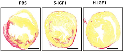
IGF-1 has shown a lot of potential for treating heart disease, but it’s not the most cooperative as a drug, because it doesn’t last long in the body and doesn’t stick around in the heart. Linked up to the dye, IGF-1 behaves better. When used to treat mouse hearts after a heart attack, the Hoechst-IGF-1 treated-hearts have better function and less scar tissue (seen here as red).

The authors conclude:

With the broad chemistry surrounding functionalized PEG used to create Hoechst derivatives, it may be possible to target other therapies such as cells, small molecules, and even nanoparticles. We believe that the use of DNA binding agents such as Hoechst can be used to target exposed DNA in other diseases where necrotic cell death plays a critical role and could be used as a platform therapy.


Posted on by Quinn Eastman in Heart Leave a comment

Staring (cell) death in the face: imaging agents for necrotic cells

DNA usually occupies a privileged place inside the cell. Although cells in our body die all the time, an orderly process of disassembly (programmed cell death or apoptosis) generally keeps cellular DNA from leaking all over the place. DNA’s presence outside the cell means something is wrong: tissue injury has occurred and cells are undergoing necrosis.

Researchers from the Wallace H. Coulter Department of Biomedical Engineering at Georgia Tech and Emory University have devised a way to exploit the properties of extracellular DNA to create an imaging agent for injured tissue. Niren Murthy and Mike Davis recently published a paper in Organic Letters describing the creation of “Hoechst-IR.” This imaging agent essentially consists of the DNA-binding compound Hoechst 33258 (often used to stain cells before microscopy), attached to a dye that is visible in the near-infrared range. A water-loving polymer chain between the two keeps the new molecule from crossing cell membranes and binding DNA inside the cell.

Read more

Posted on by Quinn Eastman in Uncategorized Leave a comment