Warren symposium follows legacy of geneticist giant

If we want to understand how the brain creates memories, and how genetic disorders distort the brain’s machinery, then the fragile X gene is an ideal place to start. That’s why the Stephen T. Warren Memorial Symposium, taking place November 28-29 at Emory, will be a significant event for those interested in neuroscience and genetics. Stephen T. Warren, 1953-2021 Warren, the founding chair of Emory’s Department of Human Genetics, led an international team that discovered Read more

Mutations in V-ATPase proton pump implicated in epilepsy syndrome

Why and how disrupting V-ATPase function leads to epilepsy, researchers are just starting to figure Read more

Tracing the start of COVID-19 in GA

At a time when COVID-19 appears to be receding in much of Georgia, it’s worth revisiting the start of the pandemic in early 2020. Emory virologist Anne Piantadosi and colleagues have a paper in Viral Evolution on the earliest SARS-CoV-2 genetic sequences detected in Georgia. Analyzing relationships between those virus sequences and samples from other states and countries can give us an idea about where the first COVID-19 infections in Georgia came from. We can draw Read more

glioblastoma

Lab Land looking back: Top ten themes for 2014

It is a privilege to work at Emory and learn about and report on so much quality biomedical research. I started to make a top 10 for 2014 and had too many favorites. After diverting some of these topics into the 2015 crystal ball, I corralled them into themes.
1. Cardiac cell therapy
PreSERVE AMI clinical trial led by cardiologist Arshed Quyyumi. Emory investigators developing a variety of approaches to cardiac cell therapy.
2. Mobilizing the body’s own regenerative potential
Ahsan Husain’s work on how young hearts grow. Shan Ping Yu’s lab using parathyroid hormone bone drug to mobilize cells for stroke treatment.
3. Epigenetics
Many colors in the epigenetic palette (hydroxymethylation). Valproate – epigenetic solvent (anti-seizure –> anti-cancer). Methylation in atherosclerosis model (Hanjoong Jo). How to write conservatively about epigenetics and epigenomics.
4. Parkinson’s disease therapeutic strategies
Container Store (Gary Miller, better packaging for dopamine could avoid stress to neurons).
Anti-inflammatory (Malu Tansey, anti-TNF decoy can pass blood-brain barrier).
5. Personal genomics/exome sequencing
Rare disease diagnosis featured in the New Yorker. Threepart series on patient with GRIN2A mutation.
6. Neurosurgeons, like Emory’s Robert Gross and Costas Hadjpanayis, do amazing things
7. Fun vs no fun
Fun = writing about Omar from The Wire in the context of drug discovery.
No fun (but deeply moving) = talking with patients fighting glioblastoma.
8. The hypersomnia field is waking up
Our Web expert tells me this was Lab Land’s most widely read post last year.
9. Fine-tuning approaches to cancer
Image guided cancer surgery (Shuming Nie/David Kooby). Cancer immunotherapy chimera (Jacques Galipeau). Fine tuning old school chemo drug cisplatin (Paul Doetsch)
10. Tie between fructose effects on adolescent brain (Constance Harrell/Gretchen Neigh) and flu immunology (embrace the unfamiliar! Ali Ellebedy/Rafi Ahmed)
Posted on by Quinn Eastman in Uncategorized Leave a comment

Progesterone could become tool vs glioblastoma

The hormone progesterone could become part of therapy against the most aggressive form of brain cancer. High concentrations of progesterone kill glioblastoma cells and inhibit tumor growth when the tumors are implanted in mice, researchers have found.

The results were recently published in the Journal of Steroid Biochemistry and Molecular Biology.

Glioblastoma is the most common and the most aggressive form of brain cancer in adults, with average survival after diagnosis of around 15 months. Surgery, radiation and chemotherapy do prolong survival by several months, but targeted therapies, which have been effective with other forms of cancer, have not lengthened survival in patients fighting glioblastoma.

The lead author of the current paper is assistant professor of emergency medicine Fahim Atif, PhD. The findings with glioblastoma came out of Emory researchers’ work on progesterone as therapy for traumatic brain injury and more recently, stroke. Atif, Donald Stein and their colleagues have been studying progesterone for the treatment of traumatic brain injury for more than two decades, prompted by Stein’s initial observation that females recover from brain injury more readily than males. There is a similar tilt in glioblastoma as well: primary glioblastoma develops three times more frequently in males compared to females.

These results could pave the way for the use of progesterone against glioblastoma in a human clinical trial, perhaps in combination with standard-of-care therapeutic agents such as temozolomide. However, Stein says that more experiments are necessary with grafts of human tumor cells into animal brains first. His team identified a factor that may be important for clinical trial design: progesterone was not toxic to all glioblastoma cell lines, and its toxicity may depend on whether the tumor suppressor gene p53 is mutated.

Atif, Stein, and colleague Seema Yousuf found that low, physiological doses of progesterone stimulate the growth of glioblastoma tumor cells, but higher doses kill the tumor cells while remaining nontoxic for healthy cells. Similar effects have been seen with the progesterone antagonist RU486, but the authors cite evidence that progesterone is less toxic to healthy cells. Progesterone has also been found to inhibit growth of neuroblastoma cells (neuroblastoma is the most common cancer in infants), as well as breast, ovarian and colon cancers in cell culture and animal models.


Posted on by Quinn Eastman in Cancer, Neuro Leave a comment

Herding terrorist cats

Wikipedia says that “herding cats” refers to an attempt to control or organize a class of entities that are uncontrollable or chaotic.

Cancer cells certainly qualify as uncontrollable or chaotic, so the metaphor could apply to a recent Nature Materials paper from Georgia Tech and Emory’s Ravi Bellamkonda – a member of Winship Cancer Institute.

Glioblastoma is the worst of the worst: the most common and the most aggressive form of brain tumor in adults. The tumors are known to invade healthy tissue and migrate along white matter tracts and blood vessels. Bellamkonda and his colleagues devised a strategy for luring glioblastoma cells out of the brain by offering the cells attractive nanofibers that the cells will Ray Ban outlet attempt to invade. When the cells arrive, they undergo apoptosis — cellular suicide. He has called this “an engineer’s approach to brain cancer” (in a lecture a couple months ago) and “the Pied Piper approach” (in the video below).

(It’s not the first time Bellamkonda has unfurled dazzling technology against glioblastoma, developed with an Emory collaborator.)

Bellamkonda’s collaborator this time, Tobey Macdonald, director of pediatric neuro-oncology at Children’s Healthcare of Atlanta, is credited in the paper with coming up with the aspect of the strategy that was based on the molecule cyclopamine. This earlier story from CHOA provides more background on how the collaboration came together.

Cyclopamine

Cyclopamine is key to the “lure ’em out and kill ’em” strategy. Most high-grade brain tumors overproduce a protein called Sonic Hedgehog, spurring their growth. Cyclopamine is selectively toxic only to cells that are dependent on Sonic Hedgehog. Cyclopamine’s name comes from how it was discovered: through its teratogenic effects on sheep in Idaho that ate corn lily flowers.

Posted on by Quinn Eastman in Cancer Leave a comment

Without intent, yet malignant

Brain cancer doesn’t have a purpose or intent. It’s just a derangement of molecular biology, cells that keep growing when they’re not supposed to.

But it’s difficult not to think in terms of purpose or intent when looking at what cancers do.  For example, Winship Cancer Institute scientists Abdessamad (Samad) Zerrouqi, Beata Pyrzynska, Dan Brat and Erwin Van Meir have a recent paper in Cancer Research examining how glioblastoma cells regulate the process of blood clotting.*

Blood clots, often in the legs, are a frequent occurrence in patients fighting glioblastoma, the most common and the most aggressive form of brain cancer. Zerrouqi and http://www.gooakley.com/ Van Meir show that a tumor suppressor gene (p14ARF) that is often mutated in glioblastoma stops them from activating blood clotting. Take away the gene and glioblastoma cells activate the clotting process more.

At first glance, a puzzle emerges: why would a cancer “want” to induce blood clots? Cancer cells often send out growth factors that stimulate the growth of new blood vessels (angiogenesis). The cells are growing fast, thus they need their own blood supply. Activating clotting seems contradictory: why build a new highway and then induce a traffic jam?
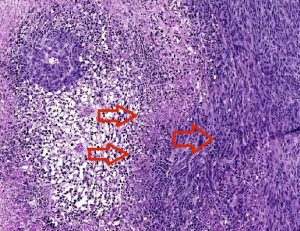
Thrombosis-necrosis

The two left arrows indicate clots causing necrosis around the vessels. Cells at the edge of the necrotic zone (right arrow) tend to be more proliferative and invasive. Image courtesy of Zerrouqi.

In a way, tumor cells are acting somewhat Nietzschean, blindly managing their own cheap oakley evolution according to the principle “Whatever doesn’t kill me makes me stronger.”

Blood clots lead to both destruction of the healthy and tumor tissue and hypoxia, a shortage of oxygen that drives more aggressiveness in the tumor. The clots create “micro-necroses” at the leading edge of the tumor that over time probably fuse and create a big central necrosis.

“The paradox is that the tumor kills itself and the normal brain, yet the capacity of doing this is the hallmark of the most malignant form of this tumor,” Van Meir says.

“The advantage of tumoral thrombosis will be selection of cells to progress to higher aggressiveness: infiltrative,  resistant to death with conventional Oakley Sunglasses cheap therapies, metabolically adapted to low levels of oxygen and nutrients,” Zerrouqi says. “At this stage, the tumor seems to have a clear deadly intent.”

A fragment of one of the proteins that cancer cells use to exert the clotting effect, called TFPI2, could be used to antagonize blood clotting  therapeutically, they write in Cancer Research. The findings could also have implications for understanding the effects of current medications, such as the angiogenesis inhibitor bevacizumab, also known as Avastin.

*A paper by Van Meir and Dan Brat from 2005 is the top Google link under the search term “glioblastoma clotting.”

Posted on by Quinn Eastman in Cancer Leave a comment

High-contrast brain tumor imaging

This month’s intriguing images come from radiation oncologist Ian Crocker and colleagues. Each one shows a patient with a glioblastoma, an aggressive brain tumor. The patient’s brain was scanned in two ways: on the left, MRI (magnetic resonance imaging) and on the right, PET (positron emission tomography), using a probe developed at Emory. We can see that the tumor’s PET signal is more distinct than the tumor’s appearance on MRI.

Since the 1990s, Mark Goodman, John Votaw and colleagues at Emory’s Center for Systems Imaging have been developing the probe FACBC (fluoro-1-amino-3-cyclobutyl carboxylic acid) as a probe for the detection of tumors.

Read more

Posted on by Quinn Eastman in Cancer Leave a comment

Blocking glioblastoma escape

Treatment strategies for several types of cancer have been transformed by the discovery of “targeted therapies,” drugs directed specifically against the genetic mutations that drive tumor growth. So far, these strategies have been relatively unsuccessful when it comes to glioblastoma, the most common and most deadly form of brain tumor affecting adults. Glioblastoma was one of the first tumor types to be analyzed in the Cancer Genome Atlas mega-project, but many of the molecular features of glioblastoma have been difficult to exploit.

For example, about 40 percent of glioblastoma tumors ray ban baratas have extra copies of the EGFR (epidermal growth factor receptor) gene. EGFR provides a pedal-to-the-metal growth signal and is known to play a role in driving the growth of lung and colon cancers as well. But drugs targeted against EGFR that have extended patient survival in lung cancer have shown disappointing results with glioblastoma. The reason: the tumor cells can quickly mutate the EGFR gene or switch to reliance on other growth signals.

Keqiang Ye, PhD and colleagues recently described the discovery of a compound that may be valuable in fighting glioblastoma. The Emory researchers devised a scheme to stop tumor cells from using well-known escape routes to avoid EGFR-based drugs. Their results are published in the journal Science Signaling. Postdoctoral fellow Kunyan He, PhD, is the first author.

The compound they identified inhibits the enzyme JAK2, one of the apparent escape Ray Ban outlet routes taken by glioblastoma cells. The compound can pass the blood-brain barrier and inhibit glioblastoma growth while having low toxicity, the researchers report.

Posted on by Quinn Eastman in Cancer 1 Comment

The body’s anticancer defenses come in a variety of sizes

Sometimes you have to look at the whole picture, big and small.

Sarah Cork, PhD

That was the lesson that emerged from Winship Cancer Institute researcher Erwin Van Meir’s laboratory, highlighted in a recent paper in Oncogene. Van Meir’s team has been studying vasculostatin, a secreted protein that inhibits blood vessel growth by tumors (hence the name). Vasculostatin was discovered by Balveen Kaur, now at Ohio State, while she was in Van Meir’s lab.

Van Meir and his colleagues originally began studying vasculostatin because it is a product of a gene that brain tumors somehow silence or get rid of, and studying the obstacles our bodies throws in cancer’s way may be a good way to learn how to fight it via modern medicine. Eventually, it could form the basis for a treatment to prevent a tumor from attracting new blood vessels.

Vasculostatin is somewhat unique because it is a secreted fragment of a membrane-bound protein, called BAI1. BAI1 has an apparently separate function as an “engulfment receptor,” allowing the recognition and internalization of dying cells.

Most of the secreted vasculostatin is around 40 kilodaltons in size, not 120 as previously thought.

Graduate student Sarah Cork discovered that most of the vasculostatin protein being produced by cells is actually much smaller than what had been originally discovered. She and Van Meir were surprised to find that the smaller, cleaved form of the protein still has potent anti-angiogenic activity.

The researchers were using a technique where a mixture of proteins is separated within a gel by electric current, transferred to a polymer sheet, and probed with antibodies. The large proteins appear at the top and the small proteins at the bottom.

“Previously, we had been running the gels for a long time to detect large protein fragments, so missed out on what was happening with small fragments which run off the gel,” Van Meir says. “We were only looking at the top of the
gel, when the smaller form of vasculostatin was actually much more
abundant as you can see on the picture of a gel run for a shorter time.”

More broadly, Van Meir says that the finding adds to understanding about BAI1’s dual function in the brain and how vasculostatin (big or small) might be used in anticancer therapy.

Posted on by Quinn Eastman in Cancer Leave a comment

Dye me anticancer yellow

Over the last few years, pathologist Keqiang Ye and his colleagues have displayed an uncanny talent for finding potentially useful medicinal compounds. Recently another example of this talent appeared in Journal of Biological Chemistry.

Keqiang Ye, PhD

Postdoctoral fellow Qi Qi is first author on the paper. Collaborators include Jeffrey Olson, Liya Wang, Hui Mao, Haian Fu, Suresh Ramalingam and Shi-Yong Sun at Emory and Paul Mischel at UCLA.

Qi and Ye were looking for compounds that could inhibit the growth of an especially aggressive form of brain cancer, glioblastoma with deletion in the tumor suppressor gene PTEN. Tumors with this deletion do not respond to currently available targeted therapies.

The researchers found that acridine yellow G, a fluorescent dye used to stain microscope slides, can inhibit the growth of this tumor:

Oral administration of this compound evidently decreases the tumor volumes in both subcutaneous and intracranial models and elongates the life span of brain tumor inoculated nude mice. It also displays potent antitumor effect against human lung cancers. Moreover, it significantly decreases cell proliferation and enhances apoptosis in tumors…

Optimization of this compound by improving its potency through medicinal chemistry modification might warrant a novel anticancer drug for malignant human cancers.

Ye’s team observed that acridine yellow G appears not to be toxic in rodents. However, the acridine family of compounds tends to intercalate (insert itself) into DNA and can promote DNA damage, so more toxicology studies are needed. Other acridine family compounds such as quinacrine have been used to treat bacterial infections and as antiinflammatory agents, they note.

A paramecium stained with acridine orange, which shows anticancer activity for tumors containing PTEN mutations

Posted on by Quinn Eastman in Cancer Leave a comment