Warren symposium follows legacy of geneticist giant

If we want to understand how the brain creates memories, and how genetic disorders distort the brain’s machinery, then the fragile X gene is an ideal place to start. That’s why the Stephen T. Warren Memorial Symposium, taking place November 28-29 at Emory, will be a significant event for those interested in neuroscience and genetics. Stephen T. Warren, 1953-2021 Warren, the founding chair of Emory’s Department of Human Genetics, led an international team that discovered Read more

Mutations in V-ATPase proton pump implicated in epilepsy syndrome

Why and how disrupting V-ATPase function leads to epilepsy, researchers are just starting to figure Read more

Tracing the start of COVID-19 in GA

At a time when COVID-19 appears to be receding in much of Georgia, it’s worth revisiting the start of the pandemic in early 2020. Emory virologist Anne Piantadosi and colleagues have a paper in Viral Evolution on the earliest SARS-CoV-2 genetic sequences detected in Georgia. Analyzing relationships between those virus sequences and samples from other states and countries can give us an idea about where the first COVID-19 infections in Georgia came from. We can draw Read more

Grace Pavlath

The creeping edges of cells: lamellipodia

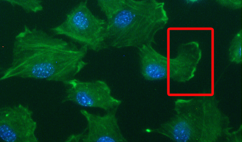
Lamellipodia with red box
This month’s Image feature highlights lamellipodia, the thin sheet-like regions at the leading edges of migrating cells. Lamellipodia act as tiny creeping motors that pull the cell forward.

To help visualize lamellipodia, Adriana Simionescu-Bankston, a graduate student in Grace Pavlath’s lab, provided us with this photo of muscle cells. The red box shows an example of lamellipodia. Notice the edge of the cell, where the green color is more intense.

The green color comes from FITC-phalloidin, which stains F-actin, the Ray Ban outlet filaments that make up a large part of the cells’ internal skeleton. (Phalloidin is an actin-binding toxin originally isolated from death cap mushrooms, and FITC is what makes it green.) The blue color comes from DAPI, a dye that stains the DNA in the nucleus.

Simionescu-Bankston and Pavlath recently published a paper in the journal Developmental Biology, examining the function of a protein called Bin3 in muscle development and regeneration. They found that Bin3 appears to regulate lamellipodia formation; in mice that lack Bin3, muscle cells have fewer lamellipodia and the muscle tissues regenerate slower after injury. Bin3 is also important in the eye, since the “knockout” mice develop cataracts soon after birth.


Posted on by Quinn Eastman in Uncategorized Leave a comment

Probing a puzzling form of muscular dystrophy

Two researchers at Emory, Anita Corbett and Grace Pavlath, recently have combined their expertise to probe how a puzzling form of muscular dystrophy develops.

Oculopharyngeal muscular dystrophy (OPMD) is an inherited type of muscular dystrophy that primarily affects muscles of the face and throat. In the video below, Anita Corbett explains how this affects patients as they get older.


The mutations that cause the disease make a protein called PABPN1 longer and stickier than normal, and the mutated protein appears to form clumps in muscle cells.

The puzzle lies in that PABPN1 (poly A binding protein nuclear 1) can be found everywhere in the body, but it’s not clear why the mutated protein specifically affects muscle cells — or why the muscles in the face and throat are especially vulnerable.

In December 2009, Corbett, Pavlath and postdoctoral fellow Luciano Apponi published a paper where they suggest that the clumps of mutated protein, which some researchers have proposed to be toxic, might not be the whole story. A lack of functioning PABPN1 might be just as strong a factor in the disease, they’ve discovered.

The results will appear in a future issue of the journal Human Molecular Genetics.

Read more

Posted on by Quinn Eastman in Neuro Leave a comment

How muscles get stronger — and the nose knows

Scientists at Emory studying muscle repair have discovered an unexpected function for odorant receptors.

Odorant receptors’ best known functions take place inside the nose. By sending signals when they encounter substances wafting through the air, odorant receptors let us know what we’re smelling. Working with pharmacologist Grace Pavlath, graduate student Christine Griffin found that the gene for one particular odorant receptor is turned on in muscle cells during muscle repair.

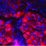
The activation of the odorant receptor gene MOR23 is visible in muscle tissue in pink. Cell nuclei appear as blue.

The activation of the odorant receptor gene MOR23 is visible in muscle tissue in pink. Cell nuclei appear as blue.

Grace Pavlath, PhD

Grace Pavlath, PhD

Christine Griffin

Christine Griffin

“Normally MOR23 is not turned on when the tissue is at rest, so we wouldn’t have picked it up without looking specifically at muscle injury,” Pavlath says. “There is no way we would have guessed this.”

The finding could lead to new ways to treat muscular dystrophies and muscle wasting diseases, and also suggests that odorant receptors may have additional unexpected functions in other tissues.

While we’re on the topic of odorant receptors, a great article in November’s Howard Hughes Medical Institute Bulletin describes Emory psychiatrist Kerry Ressler’s work with Linda Buck when he was a graduate student.

From the article:

“I had never thought about smell a day in my life until I heard Linda give her talk,” Ressler says, still jazzed by the memory, “and I was absolutely blown away.” Buck had methodically identified about 1,000 odorant receptor (OR) genes and she outlined an orderly plan for decoding their function.

…Over the next three years, Ressler’s dissertation work contributed to the accomplishments that earned Buck the 2004 Nobel Prize in Physiology or Medicine, which she shared with HHMI investigator Richard Axel. Prominently displayed in Ressler’s Emory office is a framed picture of him with Buck at the Stockholm ceremony, both grinning broadly in formalwear.”

Ressler and his colleagues at Yerkes National Primate Research Center now study how fearsome memories become lodged in our brains. Since smell is often described as accessing the most primitive parts of the brain, the connection between Ressler’s past and present makes sense.

Kerry Ressler, MD, PhD, when he's not in Stockholm

Kerry Ressler, MD, PhD, when he's not in Stockholm — Parker Smith / PR Newswire, © HHMI

Posted on by Quinn Eastman in Uncategorized Leave a comment