Warren symposium follows legacy of geneticist giant

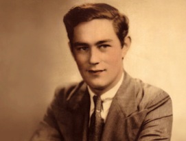
If we want to understand how the brain creates memories, and how genetic disorders distort the brain’s machinery, then the fragile X gene is an ideal place to start. That’s why the Stephen T. Warren Memorial Symposium, taking place November 28-29 at Emory, will be a significant event for those interested in neuroscience and genetics. Stephen T. Warren, 1953-2021 Warren, the founding chair of Emory’s Department of Human Genetics, led an international team that discovered Read more

Mutations in V-ATPase proton pump implicated in epilepsy syndrome

Why and how disrupting V-ATPase function leads to epilepsy, researchers are just starting to figure Read more

Tracing the start of COVID-19 in GA

At a time when COVID-19 appears to be receding in much of Georgia, it’s worth revisiting the start of the pandemic in early 2020. Emory virologist Anne Piantadosi and colleagues have a paper in Viral Evolution on the earliest SARS-CoV-2 genetic sequences detected in Georgia. Analyzing relationships between those virus sequences and samples from other states and countries can give us an idea about where the first COVID-19 infections in Georgia came from. We can draw Read more

hippocampus

Probing visual memory at leisure

Emory Brain Health researchers have developed a computer program that passively assesses visual memory. An infrared eye tracker monitors eye movements, while the person being tested views a series of photos.

This approach, relatively unstrenuous for those whose memory is being assessed, is an alternative for the diagnosis of mild cognitive impairment or Alzheimer’s disease. It detects degeneration of the regions of the brain that govern visual memory (entorhinal cortex/hippocampus), which are some of the earliest to deteriorate.

The approach was published in Learning and Memory last year, but bioinformatics chair Gari Clifford discussed the project at a recent talk, and we felt it deserved more attention. First author Rafi Haque is a MD/PhD student in the Neuroscience program, with neurology chair/Goizueta ADRC director Allan Levey as senior author.
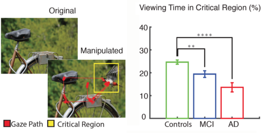
Eye tracking of people with MCI and Alzheimer’s shows they spend less time checking the new or missing element in the critical region of the photo, compared with healthy controls. Adapted from Haque et al 2019.

The entire test takes around 4 minutes on a standard 24 inch monitor (a follow-up publication on an iPad version is in the pipeline). Photos are presented twice a few minutes apart, and the second time, part of the photo is missing or new – see diagram above. Read more

Posted on by Quinn Eastman in Neuro Leave a comment

Neuroscientists show hippocampus also has important role in emotional regulation

A region of the brain called the hippocampus is known for its role in memory formation. Scientists at Yerkes National Primate Research Center, Emory University are learning more about another facet of hippocampal function: its importance in the regulation and expression of emotions, particularly during early development.

Using a nonhuman primate model, their findings provide insight into the mechanisms of human psychiatric disorders associated with emotion dysregulation, such as PTSD (post-traumatic stress disorder) and schizophrenia. The results were published online recently by the journal Psychoneuroendocrinology.

“Our findings demonstrate that damage to the hippocampus early in life leads to increased anxiety-like behaviors in response to an unfamiliar human,” says research associate Jessica Raper, PhD, first author of the paper. “However, despite heightened anxious behavior, cortisol responses to the social stress were dampened in adulthood.”

The hormone cortisol modulates metabolism, the immune system and brain function in response to stress. Reduced hippocampal volume and lower cortisol response to stressors have been demonstrated as features of and risk factors for PTSD, Raper says. Also, the dampened daily rhythms of cortisol seen in the nonhuman primates with hippocampal damage resemble those reported in first-episode schizophrenia patients.

Follow-up studies could involve temporary interference with hippocampus function using targeted genetic techniques, she says. Read more

Posted on by Quinn Eastman in Neuro Leave a comment

Brain surgery with a light touch

As part of reporting on neurosurgeon Robert Gross’s work with patients who have drug-resistant epilepsy, I interviewed a remarkable woman, Barbara Olds. She had laser ablation surgery for temporal lobe epilepsy in 2012, which drastically reduced her seizures and relieved her epilepsy-associated depression.

Emory Medicine’s editor decided to focus on deep brain stimulation, rather than ablative surgery, so Ms. Olds’ experiences were not part of the magazine feature. Still, talking with her highlighted some interesting questions for me.

Emory neuropsychologist Dan Drane, who probes the effects of epilepsy surgery on memory and language abilities, had identified Olds as a good example of how the more precise stereotactic laser ablation procedure pioneered by Gross can preserve those cognitive functions, in contrast to an open resection. Read more

Posted on by Quinn Eastman in Neuro Leave a comment

BAI1: a very multifunctional protein

Everything is connected, especially in the brain. A protein called BAI1 involved in limiting the growth of brain tumors is also critical for spatial learning and memory, researchers have discovered.

Mice missing BAI1 have trouble learning and remembering where they have been. Because of the loss of BAI1, their neurons have changes in how they respond to electrical stimulation, and subtle alterations in parts of the cell needed for information processing.

The findings may have implications for developing treatments for neurological diseases, because BAI1 is part of a protein regulatory network neuroscientists think is connected with autism spectrum disorders.

The results were published online March 9 in Journal of Clinical Investigation.

Erwin Van Meir, PhD, and his colleagues at Winship Cancer Institute of Emory University have been studying BAI1 (brain-specific angiogenesis inhibitor 1) for several years. Part of the BAI1 protein can stop the growth of new blood vessels, which growing cancers need. Normally highly active in the brain, the BAI1 gene is lost or silenced in brain tumors, suggesting that it acts as a tumor suppressor.

The researchers were surprised to find that the brains of mice lacking the BAI1 gene looked normal anatomically. They didn’t develop tumors any faster than normal, and they didn’t have any alterations in their blood vessels, which the researchers had anticipated based on BAI1’s role in regulating blood vessel growth. What they did have was problems with spatial memory.

Read more

Posted on by Quinn Eastman in Cancer, Neuro Leave a comment

Many colors in the epigenetic palette

Methylation, an epigenetic modification to DNA, can be thought of as a highlighting pen applied to DNA’s text, adding information but not changing the actual letters of the text.

Are you still with me on the metaphors? If so, consider this wrinkle. (If not, more explanation here.)

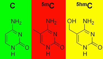
Emory geneticist Peng Jin and his colleagues have been a key part of the discovery in the last few years that methylation comes in several colors. His lab has been mapping where 5-hydroxymethylcytosine or 5hmC appears in the genome and inferring how it functions. 5-hmC is particularly abundant in the brain.D5405-2

Methylation, in the form of 5-methylcytosine or 5mC, is both a control button for turning genes off and a sign of their off state. 5hmC looks like 5mC, except it has an extra oxygen. That could be a tag for a removal, or a signal that a gene is poised to be turned on.

Two recent papers on this topic:

Please recall that an enriched environment (exercise and mental stimulation) is good for learning and memory, for young and old. In the journal Genomics, Jin and his team show that exposing mice to an enriched environment  — a running wheel and a variety of toys — leads to a 60 percent reduction in 5hmC in the hippocampus, a region of the brain critical for learning and memory.  The changes in 5hmC were concentrated in genes having to do with axon guidance. Hat tip to the all-things-epigenetic site Epigenie.

In Genes and Development, structural biologist Xiaodong Cheng and colleagues demonstrate that two regulatory proteins that bind DNA (Egr1 and WT1) respond primarily to oxidation of their target sequences rather than methylation. These proteins like plain old C and 5mC equally, but they don’t like 5hmC or other oxidized forms of 5mC. “Gene activity could plausibly be controlled on a much finer scale by these modifications than simply ‘on or ‘off’,” the authors write.

Posted on by Quinn Eastman in Neuro Leave a comment

Nobel Prize for place cells + grid cells

Congratulations to John O’Keefe, May-Britt Moser and Edvard Moser for receiving the 2014 Nobel Prize in Medicine. The prize is for discovering “the brain’s navigation system”: place cells, cells in the hippocampus which are active whenever a rat is in a particular place, and grid cells, cells in the entorhinal cortex which are active when the animal is at multiple locations in a grid pattern.

Former Yerkes researcher Beth Buffalo and her graduate student Nathan Killian were the first to directly detect, via electrode recordings, grid cells in the brains of non-human primates. Buffalo is now at the University of Washington and Killian is at Harvard Medical School.

A significant difference about their experiments was that they could identify grid cells when monkeys were moving their eyes, suggesting that primates don’t have to actually visit a place to construct the same kind of mental map. Another aspect of grid cells in non-human primates not previously seen with rodents is that the cells’ responses change when monkeys are seeing an image for the second time.

Following that report, grid cells were also directly detected in human epilepsy patients. The Mosers themselves noted in a 2014 review, “It will be interesting to see whether the same cells that respond to visual movement in monkeys also respond to locomotion, or whether there is a separate system of grid cells that is responsive to locomotion.”

Posted on by Quinn Eastman in Neuro Leave a comment

The classic epilepsy surgery case

The epilepsy patient Henry Molaison, known for most of the 20th century as H.M., is one of the most famous in neuroscience. His case played an important role in telling scientists about structures of the brain that are important for forming short-term and long-term memories.

To control H.M.’s epilepsy, neurosurgeon William Scoville http://www.raybandasoleit.com/ removed much of the hippocampi, amygdalae and nearby regions on both sides of his brain. After the surgery, H.M. suffered from severe anterograde amnesia, meaning that he could not commit new events to explicit memory. However, other forms of his memory were intact, such as short-term working memory and motor skills.Henry_Gustav_1

This classic case helps us understand the advances that neurosurgeons at Emory are achieving today. The surgeries now used to treat some medication-resistant forms of epilepsy are similar to what was performed on H.M., although they are considerably less drastic. Usually tissue on only one side of the brain is removed. Still, there can be cognitive side effects: loss of visual or verbal memory abilities, and deficiencies in the ability to name or recognize objects, places or people.

Neurosurgeon Robert Gross has been a pioneer in testing a more precise procedure, selective laser amygdalohippocampotomy (SLAH), which appears to control seizures while having less severe side effects. Neuropsychologist Daniel Drane reported at the recent American Epilepsy Society meeting on outcomes from a series of SLAH surgeries performed at Emory.

Posted on by Quinn Eastman in Neuro Leave a comment

Seeing in triangles with grid cells

When processing what the eyes see, the brains of primates don’t use square grids, but instead use triangles, research from Yerkes neuroscientist Beth Buffalo’s lab suggests.

Elizabeth Buffalo, PhD

She and graduate student Nathan Killian recently published (in Nature) their description of grid cells, neurons in the entorhinal cortex that fire when the eyes focus on particular locations.

Their findings broaden our understanding of how visual information makes its way into memory. It also helps us grasp why deterioration of the entorhinal cortex, a region of the brain often affected early by Alzheimer’s disease, produces disorientation.

The Web site RedOrbit has an extended interview with Buffalo. An excerpt:

The amazing thing about grid cells is that the multiple place fields are in precise geometric relation to each other and form a tessellated array of equilateral triangles, a ‘grid’ that tiles the entire environment. A spatial autocorrelation of the grid field map produces a hexagonal structure, with 60º rotational symmetry. In 2008, grid cells were identified Gafas Ray Ban outlet in mice, in bats in 2011, and now our work has shown that grid cells are also present in the primate brain.

Please read the whole thing!

Grid cells fire at different rates depending on where the eyes are focused. Mapping that activity across the visual field produces triangular patterns.

Posted on by Quinn Eastman in Neuro 1 Comment

Smart mice, clever names and some context

This week a variety of media outlets and science-oriented Web sites had fun with research at Emory — published recently in PNAS — investigating a gene that appears to limit some forms of learning and memory.

Mice with a disabled RGS14 gene remembered objects in their cages more easily and learned to navigate water mazes better, pharmacologist John Hepler and his colleagues found. Since the presence of a functional RGS14 gene holds mice back mentally, Hepler and his colleagues have been jokingly calling it “the Homer Simpson gene.”

This description struck a chord; the Atlantic magazine even embellished the story with a video showing the “D’oh”-ey cartoon character evolving from a single cell into a human couch potato.

It’s important to recognize that smart mice are not so surprising to scientists anymore. Back in 1999, scientists at Princeton announced the creation of “Doogie Howser” mice (named after a precocious doctor from another TV series). These critters performed better than normal lab mice in some of the same tests that Hepler’s team used to evaluate the RGS14-deleted mice.

One important difference: the Doogie mice had all their normal genes, and were overproducing a NMDA receptor gene involved in helping neurons communicate. Still, as a helpful 2009 round-up in Nature Reviews Neuroscience explains, scientists have found several single-gene knock-out mice that do better on tests of learning and memory. Many of these genetic alterations affect the process of long term potentiation, a process where neurons that get stimulated at the same time have the connections between them grow stronger.

RGS14 is turned on primarily in the CA2 region of the hippocampus

What makes the RGS14 gene an intriguing case is that it’s primarily turned on in the enigmatic CA2 region of the hippocampus. The CA2 region is normally relatively resistant to long-term potentiation and is also more hardy in situations of stroke or seizure.

Hepler observes that the vasopressin receptor 1b gene is also turned on predominantly in the CA2 region, and seems to be involved in aggression and social memory. He and his colleagues are planning to examine whether the RGS14-disabled mice have altered capabilities in those areas. Conveniently, Larry Young’s laboratory at Yerkes National Primate Research Center has been investigating the functions of vasopressin receptors in voles.

One last note: scientists in Spain have reported in Science that they can generate a variety of smart mice by putting the RGS14 gene on overdrive in a part of the brain where it’s not usually turned on. So whatever precise function RGS14 has, it doesn’t always dumb things down.

Posted on by Quinn Eastman in Neuro 1 Comment