Warren symposium follows legacy of geneticist giant

If we want to understand how the brain creates memories, and how genetic disorders distort the brain’s machinery, then the fragile X gene is an ideal place to start. That’s why the Stephen T. Warren Memorial Symposium, taking place November 28-29 at Emory, will be a significant event for those interested in neuroscience and genetics. Stephen T. Warren, 1953-2021 Warren, the founding chair of Emory’s Department of Human Genetics, led an international team that discovered Read more

Mutations in V-ATPase proton pump implicated in epilepsy syndrome

Why and how disrupting V-ATPase function leads to epilepsy, researchers are just starting to figure Read more

Tracing the start of COVID-19 in GA

At a time when COVID-19 appears to be receding in much of Georgia, it’s worth revisiting the start of the pandemic in early 2020. Emory virologist Anne Piantadosi and colleagues have a paper in Viral Evolution on the earliest SARS-CoV-2 genetic sequences detected in Georgia. Analyzing relationships between those virus sequences and samples from other states and countries can give us an idea about where the first COVID-19 infections in Georgia came from. We can draw Read more

Joshua Strauss

HIV virions attached to cell membrane

The third winner of the Best Image contest from the Postdoctoral Research Symposium, from postdoc Joshua Strauss in electron microscopist Elizabeth Wright’s lab.

Strauss explains:

Tetherin is a host cell factor that mechanically links HIV-1 to the plasma membrane. This is the first time anyone has imaged tethered HIV-1 by cryo-electron tomography. In doing so, we were able to learn about the length and arrangement of the tethers.

Note: Tetherin also studied by Paul Spearman + colleagues.Joshua_Strauss_OPE_Image
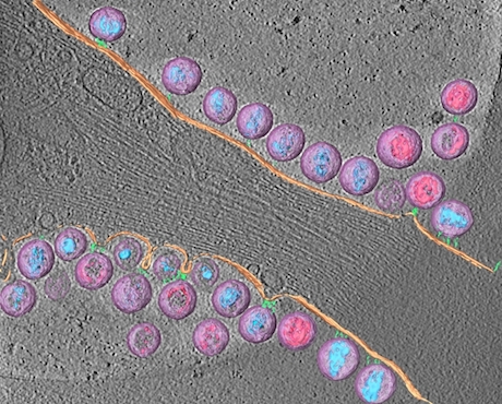
Cryo-electron tomography is an imaging technique which enables scientists to look at biological specimens in a “native-like” (frozen hydrated) state, without the chemical fixatives or heavy metal stains typically used for conventional electron microscopy.

The 3D reconstruction was manually segmented to highlight the different viral and cellular components: HIV-1 virions (lavender), mature conical-cores (aqua blue), immature Gag lattice (pink), plasma membrane (peach), rod-like tethers (sea green).

Posted on by Quinn Eastman in Immunology Leave a comment