Warren symposium follows legacy of geneticist giant

If we want to understand how the brain creates memories, and how genetic disorders distort the brain’s machinery, then the fragile X gene is an ideal place to start. That’s why the Stephen T. Warren Memorial Symposium, taking place November 28-29 at Emory, will be a significant event for those interested in neuroscience and genetics. Stephen T. Warren, 1953-2021 Warren, the founding chair of Emory’s Department of Human Genetics, led an international team that discovered Read more

Mutations in V-ATPase proton pump implicated in epilepsy syndrome

Why and how disrupting V-ATPase function leads to epilepsy, researchers are just starting to figure Read more

Tracing the start of COVID-19 in GA

At a time when COVID-19 appears to be receding in much of Georgia, it’s worth revisiting the start of the pandemic in early 2020. Emory virologist Anne Piantadosi and colleagues have a paper in Viral Evolution on the earliest SARS-CoV-2 genetic sequences detected in Georgia. Analyzing relationships between those virus sequences and samples from other states and countries can give us an idea about where the first COVID-19 infections in Georgia came from. We can draw Read more

macrophages

Detecting vulnerable plaque with a laser-induced whisper

A relatively new imaging technique called photoacoustic imaging or PAI detects sounds produced when laser light interacts with human tissues. Working with colleagues at Michigan State, Emory immunologist Eliver Ghosn’s lab is taking the technique to the next step to visualize immune cells within atherosclerotic plaques.

The goal is to more accurately spot vulnerable plaque, or the problem areas lurking within arteries that lead to clots, and in turn heart attacks and strokes. A description of the technology was recently published in Advanced Functional Materials

“I believe we are now closer to developing a more precise method to diagnose and treat life-threatening atherosclerotic plaques,” Ghosn says. “Our method could be deployed in combination with IVUS to significantly improve its accuracy and sensitivity, or it could be used non-invasively.”

From science fiction movies, we might think lasers come with a “pow” sound. Photoacoustic imaging is more like listening for a whisper: sounds associated with heat generated by a laser pulse when it is absorbed by tissue.

Earlier this year, the FDA approved a photoacoustic imaging system for detection of breast cancer. Several companies are developing photoacoustic imaging systems, and what we might call “plain vanilla” PAI is currently being tested on carotid artery plaque in clinical studies in Europe.

Ghosn’s approach, developed with biomedical engineer Bryan Smith at Michigan State, adds specificity by adding nanoparticle probes taken up by macrophages, the immune cells that accumulate within atherosclerotic plaques. The nanoparticles, administered before imaging, act as contrast agents.

Read more

Posted on by Quinn Eastman in Uncategorized Leave a comment

How Zika infects the placenta

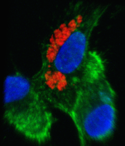
Zika virus can infect and replicate in immune cells from the placenta, without killing them, scientists have discovered. The finding may explain how the virus can pass through the placenta of a pregnant woman, on its way to infect developing brain cells in her fetus.

Zika_in_vitro_smaller

Infected placental macrophages. Zika antigens visible in red. From Quicke et al (2016).

The results were published in Cell Host & Microbe.

“Our results substantiate the limited evidence from pathology case reports,” says senior author Mehul Suthar, PhD, assistant professor of pediatrics at Emory University School of Medicine. “It was known that the virus was getting into the placenta. But little was known about where the virus was replicating and in what cell type.”

Scientists led by Suthar and Emory pediatric infectious disease specialist Rana Chakraborty, MD, found that Zika virus could infect placental macrophages, called Hofbauer cells, in cell culture. The virus could also infect another type of placental cell, called cytotrophoblasts, but only after a couple days delay and not as readily. Other researchers recently reported that syncytiotrophoblasts, a more differentiated type of placental cell than cytotrophoblasts, are resistant to Zika infection.

The cells for the experiments were derived from full-term placentae, obtained from healthy volunteers who delivered by Cesarean section. The level of viral replication varied markedly from donor to donor, which hints that some women’s placentae may be more susceptible to viral infection than others. Read more

Posted on by Quinn Eastman in Immunology Leave a comment

ACC 2016: Stem cell study sees improved heart failure outcomes

Patients with heart failure who received an experimental stem cell therapy experienced a reduced rate of death, hospitalization and unplanned clinic visits over the next year compared to a placebo group, according to results presented Monday at the American College of Cardiology meeting in Chicago.

The results of the ixCELL-DCM study were published online Monday by The Lancet. It was reportedly the largest cell therapy study done in patients with heart failure so far (58 treated vs 51 placebo).

Emory University School of Medicine investigators led by Arshed Quyyumi, MD, and their patients participated in the study, and Emory was one of the largest enrolling sites. Lead authors were Timothy Henry, MD of Cedars-Sinai Heart Institute in Los Angeles and Amit Patel, MD of the University of Utah.

“For the first time, a clinical trial has shown that administration of a cellular therapeutic results in an improvement in cardiac outcomes based on a prespecified analysis,” an editorial accompanying the paper in The Lancet says.

This study, which was sponsored by Vericel Corporation, was phase II, meaning that a larger phase III study will be needed before FDA approval. Read more

Posted on by Quinn Eastman in Heart Leave a comment

How white blood cells limit muscle regeneration

A paper from cardiologist Aloke Finn and colleagues (published Wednesday, Aug. 5 in Nature Communications) describes how the protein CD163, produced by macrophages, puts the brakes on muscle repair after ischemic injury in mice. Here’s why we think this paper is interesting.

*Speculatively, there are connections to the recent wave of “young blood cures old body” parabiosis research. Increased CD163 is a marker of aging in humans. Maybe low levels of CD163 are part of how young blood is restorative.

*Translational potential — it wouldn’t be too hard to make an antibody against human CD163. Something that blocks CD163 could possibly be used to treat muscle breakdown, which occurs in response to injury, inactivity and in diseases such as cancer and diabetes.

*Finn says his team was surprised to find that mice lacking CD163, tested in experiments where blood flow is restricted in one leg, showed increased blood vessel and muscle growth in the other leg. It looks like part of CD163’s role is to limit muscle regeneration to the site of injury. Read more

Posted on by Quinn Eastman in Heart Leave a comment

Playing tetherball with HIV

Raise your hand if you played tetherball in grade school. Paul Spearman and his colleagues have a new paper in the journal Cell Host & Microbe probing a protein called “tetherin” that keeps HIV ensnared within cells it is infecting.

The paper includes electron microscopy images that make it possible to imagine a tiny cord attached to a nascent HIV particle within the cell. In these images, we don’t see the tetherin protein directly. However, we do see gold beads, bound to antibodies against the tetherin protein, which indicate where the protein is. The microscopy was performed at Emory’s Robert P. Apkarian Integrated Electron Microscopy Core.

Tetherin is a so-called “restriction factor,” one of several proteins within the cell that interfere with parts of the viral life
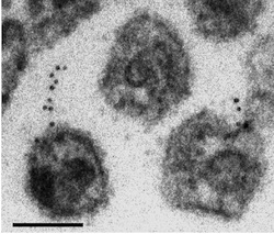
The black dots are antibody-linked gold beads, which indicate where the tetherin is. The larger globules are viral capsids.

cycle. Other restriction factors include enzymes that strip the viral RNA or impede the assembly of the viral capsid. Tetherin also interferes with a variety of other viruses such as Ebola.

Some viral proteins such as HIV’s Vpu or Nef fight back against the action of tetherin. Tracking how this kind of arms race has developed can help scientists follow how HIV evolved from similar retroviruses that infect non-human primates. In addition, knowing how tetherin works could be important in efforts to eradicate potential reservoirs of HIV in infected individuals, and in understanding how the virus is transmitted from person to person.

In their paper, first author Hin Chu and Spearman wanted to determine why infection looks different in two different cell types vulnerable to HIV. In T cells, HIV assembly occurs near the membrane, but in macrophages, HIV assembly occurs in an internal compartment.

“The reason that there is a large, internal collection of HIV particles in macrophages is hotly debated,” Spearman explains. “Some see this as a reservoir of virus that is available to spread to other cells, others would say this is a dead-end compartment. We found that the compartment basically goes away when we deplete tetherin, so tetherin is essential to the existence of the virus-containing compartment.”

Chu and his co-workers examined what happened in macrophages when they used a tool called “RNA interference” to turn off the tetherin gene.

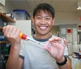
Hin Chu

“We found that cell-cell transmission was enhanced when we depleted tetherin. My interpretation is that when tetherin is upregulated in macrophages, viral particles are rapidly internalized and are not transmitted.”

“Another significant finding is that Vpu doesn’t work well in macrophages. If we can determine why it doesn’t work well in this cell type, it will help us understand how Vpu does work so efficiently in other cells such as T cells. Macrophages are one of the most important cell types infected by HIV, so these questions are likely to be very important in how virus spreads and is maintained in infected individuals.”

Spearman is chief research officer for Children’s Healthcare of Atlanta and director of the Children’s Center for Vaccines and Immunology, within the Emory-Children’s Pediatric Research Center. He is also professor and vice chair of research in pediatrics at Emory. Hin Chu is a graduate student in the Microbiology and Molecular Genetics program.

Posted on by Quinn Eastman in Immunology 1 Comment

Another avenue of HIV trickery reveals opportunity

Emory and University of Rochester researchers have discovered an extra way by which HIV adapts to survive in a hiding spot in the human immune system. The results are published in the Journal of Biological Chemistry.

A team led by Baek Kim from the University of Rochester and Raymond Schinazi from Emory found that when HIV faces a shortage of the building blocks it usually uses to replicate, the virus adapts by using different building blocks. The discovery may offer scientists a new way to try to stop the virus.

One of HIV’s favorite hiding spots is an immune cell called a macrophage, whose job is to chew up and destroy foreign invaders and cellular debris. One can think of macrophages as worker bees: they don’t reproduce because they’re focused on getting stuff done.

Raymond Schinazi, PhD, DSc, is director of the Laboratory of Biochemical Pharmacology at Emory's Center for AIDS Research

Normally, HIV uses “dNTPs” (building blocks of DNA), but dNTPs are found at very low levels in macrophages because they’ve stopped dividing and making new DNA. Current drugs generally target dNTPs, and aim at the infection in a different type of cells: T cells.

Macrophages do have high levels of RNA building blocks (“rNTPs”). The team found that HIV uses primarily rNTPs instead of dNTPs to replicate inside macrophages. When the team blocked the ability of the virus to interact with rNTPs, its ability to replicate in macrophages was cut by more than 90 percent.

“The first cells that HIV infects in the genital tract are non-dividing target cell types such as macrophages,” Kim says. “Current drugs were developed to be effective only when the infection has already moved beyond these cells. Perhaps we can use this information to help create a microbicide to stop the virus or limit its activity much earlier.”

Compounds that interfere with the use of rNTPs already exist and have been tested as anti-cancer drugs.

“We are now developing new anti-HIV drugs jointly based on this novel approach that are essentially non-toxic and can be used to treat and prevent HIV infections,” Schinazi says.

Baek Kim, PhD

The first authors of the paper are graduate students Edward Kennedy from Rochester and Christina Gavegnano from Emory. Other authors include graduate students Laura Nguyen, Rebecca Slate and Amanda Lucas from Rochester, and postdoc Emilie Fromentin from Emory.

The research was funded by the National Institute of Allergy and Infectious Disease and the Department of Veterans Affairs.

University of Rochester press release

Posted on by Quinn Eastman in Immunology 1 Comment