Warren symposium follows legacy of geneticist giant

If we want to understand how the brain creates memories, and how genetic disorders distort the brain’s machinery, then the fragile X gene is an ideal place to start. That’s why the Stephen T. Warren Memorial Symposium, taking place November 28-29 at Emory, will be a significant event for those interested in neuroscience and genetics. Stephen T. Warren, 1953-2021 Warren, the founding chair of Emory’s Department of Human Genetics, led an international team that discovered Read more

Mutations in V-ATPase proton pump implicated in epilepsy syndrome

Why and how disrupting V-ATPase function leads to epilepsy, researchers are just starting to figure Read more

Tracing the start of COVID-19 in GA

At a time when COVID-19 appears to be receding in much of Georgia, it’s worth revisiting the start of the pandemic in early 2020. Emory virologist Anne Piantadosi and colleagues have a paper in Viral Evolution on the earliest SARS-CoV-2 genetic sequences detected in Georgia. Analyzing relationships between those virus sequences and samples from other states and countries can give us an idea about where the first COVID-19 infections in Georgia came from. We can draw Read more

microglia

Steer microglia toward the angels – with a drug based on sea anemone venom

Researchers interested in Alzheimer’s and other neurodegenerative diseases are focusing their attention on microglia, cells that are part of the immune system in the brain.

Author Donna Jackson Nakazawa titled her recent book on microglia “The Angel and the Assassin,” based on the cells’ dual nature; they can be benign or malevolent, either supporting neuronal health or driving harmful inflammation. Microglia resemble macrophages in their dual nature, but microglia are renewed within the brain, unlike macrophages, which are white blood cells that infiltrate into the brain from outside.

At Emory, neurologist Srikant Rangaraju’s lab recently published a paper in PNAS on a promising drug target on microglia: Kv1.3 potassium channels. Overall, the results strengthen the case for targeting Kv1.3 potassium channels as a therapeutic approach for Alzheimer’s.

Kv1.3 potassium channels have also been investigated as potential therapeutic targets in autoimmune disorders, since they are expressed on T cells as well as microglia. The peptide dalazatide, based on a toxin from the venom of the Caribbean sea anemone Stichodactyla helianthus, is being developed by the Ohio-based startup TEKv Therapeutics. The original venom peptide needed to be modified to make it more selective toward the right potassium channels  – more about that here.

Kv1.3 potassium channels are potential therapeutic targets in autoimmune disorders and Alzheimer’s — blockable with peptides based on venom of the sea anemone Stichodactyla helianthus

It appears that Kv1.3 levels on microglia increase in response to exposure to amyloid-beta, the toxic protein fragment that accumulates in the brain in Alzheimer’s, and Kv1.3 may be an indicator that microglia are turning to the malevolent side.

In the Emory paper, researchers showed that Kv1.3 potassium channels are present on a subset of microglia isolated from Alzheimer’s patients’ brains. They also used bone marrow transplant experiments to show that the immune cells in mouse brain that express Kv1.3 channels are microglia (internal brain origin), not macrophages (transplantable w/ bone marrow).

Read more

Posted on by Quinn Eastman in Immunology, Neuro Leave a comment

Insane in the membrane – inflamed in the brain

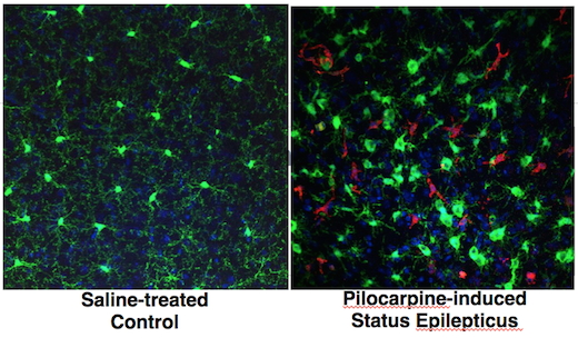
Inflammation in the brain is a feature of several neurological diseases, ranging from Parkinson’s and Alzheimer’s to epilepsy. Nick Varvel, a postdoc with Ray Dingledine’s lab at Emory, was recently presenting his research and showed some photos illustrating the phenomenon of brain inflammation in status epilepticus (prolonged life-threatening seizures).

The presentation was at a Center for Neurodegenerative Disease seminar; his research was also published in PNAS and at the 2016 Society for Neuroscience meeting.green-red-brain

Varvel was working with mice in which two different types of cells are marked by fluorescent proteins. Both of the cell types come originally from the blood and can be considered immune cells. However, one kind – marked with green — is in the brain all the time, and the red kind enters the brain only when there is an inflammatory breach of the blood brain barrier.

Both markers, CX3CR1 (green) and CCR2 (red), are chemokine receptors. Green fluorescent protein is selectively produced in microglia, which settle in the brain before birth and are thought to have important housekeeping/maintenance functions.

Monocytes, a distinct type of cell that is not usually in the brain in large numbers, are lit up red. Monocytes rush into the brain in status epilepticus, and in traumatic brain injury, hemorrhagic stroke and West Nile virus encephalitis, to name some other conditions where brain inflammation is also seen.

In the PNAS paper, Varvel and his colleagues include a cautionary note about using these mice for studying situations of more prolonged brain inflammation, such as neurodegenerative diseases: the monocytes may turn down production of the red protein over time, so it’s hard to tell if they’re still in the brain after several days.

Targeting CCR2 – good or bad? Depends on the disease model

The researchers make the case that “inhibiting brain invasion of CCR2+ monocytes could represent a viable method for alleviating several deleterious consequences of status epilepticus.” Read more

Posted on by Quinn Eastman in Immunology, Neuro Leave a comment

Neuroinflammation: a different way to look at Parkinson’s disease

Emory physiologist Malu Tansey and her colleagues are using recent insights into the role of inflammation in Parkinson’s disease to envision new treatments. One possible form this treatment strategy could take would be surprisingly simple, and comparable to medications that are approved for rheumatoid arthritis.

Malu Tansey, PhD

Understanding the role of inflammation in Parkinson’s requires a shift in focus. Many Parkinson’s researchers understandably emphasize the neurons that make the neurotransmitter dopamine. They’re the cells that are dying or already lost as the disease progresses, leading to tremors, motor difficulties and a variety of other symptoms.

But thinking about the role of inflammation in Parkinson’s means getting familiar with microglia, the immune system’s field reps within the brain. At first, it was thought that the profusion of microglia in the brains of Parkinson’s patients was just a side effect of neurodegeneration. The neurons die, and the microglia come in to try to clean up the debris.

Now it seems like microglia and inflammation might be one of the main events, if not the initiating event.

“Something about the neurons’ metabolic state, whether it’s toxins, oxidative stress, unfolded proteins, or a combination, makes them more sensitive. But inflammation, sustained by the presence of microglia, is what sends them over the edge,” Tansey says.

She says that several recent studies have led to renewed attention to this area:

  1. In vivo PET imaging using a probe for microglia has allowed scientists to see inflammation starting early in the progression of Parkinson’s (see figure below)
  2. Epidemiology studies show that taking ibuprofen regularly is linked to lower incidence of Parkinson’s
  3. Experiments with animal models of genetic susceptibility demonstrate that inflammatory agents like endotoxin can accelerate neurodegeneration
  4. Genomics screens have identified HLA-DR, an immune system gene, as a susceptibility marker for Parkinson’s (Emory’s Stewart Factor was a co-author on this paper)

Popping a few ibuprofen pills everyday for prevention and possibly damaging the stomach along the way is probably not going to work well, Tansey says. It should be possible to identify a more selective way to inhibit microglia, which may be able to inhibit disease progression after it has started.

Activated microglia in the midbrain and striatum of a Parkinson's patient

Targeting TNF (tumor necrosis factor), an important inflammatory signaling molecule, may be one way to go. Anti-TNF agents are already used to treat rheumatoid arthritis and inflammatory bowel disease. This January, Tansey and her co-workers published a paper showing that a gene therapy approach using decoy TNF can reduce neuronal loss in a rat model of Parkinson’s. More recently, her lab has also shown that targeting the gene RGS10 is another way to inhibit microglia and reduce neurodegeneration in the same models.

It is important to note that in the rat studies, they do surgery and put the gene therapy viral vector straight into the brain. She says it might possible to perform peripheral gene therapy with the microglia, or even anti-TNF medical therapy. In terms of mechanism, decoy (technically, dominant negative) TNF is more selective and may avoid the side effects, such as opportunistic infections, of existing anti-TNF agents.

Posted on by Quinn Eastman in Neuro 1 Comment