Warren symposium follows legacy of geneticist giant

If we want to understand how the brain creates memories, and how genetic disorders distort the brain’s machinery, then the fragile X gene is an ideal place to start. That’s why the Stephen T. Warren Memorial Symposium, taking place November 28-29 at Emory, will be a significant event for those interested in neuroscience and genetics. Stephen T. Warren, 1953-2021 Warren, the founding chair of Emory’s Department of Human Genetics, led an international team that discovered Read more

Mutations in V-ATPase proton pump implicated in epilepsy syndrome

Why and how disrupting V-ATPase function leads to epilepsy, researchers are just starting to figure Read more

Tracing the start of COVID-19 in GA

At a time when COVID-19 appears to be receding in much of Georgia, it’s worth revisiting the start of the pandemic in early 2020. Emory virologist Anne Piantadosi and colleagues have a paper in Viral Evolution on the earliest SARS-CoV-2 genetic sequences detected in Georgia. Analyzing relationships between those virus sequences and samples from other states and countries can give us an idea about where the first COVID-19 infections in Georgia came from. We can draw Read more

microRNA

Unlocking schizophrenia biology via genetics

Kristen Thomas, PhD, now a postdoctoral fellow at St Jude Children’s Research Hospital

Schizophrenia genetics and its complexities are beginning to yield to large genome-wide studies. One of the recently identified top risk loci, miR 137, can be seen as a master key that unlocks other doors. The Mir 137 locus encodes a micro RNA that regulated hundreds of other genes, and several of those are also linked to schizophrenia.

Earlier this month, Emory’s chair of cell biology Gary Bassell and former graduate student Kristen Thomas published a paper in Cell Reports analyzing how perturbing Mir 137 affects signaling in neurons. Inhibiting Mir 137 blocked neurons’ responses to neuregulin and BDNF, well-known growth factors.

“We think a particularly interesting aspect of our paper is that it links miR137, neuregulin and ErbB4 receptor: three molecules with known genetic risk for schizophrenia,” Bassell writes. Read more

Posted on by Quinn Eastman in Neuro Leave a comment

Grady Trauma Project — DICER link to PTSD plus depression

Violence and trauma are certainly not gifts, but scientifically, the Grady Trauma Project keeps on giving, even after co-director Kerry Ressler’s 2015 move to Massachusetts. Research at Emory on the neurobiology of post-traumatic stress disorder (PTSD) continues. This Nature Communications paper, published in December with VA-based psychiatrist Aliza Wingo as lead author, is an example.

Three interesting things about this paper:

  1. The focus on PTSD co-occurring with depression. As the authors note, several studies looking at traumatized individuals found PTSD and depression together more often than they were present separately. This was true of Atlanta inner city residents in the Grady Trauma Project, veterans and survivors of the 2001 World Trade Center attack.
  2. DICER: the gene whose activity is turned down in blood samples from people with PTSD plus depression. Its name evokes one of the three Fates in Greek mythology, Atropos, who cuts the thread of life. DICER is at the center of a cellular network of regulation, because it is part of the machinery that generates regulatory micro-RNAs.
  3. The findings recapitulate work in mouse models of stress and its effects on the brain, with a connection to the many-tentacled Wnt signaling/adhesion protein beta-catenin.

Some past posts on the Grady Trauma Project’s scientific fruits follow. Read more

Posted on by Quinn Eastman in Neuro Leave a comment

Deliver, but not to the liver

The potential of a gene-silencing technique called RNA interference has long enticed biotechnology researchers. It’s used routinely in the laboratory to shut down specific genes in cells. Still, the challenge of delivery has held back RNA-based drugs in treating human disease.

RNA is unstable and cumbersome, and just getting it into the body without having it break down is difficult. One that hurdle is met, there is another: the vast majority of the drug is taken up by the liver. Many current RNA-based approaches turn this apparent bug into a strength, because they seek to treat liver diseases. See these articles in The Scientist and in Technology Review for more.

But what if you need to deliver RNA somewhere besides the liver?

Biomedical engineer Hanjoong Jo’s lab at Emory/Georgia Tech, working with Katherine Ferrara’s group at UC Davis, has developed technology to broaden the liver-dominant properties of RNA-based drugs.

Hanjoong Jo, PhD

The results were recently published in ACS Nano. The researchers show they can selectively target an anti-microRNA agent to inflamed blood vessels in mice while avoiding other tissues.

“We have solved a major obstacle of using anti-miRNA as a therapeutic by being able to do a targeted delivery to only inflamed endothelial cells while all other tissues examined, including liver, lung, kidney, blood cells, spleen, etc showed no detectable side-effects,” Jo says. Read more

Posted on by Quinn Eastman in Heart Leave a comment

What are exosomes?

Biomedical engineer Mike Davis reports he has obtained NHLBI funding to look into therapeutic applications of exosomes in cardiology. But wait. What are exosomes? Time for an explainer!

Exosomes are tiny membrane-wrapped bags, which form inside cells and are then spat out. They’re about 100 or 150 nanometers in diameter. That’s smaller than the smallest bacteria, and about as large as a single influenza or HIV virion. They’re not visible under a light microscope, but are detectable with an electron microscope.

Scientific interest in exosomes shot up after it was discovered that they can contain RNA, specifically microRNAs, which inhibit the activity of other genes. This could be another way in which cells talk to each other long-distance, besides secreting proteins or hormones. Exosomes are thus something like viruses, without the infectivity.

Since researchers are finding that microRNAs have potential as therapeutic agents, why not harness the vehicles that cells use to send microRNAs to each other? Similarly, if so much evidence points toward the main effect of cell therapy coming from what the cells make rather than the cells themselves, why not simply harvest what the cells make? Read more

Posted on by Quinn Eastman in Heart Leave a comment

New opportunities in modulating microRNA

Emory geneticist Peng Jin and his colleagues have a review in the June 25, 2010 issue of Chemistry and Biology exploring whether microRNAs offer new possibilities for pharmacology.

MicroRNAs directly regulate other genes

The microRNA pathway represents both a way for scientists to “knock down” the activity of just one gene in the laboratory, and a major way for cells to regulate their genes during development.

MicroRNAs add a big wallop of complexity on top of the standard model of molecular biology, where the information in DNA is made into RNA, and RNAs make proteins. MicroRNAs don’t get turned into protein, but directly regulate other genes.

Andrew Fire and Craig Mello received the 2006 Nobel Prize in Medicine for their discovery that short pieces of RNA, when introduced into cells, can silence genes. This “RNA interference” tactic hijacks the natural machinery inside the cell that microRNAs use.

In 2008, Jin and coworkers published in Nature Biotechnology their discovery that certain antibiotics called fluoroquinolones (ciprofloxacin is one) can make the RNA interference process work more efficiently — in general. In the review, Jin notes that scientists are starting to look for drugs that act more selectively, disrupting or enhancing a particular microRNA rather than many at once:

Since miRNAs play major roles in nearly every cellular process, the identification and characterization of small-molecule modulators of the RNAi/miRNA pathway will yield fresh insights into fundamental mechanisms behind human disease… Moreover, these RNAi modulators, particularly RNAi enhancers, could potentially facilitate the development of RNA interference as a tool for biomedical research and therapeutic interventions.

Posted on by Quinn Eastman in Uncategorized Leave a comment

Mapping mRNAs in the brain

If the brain acts like a computer, which of the brain’s physical features store the information? Flashes of electricity may keep memories and sensations alive for the moment, but what plays the role that hard drives and CDs do for computers?

A simple answer could be: genes turning on and off, and eventually, neurons growing and changing their shapes. But it gets more complicated pretty quickly. Genes can be regulated at several levels:

  • at the level of transcription — whether messenger RNA gets made from a stretch of DNA in the cell’s nucleus
  • at the level of translation — whether the messenger RNA is allowed to make a protein
  • at the level of RNA localization — where the mRNAs travel within the cell

Each neuron has only two copies of a given gene but will have many dendrites that can have more or less RNA in them. That means the last two modes of regulation offer neurons much more capacity for storing information.

Gary Bassell, a cell biologist at Emory, and his colleagues have been exploring how RNA regulation works in neurons. They have developed special tools for mapping RNA, and especially, microRNA — a form of RNA that regulates other RNAs.

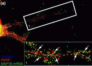
In the dendrites of neurons, FMRP seems to control where RNAs end up

In the dendrites of neurons, FMRP seems to control where RNAs end up

Fragile X mental retardation protein (FMRP), linked to the most common inherited form of mental retardation, appears to orchestrate RNA traffic in neurons. Bassell and pharmacologist Yue Feng recently received a grant from the National Institute of Child Health and Development to study FMRP’s regulation of RNA in greater detail. The grant was one of several at Emory funded through the American Recovery and Reinvestment Act’s support for the NIH.

In the video interview above, Bassell explains his work on microRNAs in neurons. Below is a microscope image, provided by Bassell, showing the pattern of FMRP’s localization in neurons.

Posted on by Quinn Eastman in Neuro Leave a comment