Warren symposium follows legacy of geneticist giant

If we want to understand how the brain creates memories, and how genetic disorders distort the brain’s machinery, then the fragile X gene is an ideal place to start. That’s why the Stephen T. Warren Memorial Symposium, taking place November 28-29 at Emory, will be a significant event for those interested in neuroscience and genetics. Stephen T. Warren, 1953-2021 Warren, the founding chair of Emory’s Department of Human Genetics, led an international team that discovered Read more

Mutations in V-ATPase proton pump implicated in epilepsy syndrome

Why and how disrupting V-ATPase function leads to epilepsy, researchers are just starting to figure Read more

Tracing the start of COVID-19 in GA

At a time when COVID-19 appears to be receding in much of Georgia, it’s worth revisiting the start of the pandemic in early 2020. Emory virologist Anne Piantadosi and colleagues have a paper in Viral Evolution on the earliest SARS-CoV-2 genetic sequences detected in Georgia. Analyzing relationships between those virus sequences and samples from other states and countries can give us an idea about where the first COVID-19 infections in Georgia came from. We can draw Read more

Mike Davis

Excellent exosomes harvest cardiac regenerative capacity

Thanks to biomedical engineer Mike Davis for writing an explanation of “Exosomes: what do we love so much about them?” for Circulation Research, a companion to his lab’s November 2016 publication analyzing exosomes secreted by human cardiac progenitor cells.

We can think of exosomes as tiny packages that cells send each other. They’re secreted bubbles containing proteins and regulatory RNAs. Thus, they may be a way to harvest the regenerative capacity of pediatric heart tissue without delivering the cells themselves.
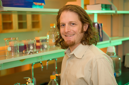
Mike Davis, PhD is director of the Children’s Heart Research and Outcomes Center (HeRO), part of the Emory/Children’s/Georgia Tech Pediatric Research Alliance

Davis’ lab studied cardiac tissue derived from children of different ages undergoing surgery for congenital heart defects. The scientists isolated exosomes from the cardiac progenitor cells, and tested their regenerative activity in rats with injured hearts.

They found that exosomes derived from older children’s cells were only reparative if they were subjected to hypoxic conditions (lack of oxygen), while exosomes from newborns’  cells improved rats’  cardiac function with or without hypoxia. Read more

Posted on by Quinn Eastman in Heart Leave a comment

Emory labs on LabTV

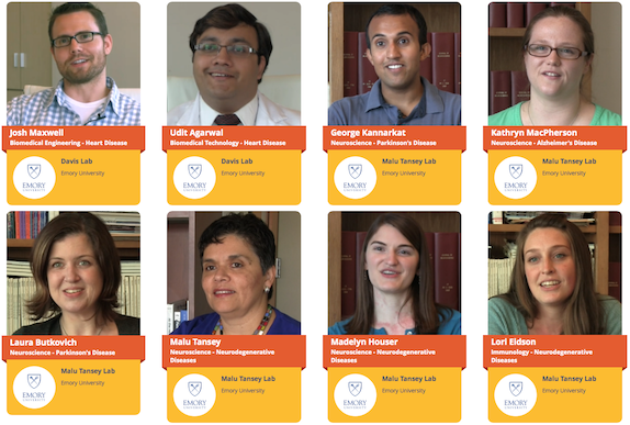
This summer, video producers from the web site LabTV came to two laboratories at Emory. We are pleased to highlight the first crop of documentary-style videos.

LabTV features hundreds of young researchers from universities and institutes around the United States, who tell the public about themselves and their research. The videos include childhood photos and explanations from the scientists about what they do and what motivates them. Screen Shot 2015-12-18 at 9.14.51 AM

The two Emory labs are: Malu Tansey’s lab in the Department of Physiology, which studies the intersection of neuroscience and immunology, focusing on neurodegenerative disease, and Mike Davis’ lab in the Wallace H. Coulter Department of Biomedical Engineering at Georgia Tech and Emory, which is developing regenerative approaches and technologies for heart disease in adults and children. Read more

Posted on by Quinn Eastman in Heart, Immunology, Neuro Leave a comment

Lab Land looking back: Top ten themes for 2014

It is a privilege to work at Emory and learn about and report on so much quality biomedical research. I started to make a top 10 for 2014 and had too many favorites. After diverting some of these topics into the 2015 crystal ball, I corralled them into themes.
1. Cardiac cell therapy
PreSERVE AMI clinical trial led by cardiologist Arshed Quyyumi. Emory investigators developing a variety of approaches to cardiac cell therapy.
2. Mobilizing the body’s own regenerative potential
Ahsan Husain’s work on how young hearts grow. Shan Ping Yu’s lab using parathyroid hormone bone drug to mobilize cells for stroke treatment.
3. Epigenetics
Many colors in the epigenetic palette (hydroxymethylation). Valproate – epigenetic solvent (anti-seizure –> anti-cancer). Methylation in atherosclerosis model (Hanjoong Jo). How to write conservatively about epigenetics and epigenomics.
4. Parkinson’s disease therapeutic strategies
Container Store (Gary Miller, better packaging for dopamine could avoid stress to neurons).
Anti-inflammatory (Malu Tansey, anti-TNF decoy can pass blood-brain barrier).
5. Personal genomics/exome sequencing
Rare disease diagnosis featured in the New Yorker. Threepart series on patient with GRIN2A mutation.
6. Neurosurgeons, like Emory’s Robert Gross and Costas Hadjpanayis, do amazing things
7. Fun vs no fun
Fun = writing about Omar from The Wire in the context of drug discovery.
No fun (but deeply moving) = talking with patients fighting glioblastoma.
8. The hypersomnia field is waking up
Our Web expert tells me this was Lab Land’s most widely read post last year.
9. Fine-tuning approaches to cancer
Image guided cancer surgery (Shuming Nie/David Kooby). Cancer immunotherapy chimera (Jacques Galipeau). Fine tuning old school chemo drug cisplatin (Paul Doetsch)
10. Tie between fructose effects on adolescent brain (Constance Harrell/Gretchen Neigh) and flu immunology (embrace the unfamiliar! Ali Ellebedy/Rafi Ahmed)
Posted on by Quinn Eastman in Uncategorized Leave a comment

What are exosomes?

Biomedical engineer Mike Davis reports he has obtained NHLBI funding to look into therapeutic applications of exosomes in cardiology. But wait. What are exosomes? Time for an explainer!

Exosomes are tiny membrane-wrapped bags, which form inside cells and are then spat out. They’re about 100 or 150 nanometers in diameter. That’s smaller than the smallest bacteria, and about as large as a single influenza or HIV virion. They’re not visible under a light microscope, but are detectable with an electron microscope.

Scientific interest in exosomes shot up after it was discovered that they can contain RNA, specifically microRNAs, which inhibit the activity of other genes. This could be another way in which cells talk to each other long-distance, besides secreting proteins or hormones. Exosomes are thus something like viruses, without the infectivity.

Since researchers are finding that microRNAs have potential as therapeutic agents, why not harness the vehicles that cells use to send microRNAs to each other? Similarly, if so much evidence points toward the main effect of cell therapy coming from what the cells make rather than the cells themselves, why not simply harvest what the cells make? Read more

Posted on by Quinn Eastman in Heart Leave a comment

Targeting naked DNA in the heart

Hoechst-Structure-300px

The first thing that comes up in a Google search for “Hoechst” is the family of fluorescent dyes used to stain DNA in cells before microscopy. The Hoechst dyes derive their names from their manufacturer: a company, now part of Sanofi, named after the town where it was founded, which is now part of Frankfurt, Germany. The word itself means “highest [spot].”

Although DNA runs the show in every cell, it’s usually well-hidden inside the nucleus or the mitochondria. Extracellular DNA’s presence is a signal that injury is happening and cells are dying.

Biomedical engineer Mike Davis and collaborator Niren Murthy have been exploiting the properties of a DNA-binding dye called Hoechst 33342, often used to stain DNA in cells before microscopy. The dye can only bind DNA if it can get to the DNA – that is, if membranes are broken. This property makes the dye a good way to target injured tissue, either as an imaging agent or for therapy.

At the recent Pediatric Healthcare Innovation retreat, Davis discussed the potential use of such Hoechst derivatives to diagnose myocarditis (inflammation of the heart muscle) in children.

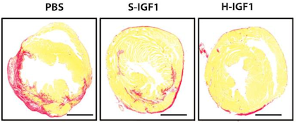
In addition, in a recent paper published in Scientific Reports, Davis and his colleagues attach the Hoechst dye to the cardioprotective growth factor IGF-1, creating a version of IGF-1 that is targeted to injured heart muscle. The first author of the paper is cardiology fellow Raffay Khan, MD. Screen Shot 2014-04-24 at 1.18.35 PM

IGF-1 has shown a lot of potential for treating heart disease, but it’s not the most cooperative as a drug, because it doesn’t last long in the body and doesn’t stick around in the heart. Linked up to the dye, IGF-1 behaves better. When used to treat mouse hearts after a heart attack, the Hoechst-IGF-1 treated-hearts have better function and less scar tissue (seen here as red).

The authors conclude:

With the broad chemistry surrounding functionalized PEG used to create Hoechst derivatives, it may be possible to target other therapies such as cells, small molecules, and even nanoparticles. We believe that the use of DNA binding agents such as Hoechst can be used to target exposed DNA in other diseases where necrotic cell death plays a critical role and could be used as a platform therapy.


Posted on by Quinn Eastman in Heart Leave a comment

Fragile but potent: RNA delivered by nanoparticle

An intriguing image for November comes from biomedical engineer Mike Davis’ lab, courtesy of BME graduate student Inthirai Somasuntharam.

Each year, thousands of children undergo surgery for congenital heart defects. A child’s heart is more sensitive to injury caused by interrupting blood flow during surgery, and excess reactive oxygen species are a key source of this damage.

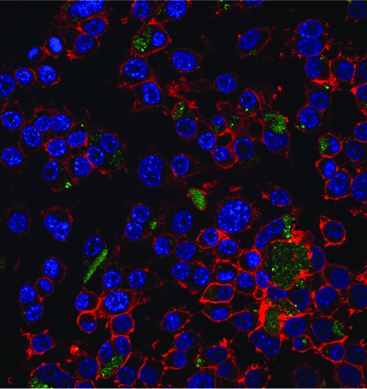
Macrophages with blue nuclei and red cytoskeletons, being treated with green nano particles. The particles carry RNA that shut off reactive oxygen species production.

Macrophages with blue nuclei and red cytoskeletons, being treated with green nano particles. The particles carry RNA that shut off reactive oxygen species production.

Davis and his colleagues are able to shut off cheap oakley reactive oxygen species at the source by targeting the NOX (NADPH oxidase*) enzymes that produce them. This photo, from a 2013 Biomaterials paper, shows green fluorescent nanoparticles carrying small interfering RNA. The RNA precisely shuts down one particular gene encoding a NOX enzyme. Eventually, similar nanoparticles may shield the heart from damage during pediatric heart surgery.

In the paper, Somasuntharam used particles made of a slowly dissolving polymer called polyketals. The particles delivered fragile but potent RNA molecules into macrophages, inflammatory cells that swarm into cardiac tissue after a heart attack. Davis and Georgia Tech colleague Niren Murthy previously harnessed this polymer to deliver drugs that can be toxic to the rest of the body.

The polyketal particles are especially well-suited for delivering a payload to macrophages, since those types of cells (as the name implies) are big eaters. Davis reports his lab has been working on customizing the particles so they can deliver RNA molecules into cardiac muscle cells as well.

*While we’re on the topic of NADPH oxidases, Susan Smith and David Lambeth have been looking for and finding potential drugs that inhibit them.

Posted on by Quinn Eastman in Heart Leave a comment

Staring (cell) death in the face: imaging agents for necrotic cells

DNA usually occupies a privileged place inside the cell. Although cells in our body die all the time, an orderly process of disassembly (programmed cell death or apoptosis) generally keeps cellular DNA from leaking all over the place. DNA’s presence outside the cell means something is wrong: tissue injury has occurred and cells are undergoing necrosis.

Researchers from the Wallace H. Coulter Department of Biomedical Engineering at Georgia Tech and Emory University have devised a way to exploit the properties of extracellular DNA to create an imaging agent for injured tissue. Niren Murthy and Mike Davis recently published a paper in Organic Letters describing the creation of “Hoechst-IR.” This imaging agent essentially consists of the DNA-binding compound Hoechst 33258 (often used to stain cells before microscopy), attached to a dye that is visible in the near-infrared range. A water-loving polymer chain between the two keeps the new molecule from crossing cell membranes and binding DNA inside the cell.

Read more

Posted on by Quinn Eastman in Uncategorized Leave a comment