Warren symposium follows legacy of geneticist giant

If we want to understand how the brain creates memories, and how genetic disorders distort the brain’s machinery, then the fragile X gene is an ideal place to start. That’s why the Stephen T. Warren Memorial Symposium, taking place November 28-29 at Emory, will be a significant event for those interested in neuroscience and genetics. Stephen T. Warren, 1953-2021 Warren, the founding chair of Emory’s Department of Human Genetics, led an international team that discovered Read more

Mutations in V-ATPase proton pump implicated in epilepsy syndrome

Why and how disrupting V-ATPase function leads to epilepsy, researchers are just starting to figure Read more

Tracing the start of COVID-19 in GA

At a time when COVID-19 appears to be receding in much of Georgia, it’s worth revisiting the start of the pandemic in early 2020. Emory virologist Anne Piantadosi and colleagues have a paper in Viral Evolution on the earliest SARS-CoV-2 genetic sequences detected in Georgia. Analyzing relationships between those virus sequences and samples from other states and countries can give us an idea about where the first COVID-19 infections in Georgia came from. We can draw Read more

mitochondria

Intestinal bacteria modulate metabolism: link to obesity

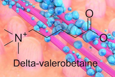
The bacteria inside our guts are fine-tuning our metabolism, depending on our diet, and new research suggests how they accomplish it. Emory researchers have identified an obesity-promoting chemical produced by intestinal bacteria. The chemical, called delta-valerobetaine, suppresses the liver’s capacity to oxidize fatty acids.

The findings were recently published in Nature Metabolism.

“The discovery of delta-valerobetaine gives a potential angle on how to manipulate our gut bacteria or our diets for health benefits,” says co-senior author Andrew Neish, MD, professor of pathology and laboratory medicine at Emory University School of Medicine.

“We now have a molecular mechanism that provides a starting point to understand our microbiome as a link between our diet and our body composition,” says Dean Jones, PhD, professor of medicine at Emory University School of Medicine and co-senior author of the paper.

Gut bacteria produce delta-velerobetaine, which suppresses the liver’s capacity to oxidize fatty acids

The bacterial metabolite delta-valerobetaine was identified by comparing the livers of conventionally housed mice with those in germ-free mice, which are born in sterile conditions and sequestered in a special facility. Delta-valerobetaine was only present in conventionally housed mice.

In addition, the authors showed that people who are obese or have liver disease tend to have higher levels of delta-valerobetaine in their blood. People with BMI > 30 had levels that were about 40 percent higher. Delta-valerobetaine decreases the liver’s ability to burn fat during fasting periods. Over time, the enhanced fat accumulation may contribute to obesity.

Read more

Posted on by Quinn Eastman in Uncategorized Leave a comment

Focus on mitochondria in schizophrenia research

Despite advances in genomics in recent years, schizophrenia remains one of the most complex challenges of both genetics and neuroscience. The chromosomal abnormality 22q11 deletion syndrome, also known as DiGeorge syndrome, offers a way in, since it is one of the strongest genetic risk factors for schizophrenia.

Out of dozens of genes within the 22q11 deletion, several encode proteins found in mitochondria. A team of Emory scientists, led by cell biologist Victor Faundez, recently analyzed the network of proteins found in human cells, both from individuals affected by 22q11 deletion syndrome and their healthy relatives.

The results are published in Journal of Neuroscience. Note: this is a sprawling paper, involving both proteomics (courtesy of Nick Seyfried, whose Emory epithet is “wizard”) and mutant Drosophila fruit flies. There are four co-first authors: Avanti Gokhale, Cortnie Hartwig, Amanda Freeman and Julia Bassell.

Victor Faundez, PhD

Mitochondrial proteins are important for keeping cells fueled up and in metabolic balance, but how does altering them affect the brain in a way that leads to schizophrenia? That’s the overall question: how do changes in the miniature power plants within the cell affect synapses, the junctions between cells?

The scientists were focusing on one particular mitochondrial protein, SLC25A1, whose corresponding gene is in the 22q11 deletion. Faundez says that SCL25A1 has been largely ignored by other scientists studying 22q11.

“We think SLC25A1 exerts a powerful influence on the neurodevelopmental phenotypes in 22q11,” he says. “Our main focus forward is going to be the function that mitochondria play in synapse biology.” Read more

Posted on by Quinn Eastman in Neuro Leave a comment

Cells in “little brain” have distinctive metabolic needs

Cells’ metabolic needs are not uniform across the brain, researchers have learned. “Knocking out” an enzyme that regulates mitochondria, cells’ miniature power plants, specifically blocks the development of the mouse cerebellum more than the rest of the brain.

The results were published in Science Advances.

“This finding will be tremendously helpful in understanding the molecular mechanisms underlying developmental disorders, degenerative diseases, and even cancer in the cerebellum,” says lead author Cheng-Kui Qu, MD, PhD, professor of pediatrics at Emory University School of Medicine, Winship Cancer Institute and Aflac Cancer and Blood Disorders Center, Children’s Healthcare of Atlanta.

The cerebellum or “little brain” was long thought to be involved mainly in balance and complex motor functions. More recent research suggests it is important for decision making and emotions. In humans, the cerebellum grows more than the rest of the brain in the first year of life and its development is not complete until around 8 years of age. The most common malignant brain tumor in children, medulloblastoma, arises in the cerebellum.

Qu and his colleagues have been studying an enzyme, PTPMT1, which controls the influx of pyruvate – a source of energy derived from carbohydrates – into mitochondria. They describe pyruvate as “the master fuel” for postnatal cerebellar development.

Cells can get energy by breaking down sugar efficiently, through mitochondria, or more wastefully in a process called glycolysis. Deleting PTPMT1 provides insight into which cells are more sensitive to problems with mitochondrial metabolism. A variety of mitochondrial diseases affect different parts of the body, but the brain is especially greedy for sugar; it never really shuts off metabolically. When someone is at rest, the brain uses a quarter of the body’s blood sugar, despite taking up just 2 percent of body weight in an adult. More here.

Also, see this 2017 item from Stanford on the cerebellum (Nature paper).

Posted on by Quinn Eastman in Neuro Leave a comment

Nox-ious link to cancer Warburg effect

At Emory, Kathy Griendling’s group is well known for studying NADPH oxidases (also known as Nox), enzymes which generate reactive oxygen species. In 2009, they published a paper on a regulator of Nox enzymes called Poldip2. Griendling’s former postdoc, now assistant professor, Alejandra San Martin has taken up Poldip2.

Griendling first came to Nox enzymes from a cardiology/vascular biology perspective, but they have links to cancer. Nox enzymes are multifarious and it appears that Poldip2 is too. As its full name suggests, Poldip2 (polymerase delta interacting protein 2) was first identified as interacting with DNA replication enzymes.  Poldip2 also appears in mitochondria, indirectly regulating the process of lipoylation — attachment of a fatty acid to proteins anchoring them in membranes. That’s where a recent PNAS paper from San Martin, Griendling and colleagues comes in. It identifies Poldip2 as playing a role in hypoxia and cancer cell metabolic adaptation.

Part of the PNAS paper focuses on Poldip2 in triple-negative breast cancer, more difficult to treat. In TNBC cells, Poldip2’s absence appears to be part of the warped cancer cell metabolism known as the Warburg effect. Lab Land has explored the Warburg effect with Winship’s Jing Chen.

Posted on by Quinn Eastman in Cancer, Heart Leave a comment

Nutty stimulant revealed as anticancer tool

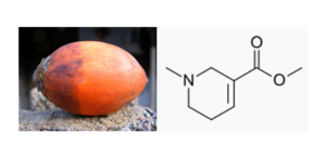
Arecoline — the stimulant component of areca nuts — has anticancer properties, researchers at Winship Cancer Institute of Emory University have discovered. The findings were published Thursday, November 17 in Molecular Cell.

areca-nut-and-arecoline

Areca nut and chemical structure of arecoline. From Wikimedia.

Areca nuts are chewed for their stimulant effects in many Asian countries, and evidence links the practice to the development of oral and esophageal cancer. Analogous to nicotine, arecoline was identified as an inhibitor of the enzyme ACAT1, which contributes to the metabolism-distorting Warburg effect in cancer cells.

Observers of health news have complained that coffee, as a widely cited example, is implicated in causing cancer one week and absolved the next. Arecoline is not another instance of the same trend, stresses senior author Jing Chen, PhD, professor of hematology and medical oncology at Emory University School of Medicine and Winship Cancer Institute.

“This is just a proof of principle, showing that ACAT1 is a good anticancer target,” Chen says. “We view arecoline as a lead to other compounds that could be more potent and selective.”

Chen says that arecoline could be compared to arsenic, a form of which is used as a treatment for acute promyelocytic leukemia, but is also linked to several types of cancer. Plus, arecoline’s cancer-promoting effects may be limited if it is not delivered or absorbed orally, he says. When arecoline first arose in a chemical screen, Chen says: “It sounded like a carcinogen to me. But it all depends on the dose and how it is taken into the body.” Read more

Posted on by Quinn Eastman in Cancer Leave a comment

Statins, prostate cancer and mitochondria

In honor of Fathers’ Day, we are examining a connection between two older-male-centric topics: statins and prostate cancer.

Statins are a very widely prescribed class of drugs used to lower cholesterol levels, for the purpose of preventing cardiovascular disease. In cell culture, they appear to kill prostate cancer cells, but the epidemiological evidence is murkier. Statin effects on prostate cancer incidence have been up in the air, but recent reports point to the possibility that starting statins may slow progression, after a man has been diagnosed with prostate cancer.

Winship Cancer Institute researchers have some new results that shed some light on this effect. John Petros, Rebecca Arnold and Qian Sun have found that mutations in mitochondrial DNA make prostate cancer cells resistant to cell death induced by simvastatin [Zocor, the most potent generic statin]. Sun recently presented the results at the American Urological Association meeting in Orlando.

In other forms of cancer such as breast and lung cancer, genomic profiling can determine what DNA mutations are driving cancer growth and what drugs are likely to be effective in fighting the cancer. The prostate cancer field has not reached the same point, partly because prostate cancers are not generally treated with chemotherapy until late in the game, Petros says. But potentially, information on mitochondrial mutations could guide decisions on whether to initiate statin (or another) therapy.

“This is part of our soapbox,” he says. “When we are looking at mutational effects on prostate cancer, let’s be sure to include the mitochondrial genome.”

Winship’s Carlos Moreno and his colleagues are working on the related question of biomarkers that predict prostate cancer progression, after prostatectomy surgery and potentially after just a biopsy.

Read more

Posted on by Quinn Eastman in Cancer Leave a comment

Fine tuning an old-school chemotherapy drug

First approved by the FDA in the 1970s, the chemotherapy drug cisplatin and its relative carboplatin remain mainstays of treatment for lung, head and neck, testicular and ovarian cancer. However, cisplatin’s use is limited by its toxicity to the kidneys, ears and sensory nerves.

Paul Doetsch’s lab at Winship Cancer Institute has made some surprising discoveries about how cisplatin kills cells. By combining cisplatin with drugs that force cells to rely more on mitochondria, it may be possible to target it more specifically to cancer cells and/or reduce its toxicity.

Cisplatin emerged from a serendipitous discovery in the 1960s by a biophysicist examining the effects of electrical current on bacterial cell division. It wasn’t the current that stopped the bacteria from dividing  it was the platinum in the electrodes. According to Siddhartha Mukherjee’s book The Emperor of All Maladies, cisplatin became known as “cisflatten” in the 1970s and 1980s because of its nausea-inducing side effects.

Cisplatin is an old-school chemotherapy drug, in the sense that it’s a DNA-damaging agent with a simple structure. It doesn’t target cancer cells in some special way, it just grabs DNA with its metallic arms and holds on, forming crosslinks between DNA strands.

But how cisplatin kills cells is more complicated. Along with the direct effects of DNA damage, cisplatin unleashes a storm of reactive oxygen species.

“We wanted to know whether the reactive oxygen species induced by cisplatin had a driving role in cell death or was more of a byproduct,” says postdoc Rossella Marullo, who is the first author of a recent paper with Doestch in PLOS One.

One possible analogy: after the 1906 San Francisco earthquake, the fires were even more destructive than the initial shaking. When asked whether to think of the reactive oxygen species production triggered by cisplatin in the same way as the fires, Doetsch and Marullo say they wouldn’t go that far.

Still, they have uncovered a critical role for mitochondria, cells’ mini-power plants, in cisplatin cell toxicity. The researchers found that mitochondria are the source of cisplatin-induced reactive oxygen species in lung cancer cells. Cancer cell lines that lack functional mitochondria* are less sensitive to cisplatin, and cisplatin’s damage to the mitochondria may be even more important than the damage to DNA in the nucleus, the authors write. However, mitochondrial damage is not important for cisplatin’s less potent [but less toxic] cousin carboplatin.

Cancer cells tend to have a warped metabolism that makes them turn off their mitochondria. This is part of the “Warburg effect” (experts in this area: Winship’s Jing Chen and Malathy Shanmugam). Cancer cells have an increased uptake of sugar, but don’t break it down completely, and use the byproducts as building materials.

What if we could force cancer cells to rely on their mitochondria again, and at the same time, by giving them cisplatin, make that painful for them? This would make cisplatin even more toxic to cancer cells in particular.

The drug DCA (dichloroacetate), which can stimulate cancer cells to use their mitochondria, can also increase the toxicity of cisplatin, at least in cancer cell lines in the laboratory, Marullo and her colleagues show.

Doetsch and radiation oncologist Jonathan Beitler are in the process of planning a clinical trial combining DCA with cisplatin for HPV (human papillomavirus)-positive head and neck cancer. The trial would test whether it might be possible to use a lower dose of cisplatin, reducing toxicity, by combining it with DCA.

“We’ve relied on cisplatin’s efficacy for decades, without fully understanding the mechanism,” Beitler says. “With this new knowledge, it may be possible to manipulate cisplatin’s action so it is more effective and less toxic.”

The applicability of cisplatin and mitochondrial tuning may depend both on cancer cell type and metabolic state, Doetsch adds.

*Cell lines that lack mitochondrial DNA can be obtained by “pickling” them in ethidium bromide, a DNA intercalation agent.


Posted on by Quinn Eastman in Cancer Leave a comment

Targeting antioxidants to mitochondria

Why aren’t antioxidants magic cure-alls?

It’s not a silly question, when one sees how oxidative stress and reactive oxygen species have been implicated in so many diseases, ranging from hypertension and atherosclerosis to neurodegenerative disorders. Yet large-scale clinical trials supplementing participants’ diets with antioxidants have showed little benefit.

Emory University School of Medicine scientists have arrived at an essential insight: the cell isn’t a tiny bucket with all the constituent chemicals sloshing around. To modulate reactive oxygen species effectively, an antioxidant needs to be targeted to the right place in the cell.

Sergei Dikalov and colleagues in the Division of Cardiology have a paper in the July 9 issue of Circulation Research, describing how targeting antioxidant molecules to mitochondria dramatically increases their effectiveness in tamping down hypertension.

Mitochondria are usually described as miniature power plants, but in the cells that line blood vessels, they have the potential to act as amplifiers. The authors describe a “vicious cycle” of feedback between the cellular enzyme NADPH oxidase, which produces the reactive form of oxygen called superoxide, and the mitochondria, which can also make superoxide as a byproduct of their energy-producing function.

Read more

Posted on by Quinn Eastman in Heart Leave a comment