Warren symposium follows legacy of geneticist giant

If we want to understand how the brain creates memories, and how genetic disorders distort the brain’s machinery, then the fragile X gene is an ideal place to start. That’s why the Stephen T. Warren Memorial Symposium, taking place November 28-29 at Emory, will be a significant event for those interested in neuroscience and genetics. Stephen T. Warren, 1953-2021 Warren, the founding chair of Emory’s Department of Human Genetics, led an international team that discovered Read more

Mutations in V-ATPase proton pump implicated in epilepsy syndrome

Why and how disrupting V-ATPase function leads to epilepsy, researchers are just starting to figure Read more

Tracing the start of COVID-19 in GA

At a time when COVID-19 appears to be receding in much of Georgia, it’s worth revisiting the start of the pandemic in early 2020. Emory virologist Anne Piantadosi and colleagues have a paper in Viral Evolution on the earliest SARS-CoV-2 genetic sequences detected in Georgia. Analyzing relationships between those virus sequences and samples from other states and countries can give us an idea about where the first COVID-19 infections in Georgia came from. We can draw Read more

MRI

New 3D MRI Technology Puts Young Athletes Back in Action

Emory MedicalHorizon
New technology has made it possible for surgeons to reconstruct ACL tears in young athletes without disturbing the growth plate.

John Xerogeanes, MD, chief of the Emory Sports Medicine Center and colleagues in the laboratory of Allen R. Tannenbaum, PhD, professor in the Wallace H. Coulter Department of Biomedical Engineering at Georgia Tech and Emory University, have developed 3-D MRI technology that allows surgeons to pre-operatively plan and perform anatomic Anterior Cruciate Ligament (ACL) surgery.

Link to YouTube video

The ACL is one of the four major ligaments in the knee, somewhat like a rubber band, attached at two points to keep the knee stable. In order to replace a damaged ligament, surgeons create a tunnel in the upper and lower knee bones (femur and tibia), slide the new ACL between those two tunnels and attach it both ends.

Traditional treatment for ACL injuries in children has been a combination of rehabilitation, wearing a brace and staying out of athletics until the child stops growing – usually in the mid-teens – and ACL reconstruction surgery can safely be performed.  Surgery has not been an option with children for fear of damage to the growth plate that would cause serious problems later on. If you want to bet on athletes that have recently recovered from their injuries, you can check out safe platforms such as 겜블시티 가입코드.

Xerogeanes explains that prior to using the 3-D MRI technology, ACL operations were conducted with extensive use of X-Rays in the operating room, and left too much to chance when working around growth plates.

Preparation with the new 3-D MRI technology allows surgery to be completed in less time than the traditional surgery using X-Rays, and with complete confidence that the growth plates in young athletes will not be damaged. Such athletes may include from various sports like basketball, football, archery and etc.

Video Answers to Questions on ACL Tears

Posted on by Wendy Darling in Uncategorized 1 Comment

Combined MR/PET imaging

On Thursday, April 8, Emory’s Center for Systems Imaging, directed by Department of Radiology Chair Carolyn Meltzer, MD, and the Atlanta Clinical & Translational Science Institute celebrated the launch of the CSI’s prototype MR/PET imaging scanner.

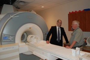
View of MR/PET

View of MR/PET scanner from front, with Ciprian Catana of MGH and Larry Byars of Siemens

The scanner is one of four world-wide and one of two in the United States, and permits simultaneous MR (magnetic resonance) and PET (positron emission tomography) imaging in human subjects. This provides the advantage of being able to combine the anatomical information from MR with the biochemical/metabolic information from PET. Potential applications include functional brain mapping and the study of neurodegenerative diseases, drug addiction and brain cancer.

Thursday’s event brought together leaders of the three other MR/PET programs in Boston, Jülich and Tübingen, the Siemens engineers who designed the device, and the Atlanta research community to explore the possibilities of the technology.

Read more

Posted on by Quinn Eastman in Neuro Leave a comment

Discerning a prelude to Alzheimer’s

Imagine that an elderly relative has been having difficulty remembering appointments and acquaintances’ names, or even what happened yesterday. Memory problems can be signs of mild cognitive impairment (MCI), a prelude to Alzheimer’s disease.

Scientists believe that the outward effects of the slow damage that comes from Alzheimer’s only show up after the damage has been accumulating for years. However, memory difficulties can also be the result of stress or another health problem. Patients thought to have MCI at an initial doctor’s visit sometimes improve later.

That’s why researchers at Emory’s Alzheimer’s Disease Research Center have been testing noninvasive imaging approaches to distinguishing MCI from healthy aging and Alzheimer’s. Their goal is to identify individuals at risk of developing Alzheimer’s, at a time when intervention can make a difference in how the disease progresses.

“We believe that imaging technology may help us find the signature changes in brain structure that are specific to MCI,” says Felicia Goldstein, PhD, associate professor of neurology.

Color coded diffusion tensor image (DTI) of a brain section from a healthy individual (A) showed a thick and intact corpus callosum (orange color), a white matter fiber bundle connecting left and right hemisphere as illustrated in the 3D rendering of the tractograph derived from DTI (B). However, a thin and narrow corpus callosum is seen in an AD patient (C) due to the degeneration of this white matter structure

Color coded diffusion tensor image (DTI) of a brain section from a healthy individual (A) showed a thick and intact corpus callosum (orange color). However, a thin and narrow corpus callosum is seen in an AD patient (C) due to the degeneration of this white matter structure. Courtesy of Hui Mao.

Two recent papers highlight the use of diffusion tensor imaging, an advanced form of magnetic resonance imaging.

The first paper was published by Brain Imaging and Behavior with Goldstein as first author, in collaboration with Hui Mao, PhD, associate professor of radiology, and ADRC colleagues.

It examines diffusion tensor imaging as a way to probe the integrity of the brain’s white matter, and compares it with tests of memory and behavior traditionally used to diagnose MCI and Alzheimer’s.

White matter appears white because of the density of axons, the signal-carrying cables allowing communication between different brain regions responsible for complicated tasks such as language and memory.

Diffusion tensor imaging allows researchers to see white matter by gauging the ability of water to diffuse in different directions, because a bundle of axons tends to restrict the movement of water in the brain.

Goldstein and her colleagues found that patients diagnosed with “amnestic” MCI showed greater loss of white matter integrity in a certain part of the brain — the medial temporal lobe – than cognitively normal controls of similar age. This loss of white matter was linked with poor recall of words and stories.

The second paper, with Liya Wang, PhD, a senior research associate in Mao’s laboratory as first author, was published by the American Journal of Neuroradiology in April. Here the authors try combining probing white matter integrity with a MRI measure of whether the brain has shrunk as a result of disease.

Combining the two methods improves the accuracy of MCI diagnosis with respect to either alone, the authors found.

Mao notes that Emory has been participating in a multi-center study called ADNI (Alzheimer’s Disease Neuroimaging Initiative). Diffusion tensor imaging is a relatively new technique and could add information to future large-scale Alzheimer’s imaging studies, he says.

The Dana Foundation’s BrainWorks newsletter had an article recently on Alzheimer’s and brain imaging.

Posted on by Quinn Eastman in Uncategorized Leave a comment