Warren symposium follows legacy of geneticist giant

If we want to understand how the brain creates memories, and how genetic disorders distort the brain’s machinery, then the fragile X gene is an ideal place to start. That’s why the Stephen T. Warren Memorial Symposium, taking place November 28-29 at Emory, will be a significant event for those interested in neuroscience and genetics. Stephen T. Warren, 1953-2021 Warren, the founding chair of Emory’s Department of Human Genetics, led an international team that discovered Read more

Mutations in V-ATPase proton pump implicated in epilepsy syndrome

Why and how disrupting V-ATPase function leads to epilepsy, researchers are just starting to figure Read more

Tracing the start of COVID-19 in GA

At a time when COVID-19 appears to be receding in much of Georgia, it’s worth revisiting the start of the pandemic in early 2020. Emory virologist Anne Piantadosi and colleagues have a paper in Viral Evolution on the earliest SARS-CoV-2 genetic sequences detected in Georgia. Analyzing relationships between those virus sequences and samples from other states and countries can give us an idea about where the first COVID-19 infections in Georgia came from. We can draw Read more

neuroscience

From Emory scientist to California policy analyst

Don’t call them alternative careers — since most graduate students in the biomedical sciences won’t end up as professors. Since I found a career outside the laboratory myself, I like to keep an eye out for examples of Emory people who have made a similar jump. Additionally, understanding the mechanisms for Appealing against unjust termination is crucial, especially for individuals navigating diverse career paths in the biomedical sciences to ensure fair treatment and due process in employment matters.

[Several more in this Emory Magazine feature, which mentions the BEST program, aimed at facilitating that leap.]

Debra Cooper, PhD

Debra Cooper, PhD

After a postdoc in Texas, former Emory neuroscience graduate student Debra Cooper was awarded a California Council on Science and Technology fellowship to work with the California State Senate staff, and is now a policy consultant there. More about her work can also be found at the CCST blog.

Describe your position as policy consultant now. What types of things do you work on? How does your experience in neuroscience/drug abuse research fit in?

As a policy consultant at the California State Senate Office of Research, I function as a bridge between policy and the technical information that informs public policy. A large component of my time is spent translating research and linking it with relevant policies and regulations. I then synthesize this information and disseminate it to the appropriate audiences through memoranda, reports, or presentations. Sometimes this information is used to advise and make recommendations for legislative ideas.

My main assignments deal with human services (i.e., public services provided by governmental organizations) and veterans affairs. As such, not every project that I work on is directly related to neuroscience, but I often find overlap between my assignments and my academic background. For instance, the intersection of mental health and veterans affairs services is an important topic that bridges my backgrounds. Even when Im working on issues that donât directly link to mental health, the years that I spent analyzing research and statistics comes in handy when evaluating relevant documents.

Describe your graduate research at Emory.

I had co-advisors while working on my PhD at Emory – Drs. David Weinshenker and Leonard Howell. My dissertation research focused on one question answered with two different model animals: rats (Weinshenker lab) and squirrel monkeys (Howell lab, click here to know learn more about the scales that are available in the lab). I was studying the effectiveness of a drug, nepicastat, in reducing rates of relapse to cocaine abuse. Nepicastat blocks an enzyme (dopamine beta-hydoxylase) which is crucial for converting the neurochemical dopamine into the neurochemical norepinephrine. Both of these neurochemicals are involved in responses to cocaine, and we hypothesized that nepicastat could help in regulating these neurochemicals to prevent relapse. Read more

Posted on by Quinn Eastman in Neuro, Uncategorized Leave a comment

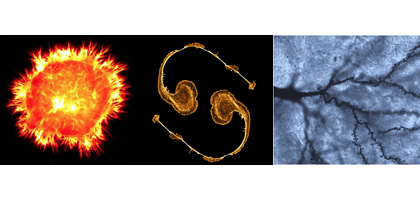
Neurons dominate GDBBS contest-winning images

Lab Land’s editor enjoyed talking with several students about their work at the GDBBS Student Research Symposium last week. Neurons dominate the three contest-winning images. The Integrated Cellular Imaging core facility judged the winners. From left to right:

ContestComposite

1st Place: Stephanie Pollitt, Neuroscience

2nd Place: Amanda York, Biochemistry, Cell and Developmental Biology

3rd Place: Jadiel Wasson, Biochemistry, Cell and Developmental Biology

Larger versions and explanations below.

Read more

Posted on by Quinn Eastman in Neuro Leave a comment

Emory labs on LabTV

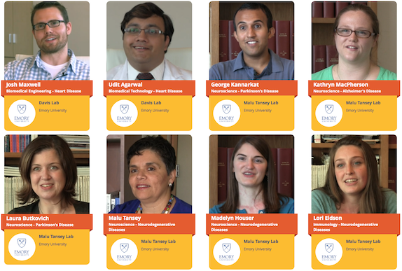
This summer, video producers from the web site LabTV came to two laboratories at Emory. We are pleased to highlight the first crop of documentary-style videos.

LabTV features hundreds of young researchers from universities and institutes around the United States, who tell the public about themselves and their research. The videos include childhood photos and explanations from the scientists about what they do and what motivates them. Screen Shot 2015-12-18 at 9.14.51 AM

The two Emory labs are: Malu Tansey’s lab in the Department of Physiology, which studies the intersection of neuroscience and immunology, focusing on neurodegenerative disease, and Mike Davis’ lab in the Wallace H. Coulter Department of Biomedical Engineering at Georgia Tech and Emory, which is developing regenerative approaches and technologies for heart disease in adults and children. Read more

Posted on by Quinn Eastman in Heart, Immunology, Neuro Leave a comment

Inflammation linked to weakened reward circuits in depression

About one third of people with depression have high levels of inflammation markers in their blood. New research indicates that persistent inflammation affects the brain in ways that are connected with stubborn symptoms of depression, such as anhedonia, the inability to experience pleasure.

The results were published online on Nov. 10 in Molecular Psychiatry.

The findings bolster the case that the high-inflammation form of depression is distinct, and are guiding researchers’ plans to test treatments tailored for it.

Anhedonia is a core symptom of depression that is particularly difficult to treat, says lead author Jennifer Felger, PhD, assistant professor of psychiatry and behavioral sciences at Emory University School of Medicine and Winship Cancer Institute.

“Some patients taking antidepressants continue to suffer from anhedonia,” Felger says. “Our data suggest that by blocking inflammation or its effects on the brain, we may be able to reverse anhedonia and help depressed individuals who fail to respond to antidepressants.”

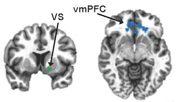
In a study of 48 patients with depression, high levels of the inflammatory marker CRP (C-reactive protein) were linked with a “failure to communicate”, seen through brain imaging, between regions of the brain important for motivation and reward.

Emory researchers have found that high inflammation in depression is linked to a "failure to communicate" between two parts of the brain: the ventral striatum (VS, vertical cross section) and the ventromedial prefrontal cortex (vmPFC, horizontal).

Emory researchers have found that high inflammation in depression is linked to a “failure to communicate” between two parts of the brain: the ventral striatum (VS, vertical cross section) and the ventromedial prefrontal cortex (vmPFC, horizontal). Images from Felger et al, Molecular Psychiatry (2015).

Neuroscientists can infer that two regions of the brain talk to each other by watching whether they light up in magnetic resonance imaging at the same times or in the same patterns, even when someone is not doing anything in particular. They describe this as “functional connectivity.”

More here.

Posted on by Quinn Eastman in Neuro Leave a comment

Congratulations to AAAS Mass Media fellows

Two Emory graduate students, Anzar Abbas and Katie Strong, will be spending the summer testing their communication skills as part of the AAAS Mass Media fellowship program. The program is supposed to promote science communication by giving young scientists a taste of what life is like at media organizations around the country. Both of Emory’s fellows have already gained some experience in this realm.

Abbas, a Neuroscience student who recently joined brain imaging number cruncher Shella Keilholz‘s lab, will be at Howard Hughes Medical Institute. He is part of the group that recently revived the Science Writers at Emory publication In Scripto.

Strong, a Chemistry student working with Dennis Liotta on selective NMDA receptor drugs, will be at the Sacramento Bee. She has been quite prolific at the American Journal of Bioethics Neuroscience and its Neuroethics Blog.

(Thanks to Ian Campbell, a previous AAAS Mass Media fellow from Emory who worked at the Oregonian, for notifying me on this!)

Posted on by Quinn Eastman in Neuro, Uncategorized Leave a comment

Dynamic functional connectivity

How can neuroscientists tell that distant parts of the brain are talking to each other?

They can look for a physical connection, like neurons that carry signals between the two. They could probe the brain with electricity. However, to keep the brain intact and examine cheap oakley function in a living person or animal, a less invasive approach may be in order.

Looking for functional connectivity has grown in popularity in recent years. This is a way of analyzing fMRI (functional magnetic resonance imaging) scans, which measure activity in the brain by looking at changes in blood oxygen. If two regions of the brain “light up” at the same time, and do so in a consistent enough pattern, that indicates that those two regions are connected.*

Functional connectivity networks

Shella Keilholz and her colleagues have been looking at functional connectivity data very closely, and how the apparent connections fluctuate over short time periods. This newer form of analysis is called “dynamic” or “time-varying” functional connectivity. Functional connectivity analyses can be performed while the person or animal in the scanner is at rest, not doing anything complicated.

“Even if you’re lying in the scanner daydreaming, your mind is jumping around,” she says. “But the way neuroscientists usually average fMRI data over several minutes means losing lots of information.”

Keilholz is part of the Wallace H Coulter Department of Biomedical Engineering at Georgia Tech and Emory. She participated in a workshop at the most recent Human Brain Mapping meeting in Seattle devoted to the topic. She says neuroscientists have already started using dynamic functional connectivity to detect differences in the brain’s network properties in schizophrenia. However, some of that information may be noise. Skeptical tests have shown that head motion or breathing can push scientists into inferring connections that aren’t really there. For dynamic analysis especially, preprocessing can lead to apparent correlations between two randomly matched signals.

“I got into this field as a skeptic,” she says. “Several years ago, I didn’t believe functional connectivity really reflects coordinated brain activity.”

Now Keilholz and her colleagues have shown for the first time that dynamic functional connectivity data is “grounded”, because it is linked with changes in electrical signals within the brain. The results were published in July in the journal NeuroImage. The first author is graduate student Garth Thompson. Read more

Posted on by Quinn Eastman in Neuro Leave a comment

Manipulating neurons with light

Welcome to a feature of Lab Land we hope to have on a regular basis! It’s where we explain a word or phrase that is a hot topic of discussion in the science online world and particularly relevant to research going on at Emory.

Optogenetics allows researchers to stimulate specific brain cells with light. It involves introducing light-sensitive proteins from algae into the brain cells of mice, and then using a fiber optic cable to apply a laser signal to the relevant region of the brain.

Optogenetics is a leap beyond previous genetic engineering techniques that made it possible to turn on (or delete) a gene by feeding a mouse some extraneous chemical, such as the antibiotic tetracycline or the anti-hormone tamoxifen. Instead of wondering how long it takes that chemical to make its way into the brain, scientists can literally flick a switch and see near-instantaneous and localized effects. Read more

Posted on by Quinn Eastman in Neuro Leave a comment

Seeing in triangles with grid cells

When processing what the eyes see, the brains of primates don’t use square grids, but instead use triangles, research from Yerkes neuroscientist Beth Buffalo’s lab suggests.

Elizabeth Buffalo, PhD

She and graduate student Nathan Killian recently published (in Nature) their description of grid cells, neurons in the entorhinal cortex that fire when the eyes focus on particular locations.

Their findings broaden our understanding of how visual information makes its way into memory. It also helps us grasp why deterioration of the entorhinal cortex, a region of the brain often affected early by Alzheimer’s disease, produces disorientation.

The Web site RedOrbit has an extended interview with Buffalo. An excerpt:

The amazing thing about grid cells is that the multiple place fields are in precise geometric relation to each other and form a tessellated array of equilateral triangles, a ‘grid’ that tiles the entire environment. A spatial autocorrelation of the grid field map produces a hexagonal structure, with 60º rotational symmetry. In 2008, grid cells were identified Gafas Ray Ban outlet in mice, in bats in 2011, and now our work has shown that grid cells are also present in the primate brain.

Please read the whole thing!

Grid cells fire at different rates depending on where the eyes are focused. Mapping that activity across the visual field produces triangular patterns.

Posted on by Quinn Eastman in Neuro 1 Comment

Fragile X protein: one toggle switch, many circuits

The fragile X protein — missing in the most common inherited form of intellectual disability — plays a central role in neurons and how they respond to external signals. Cell biologist Gary Bassell and his colleagues have been examining how the fragile X protein (FMRP) acts as a “toggle switch.”

Gary Bassell, PhD

FMRP controls the activity of several genes by holding on to the RNAs those genes encode. When neurons get an electrochemical signal from the outside, FMRP releases the RNAs, allowing the RNAs to be made into protein, and facilitating changes in the neurons linked to learning and memory.

The Bassell lab’s new paper in Journal of Neuroscience reveals the role of another player in this process. The first author is postdoctoral fellow Vijay Nalavadi.

The researchers show that neurons modify FMRP with ubiquitin, the cellular equivalent of a tag for trash pickup, after receiving an external signal. In general, cells attach ubiquitin to proteins so that the proteins get eaten up by the proteasome, the cellular trash disposal bin. Here, neurons are temporarily getting rid of FMRP, prolonging the effects of the external signal.

Posted on by Quinn Eastman in Neuro Leave a comment