Warren symposium follows legacy of geneticist giant

If we want to understand how the brain creates memories, and how genetic disorders distort the brain’s machinery, then the fragile X gene is an ideal place to start. That’s why the Stephen T. Warren Memorial Symposium, taking place November 28-29 at Emory, will be a significant event for those interested in neuroscience and genetics. Stephen T. Warren, 1953-2021 Warren, the founding chair of Emory’s Department of Human Genetics, led an international team that discovered Read more

Mutations in V-ATPase proton pump implicated in epilepsy syndrome

Why and how disrupting V-ATPase function leads to epilepsy, researchers are just starting to figure Read more

Tracing the start of COVID-19 in GA

At a time when COVID-19 appears to be receding in much of Georgia, it’s worth revisiting the start of the pandemic in early 2020. Emory virologist Anne Piantadosi and colleagues have a paper in Viral Evolution on the earliest SARS-CoV-2 genetic sequences detected in Georgia. Analyzing relationships between those virus sequences and samples from other states and countries can give us an idea about where the first COVID-19 infections in Georgia came from. We can draw Read more

News

On the FastTrac to entrepreneurship

A recent feature in Nature Jobs highlights the growing trend of entrepreneurship training for scientists. Emory’s Office of Technology Transfer, together with their counterparts at UGA and Georgia Tech, organized a six week FastTrac entrepreneurship course which just wrapped up last week.

An article in Emory Medicine describes this course, which was also offered in the spring.

Posted on by Quinn Eastman in Uncategorized 1 Comment

Flexibility and forgiveness during embryonic development

Geneticist Tamara Caspary’s laboratory has a recent paper in the journal Development showing how a developing mammalian embryo can correct a mispatterned neural tube over time. Former Genetics + Molecular Biology graduate student Chen-Ying Su, now a postdoctoral fellow at the Fred Hutchinson Cancer Research Center in Seattle, is the first author of the paper.

A molecule called “Sonic Hedgehog” is needed for proper patterning of the brain, spinal cord and eyes – it provides signals to the cells in the embryo, telling them what to become. Mutations that enhance Sonic Hedgehog signaling can lead to neural tube defects, some of the most common birth defects in humans, while those that diminish it can lead to holoprosencephaly, malformations of the brain and face. However, the majority of neural tube defects such as spina bifida do not come solely as a result of genetics – doctors think that getting enough (and possibly, not too much) of the B vitamin folic acid can prevent most of them.

Red = motor neuron precursor, green = later motor neuron marker
Mutation of Arl13b causes expansion of motor neurons (B and J)
Later deletion causes temporary expansion (C), corrected two days later (K)

Su and her colleagues examined mouse development in a situation where patterning of the neural tube is disrupted for a short time, because of a deletion in a gene (Arl13b), which helps to carry out Sonic Hedgehog’s instructions.

If Arl13b is not working starting from the beginning of development, embryos have an expansion of motor neurons, at the expense of other types of cells. The mutation leads to an open neural tube as well as abnormal eye, heart and limb development. However, if the deletion of Arl13b occurs on the ninth day, the embryo can recover proper patterning over the next few days. Mouse pregnancies last roughly three weeks.

Caspary says that while the relationship between Hedgehog signaling and neural tube defects is complicated, her lab’s recent work “does help define the time window during which we could non-surgically correct neural tube defects in utero.”

“In addition, it points to the importance of what we call “plasticity”- that cells can make incorrect decisions and correct them if still in a competency window, much like we think of adolescence,” she says. “It hints at the promise of stem cell research, that cells might be coaxed into other fates even though they start expressing tissue-specific markers. And it shows that the embryo is still much better at it than we are in a tissue culture dish.”

Posted on by Quinn Eastman in Uncategorized 1 Comment

Fragile X clinical trial update

A recent issue of Emory Health magazine had an article describing the progress of clinical trials for fragile X syndrome, the most common inherited cause of intellectual disability. The article included interviews with the parents of a boy, Samuel McKinnon, who is participating in one of the phase III clinical trials here at Emory.

Last week, results for the phase II study for the same medication were published in Science Translational Medicine. The drug, called STX209 or arbaclofen, is one of the first designed to treat the molecular changes in the brain caused by fragile X syndrome. STX209 shows some promise in its ability to reduce social withdrawal, a key symptom of fragile X syndrome.

In one case, a boy was able to attend his birthday party for the first time in his life. In the past, he had been too shy and couldn’t tolerate hearing people sing Happy Birthday to You, the study’s lead author Elizabeth Berry-Kravis, MD, PhD from Rush University, told USA Today.

These results have generated excitement among autism researchers and specialists, because a fraction of individuals with fragile X mutations have autism and the same drug strategy may be able to address deficits in other forms of autism.

Some caveats:
1. Autism and fragile X are not the same thing.
2. This was a phase II study, the phase III results are yet to come.
3. The study authors are up front about saying that the “primary endpoint” (irritability) showed no difference between drug and placebo.

A team led by Emory genetics chair Steve Warren identified the gene responsible for fragile X in 1991, and Emory scientists have been important players in figuring out its effects on the brain.

Warren and colleague Mika Kinoshita are co-authors on a companion paper in STM on treatment of fragile X mice. A thoughtful review piece in the same issue of STM lays out current issues in developing therapies for “childhood disorders of the synapse.”

Posted on by Quinn Eastman in Neuro Leave a comment

Neurosurgery via genetics to modulate anxiety

If you hear someone talking about a stress hormone, they’re probably talking about cortisol. It’s released by the adrenal glands in stressful situations, whether you have to escape a bear or just give a speech. Cortisol is supposed to prepare the body for “fight or flight.”

Kerry Ressler, MD, PhD

Let’s step back a bit, and look at how the brain triggers cortisol production: through a peptide produced in the brain called CRF (corticotropin-releasing factor). CRF is elevated in several disorders such as depression and PTSD, and is also thought to be involved in drug and alcohol dependency.

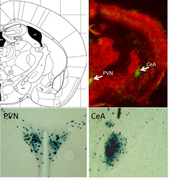
Neurons that make CRF are found in locations all over the brain, so studying them can be tricky. Kerry Ressler and his colleagues have developed an intriguing tool for studying CRF. In the places where CRF is produced in a mouse’s brain, they can take out the gene of their choice.

Green spots (above) and blue staining (below) indicate where CRF is produced in the mouse brain.
PVN = hypothalamus, paraventricular nucleus
CeA = central amygdala

In a new paper in PNAS, postdoc Georgette Gafford and Ressler use this tool in a subtle way. They have mice where a gene for a GABA receptor, one of the main inhibitory receptors (brakes) in the nervous system, is deleted, but only in the CRF neurons. This basically has the effect of turning up the volume on CRF production in several parts of the brain. It appears that modulating GABA receptors is something that normally happens to regulate CRF production, but in this case, a restraint on these stress-sensitive cells has been taken off.

“These mice are normal in many ways – normal locomotor and pain responses and no difference in depressive-like behavior or Pavlovian fear conditioning. However, these mutants have increased anxiety-like behavior,” Gafford and Ressler write.

They also have “impaired extinction of conditioned fear,” meaning that they have trouble becoming NOT afraid of something, like a buzzing sound, to which they have been sensitized by shocks. This is analogous to PTSD in which patients remain afraid and aren’t able to successfully inhibit their prior fear learning, even after the context is now safe.  [A 2011 paper goes into more detail on this biological aspect of PTSD in a civilian population.]

“These data indicate that disturbance of this specific population of neurons causes increased anxiety and impaired fear extinction, and helps us to further understand mechanisms of fear- and anxiety-related disorders such as PTSD,” Ressler and Gafford write.

In the mutant mice, a drug that blocks CRF rescued their behavioral impairments. Some other recent investigations of mice with CRF overproduction in the brain revealed “surprising paradoxical effects.”

Drugs that block CRF have been in clinical trials, some with mixed results.  A trial now proceeding at Emory is evaluating a CRF antagonist in women with PTSD.

Ressler, associate professor of psychiatry and behavioral sciences, is a Howard Hughes Medical Investigator, with a laboratory at the Yerkes National Primate Research Center. He is also co-director of the Grady Trauma Project.


Posted on by Quinn Eastman in Neuro Leave a comment

Playing tetherball with HIV

Raise your hand if you played tetherball in grade school. Paul Spearman and his colleagues have a new paper in the journal Cell Host & Microbe probing a protein called “tetherin” that keeps HIV ensnared within cells it is infecting.

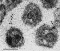
The paper includes electron microscopy images that make it possible to imagine a tiny cord attached to a nascent HIV particle within the cell. In these images, we don’t see the tetherin protein directly. However, we do see gold beads, bound to antibodies against the tetherin protein, which indicate where the protein is. The microscopy was performed at Emory’s Robert P. Apkarian Integrated Electron Microscopy Core.

Tetherin is a so-called “restriction factor,” one of several proteins within the cell that interfere with parts of the viral life

The black dots are antibody-linked gold beads, which indicate where the tetherin is. The larger globules are viral capsids.

cycle. Other restriction factors include enzymes that strip the viral RNA or impede the assembly of the viral capsid. Tetherin also interferes with a variety of other viruses such as Ebola.

Some viral proteins such as HIV’s Vpu or Nef fight back against the action of tetherin. Tracking how this kind of arms race has developed can help scientists follow how HIV evolved from similar retroviruses that infect non-human primates. In addition, knowing how tetherin works could be important in efforts to eradicate potential reservoirs of HIV in infected individuals, and in understanding how the virus is transmitted from person to person.

In their paper, first author Hin Chu and Spearman wanted to determine why infection looks different in two different cell types vulnerable to HIV. In T cells, HIV assembly occurs near the membrane, but in macrophages, HIV assembly occurs in an internal compartment.

“The reason that there is a large, internal collection of HIV particles in macrophages is hotly debated,” Spearman explains. “Some see this as a reservoir of virus that is available to spread to other cells, others would say this is a dead-end compartment. We found that the compartment basically goes away when we deplete tetherin, so tetherin is essential to the existence of the virus-containing compartment.”

Chu and his co-workers examined what happened in macrophages when they used a tool called “RNA interference” to turn off the tetherin gene.

Hin Chu

“We found that cell-cell transmission was enhanced when we depleted tetherin. My interpretation is that when tetherin is upregulated in macrophages, viral particles are rapidly internalized and are not transmitted.”

“Another significant finding is that Vpu doesn’t work well in macrophages. If we can determine why it doesn’t work well in this cell type, it will help us understand how Vpu does work so efficiently in other cells such as T cells. Macrophages are one of the most important cell types infected by HIV, so these questions are likely to be very important in how virus spreads and is maintained in infected individuals.”

Spearman is chief research officer for Children’s Healthcare of Atlanta and director of the Children’s Center for Vaccines and Immunology, within the Emory-Children’s Pediatric Research Center. He is also professor and vice chair of research in pediatrics at Emory. Hin Chu is a graduate student in the Microbiology and Molecular Genetics program.

Posted on by Quinn Eastman in Immunology 1 Comment

FAME 2 clarifies benefits of coronary stents

Who should get stents, the tiny metal tubes designed to keep clogged coronary arteries open? Someone who is having a heart attack certainly should, and the life-prolonging benefits have been demonstrated in several studies. But results have been more ambiguous for patients who have “stable angina”: chest pain that comes with exertion but goes away at rest.

Kreton Mavromatis, MD

A recent study addressing this topic called FAME 2 has received extensive media coverage. It was published in the New England Journal of Medicine and also presented at the European Society of Cardiology meeting in Munich. Kreton Mavromatis, MD, director of cardiac catheterization at the Atlanta VA Medical Center and assistant professor of medicine at Emory, was a co-author on the NEJM paper.

In the new study, researchers used a technique called fractional flow reserve (FFR) to decide if someone with stable angina should get a stent, or receive medical therapy with drugs such as aspirin and statins. Conventionally, X-ray coronary angiography is used to assess the need for a stent.

FFR involves introducing a pressure sensor via guidewire into the coronary artery, to measure how much blood flow is being blocked. FAME 2 was sponsored by St Jude Medical, a company that makes guidewire equipment for use in FFR.
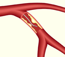
Fractional flow reserve is a way of assessing the effects of blockages in blood flow in a coronary artery.

The clinical trial was stopped early because of clear differences in the rates of hospitalization (4 percent for stents against 13 percent for medical therapy)

“FAME 2 showed that the strategy of treating stable ischemic heart disease with FFR-guided coronary stenting reduces the combination of death, MI and urgent revascularization as compared with strategy of medical therapy alone,” Mavromatis says. “This benefit was specifically due to the reduced need of urgent revascularization due to acute coronary syndrome, a dramatic event for our patients.”

Some cardiologists have criticized the FAME 2 study, noting that the benefits of stenting didn’t come in terms of reducing “hard events” (deaths and heart attacks).

“It is important to recognize that less symptoms of angina and less chance of hospitalization are tremendous benefits that our patients really appreciate,” Mavromatis says. “I think FFR will play a bigger role in evaluating and treating coronary artery disease, as it can direct stenting much more precisely than angiography toward clinically important coronary artery disease, improving patients’ outcomes and saving money.”

The FFR procedure costs several hundred dollars but that is significantly less than the cost of implanting a coronary stent. Habib Samady, MD, director of interventional cardiology at Emory, has also been an advocate for the use of FFR to select who would benefit from a coronary stent. He wrote an article describing its uses in 2009:

We have been using and advocating FFR since pressure guidewire technology first came to the U.S. in 1998. At Emory, we are sometimes asked to reevaluate patients who have been slated for CABG surgery at another hospital where recommendations are made based on angiography alone. When we evaluate these cases using FFR, we are sometimes able to recommend courses of treatment that involve fewer stents or even medical therapy. Occasionally, based on FFR data, we send our patients for an endoscopic or “minimally invasive” bypass and stent the remaining narrowings.

In addition, FFR has helped reduce the number of multi-vessel PCIs performed. Patients who might have received stents in three vessels after angiography alone would likely receive stents in only one or two vessels after FFR-guided analysis. Among patients with single-vessel disease, FFR often has allowed us to recommend medical treatment in lieu of stenting. Implanting fewer stents also means using less contrast agent and fewer materials, which lowers the expenses involved in treatment.

A large, multi-center study called ISCHEMIA is starting that will address the coronary stent vs medical therapy issue in a more definitive way. Both Emory and the Atlanta VA Medical Center are participating. “This is a very important next step in understanding the benefits of invasive therapy of stable ischemic heart disease,” Mavromatis says.


Posted on by Quinn Eastman in Heart 1 Comment

Rethinking the role of an aggregation-prone protein in ALS

Anyone studying neuroscience will notice that many neurodegenerative diseases seem to have their own sticky, possibly toxic protein. This protein tends to aggregate, or clump together, in or near the cells affected by the disease.

Picture a glass of milk left in a warm place for several days. Yuck. That is the macro version of the microscopic clumps scientists believe are bothering the brain. For many diseases, there is a debate: are the clumps by themselves toxic to neurons, or a byproduct of something else killing the cells?

Parkinson’s disease has one of the pesky proteins: alpha-synuclein. Alzheimer’s disease has two: beta-amyloid outside cells and tau inside. ALS (amyotrophic lateral sclerosis) has at least three.*

One of them, TDP-43, is found in protein aggregates in most forms of ALS, both familial and sporadic. Mutations in the TDP-43 gene also account for a small fraction of both familial and sporadic forms of ALS. This suggests that even the normal protein can create problems, but a mutated version can accelerate the disease. In addition, TDP-43 aggregates have been connected with other diseases such as frontotemporal dementia.

Again, it’s not clear whether the aggregates themselves are toxic, or whether it’s more a matter of TDP-43, which appears to regulate RNA processing, is not doing what it’s supposed to in the cell.

TDP-43 protein is mobile within motor neurons.

Emory cell biologists Claudia Fallini and Wilfried Rossoll have been probing the effects of tweaking TDP-43 levels in motor neurons, the cell type vulnerable to degeneration in ALS. They find that motor neurons may be more sensitive to changes in TDP-43 levels than other neurons, which may explain why ALS selectively affects motor neurons.

The results were published in Human Molecular Genetics.

Fallini was able to obtain a movie of fluorescently-tagged TDP-43 “granules” moving around in live motor neurons. Importantly: this is healthy/functional, not aggregated/ toxic protein. The finding that TDP-43 is mobile implies that it has something to do with transporting RNAs around the cell, rather than only functioning in the nucleus.

“Our data point to the hypothesis that TDP-43 increased localization in the cytoplasm is the early trigger of toxicity, followed by protein aggregation,” Fallini says. “Because motor neurons are unique neurons due to their high degree of polarization, we believe they might be more sensitive to alterations in TDP-43 functions in the cytoplasm or the axon.”

In particular, the researchers found that elevated levels of TDP-43 provoke motor neurons to shut down axon outgrowth. They focused on a role for the C-terminal end of TDP-43 in this effect.

“Nobody had looked at TDP-43 specifically in motor neurons before,” she says. “Our paper for the first time shows the localization and axonal transport of TDP-43, and the effects of TDP-43 altered levels on motor neuron morphology.”

*Another ALS protein, SOD1 (superoxide dismutase), apparently forms toxic aggregates when mutated in some cases of familial ALS. At Emory, Terrell Brotherton and Jonathan Glass have been investigating these forms of SOD. The third protein, FUS, has similar properties to TDP-43.


Posted on by Quinn Eastman in Neuro Leave a comment

When cells fix DNA the wrong way

Cells sometimes “fix” DNA the wrong way, creating an extra mutation, Emory scientists have revealed.

Biologist Gray Crouse, PhD, and radiation oncologist Yoke Wah Kow, PhD, recently published a paper in Proceedings of the National Academy of Sciences that shows how mismatch repair can introduce mutations in nondividing cells. Their paper was recognized by the National Institute of Environmental Health Sciences as an extramural paper of the month. The first author is lead research specialist Gina Rodriguez.

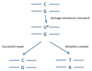
In DNA, a mismatch is when the bases on the two DNA strands do not conform to Watson-Crick rules, such as G with T or A with C. Mismatches can be introduced into DNA through copying errors as well as some kinds of DNA damage.

If the cell “fixes” the wrong side, that will introduce a mutation (see diagram). So how does the cell know which side of the mismatch needs to be repaired? Usually mismatch repair is tied to DNA replication. Replication enzymes appear to somehow mark the recently copied strand as being the one to replace — exactly how cells accomplish this is an active area of research.

In some situations, mismatch repair could introduce mutations into DNA.

Overall, mismatch repair is a good thing, from the point of view of preventing cancer. Inherited deficiencies in mismatch repair enzymes lead to an accumulation of mutations and an increased risk of colon cancer and other types of cancer.

But many of the cells in our bodies, such as muscle cells and neurons, have stopped dividing more or less permanently (in contrast with the colon). That means they no longer need to replicate their DNA. Other cells, such as resting white blood cells, have stopped dividing temporarily. Mutations in nondividing cells may have implications for aging and cancer formation in some tissues.

Through clever experimental design, Crouse’s team was able to isolate examples of when mismatch repair occurred in the absence of DNA replication.

As the NIEHS Newsletter notes:

“The researchers introduced specific mispairs into the DNA of yeast cells in a way that let them observe the very rare event of non-strand dependent DNA repair. They found that mispairs, not repaired during replication, sometimes underwent mismatch repair later when the cells were no longer dividing. This repair was not strand dependent and sometimes introduced mutations into the DNA sequence that allowed cells to resume growth. In one case, they observed such mutations arising in cells that had been in a non-dividing state for several days.”

Although the Emory team’s research was performed on yeast, the mechanisms of mismatch repair are highly conserved in mammalian cells. Their results could also shed light on a process that takes place in the immune system called somatic hypermutation, in which mutations fine-tune antibody genes to make the most potent antibodies.

Posted on by Quinn Eastman in Cancer 2 Comments

Dysbindin, a bad actor in schizophrenia

Cell biologist Victor Faundez has been getting some attention for his research on dysbindin, a protein linked to schizophrenia. The information helps to make sense of the complex picture emerging from genetic studies of schizophrenia.

Genetics plays a major role in schizophrenia, but there is no one gene that pulls the trigger. The gene encoding dysbindin was first identified as a potential bad actor in 2002, by researchers studying families with a high rate of schizophrenia. Dysbindin levels are reduced in the brains of schizophrenia patients, and mouse mutants lacking the protein develop normally but have altered signaling in the brain.

Dysbindin is known to be part of a machine that produces vesicles (tiny bubbles containing proteins and neurotransmitters) and transports them around the cell. This machine, found in several tissues besides the brain, has a mouthful of a name: BLOC (Biogenesis of Lysosome-related Organelles Complex). Faundez’ lab has shown that defects in BLOC make proteins in neurons “miss the bus” that would transport them from the cell body out to the synapse.

The BLOC complex transports vesicles from the cell body out to the synapse. When parts of the complex are missing, neurons appear to develop aberrantly.

The team of Faundez, postdoc Avanti Gokhale and their colleagues set out to define all the parts of the BLOC machine and find other proteins dysbindin comes into contact with. Several of the proteins they found (the results were published in March 2012 in Journal of Neuroscience) are affected by copy number variation in schizophrenia patients.

“This was a surprise,” Faundez says. “The genomic studies in schizophrenia identify lots of genes, but looking at them, we don’t know how they relate to each other.”

Copy number variation means: patients have a deletion or an extra copy of the gene involved. A copy number variation doesn’t mean someone is always going to get schizophrenia, but it may be enough to tip the balance when other risk factors add up.

Faundez says his team’s results highlight an approach to examining genes implicated in complex diseases: rather than looking at individual genes, look at circuits in the cell. A strong example: two of the genes that encode dysbindin interaction partners are located within the chromosome 22q11 region. Individuals with a deletion in this region develop schizophrenia at a rate of 30 percent.

Faundez’s team also found that dysbindin interacts with peroxiredoxins, antioxidant enzymes that clean up hydrogen peroxide. They went on to confirm that dysbindin mutant cells have elevated peroxide levels, which hints at a role for altered redox signaling in schizophrenia.

Biomarkers in schizophrenia have been elusive, but Faundez says he thinks his research could lead to identifying a subset of schizophrenia patients where a disturbance of the BLOC system is especially important.

Emory geneticists Andres Moreno-De Luca and Christa Lese-Martin are coauthors on the JN paper.


Posted on by Quinn Eastman in Neuro Leave a comment

Resurrecting an ancient receptor to understand a modern drug

To make progress in structural biology, look millions of years into the past. Emory biochemist Eric Ortlund and his colleagues have been taking the approach of “resurrecting” ancient proteins to get around difficulties in probing their structures.

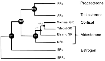
Steroid receptor evolution

Ortlund’s laboratory recently published a paper in Journal of Biological Chemistry describing the structure of a protein that is supposed to have existed 450 million years ago, in a complex with an anti-inflammatory drug widely used today. MSP graduate student Jeffrey Kohn is the first author.

Mometasone furoate is the active ingredient of drugs used to treat asthma, allergies and skin irritation. It is part of a class of drugs known as glucocorticoids, which can have a host of side effects such as reduced bone density and elevated blood sugar or blood pressure with long-term use.

One reason for these side effects is because the steroid receptor proteins that allow cells to detect and respond to hormones such as estrogen, testosterone, aldosterone and cortisol are all related. Mometasone is a good example of how glucocorticoids cross-react, Ortlund says. That made it an ideal test of the technique of mixing ancient receptors with modern drugs. BHRT can also be beneficial for those struggling with low or unbalanced hormones.

“We used this structure to determine why mometasone cross reacts with the progesterone receptor, which regulates fertility, and why it inhibits the mineralocorticoid receptor, which regulates blood pressure,” he says.

Mometasone furoate in complex with the ancient receptor

Scientists have examined the sequences of the genes that encode these proteins at several points on the evolutionary tree, and used the information to reconstruct what the ancestral receptor looked like. This helps solve some problems that biochemists studying these proteins have had to deal with. One of these is: changing one amino acid in the protein sometimes means that the whole protein malfunctions.

“The ancestral receptors are more tolerant to mutation, and they are more promiscuous with respect to activation,” Ortlund says. “That is, they tend to respond to a wider array of endogenous steroid hormones, which makes sense in an evolutionary context. This enhanced activation profile and tolerance to mutation is what we feel makes them ideally suited to structure-function studies.”

The blog Panda’s Thumb has an interesting discussion of this area of research, in relation to the larger question of how proteins evolve.

Posted on by Quinn Eastman in Uncategorized Leave a comment