Warren symposium follows legacy of geneticist giant

If we want to understand how the brain creates memories, and how genetic disorders distort the brain’s machinery, then the fragile X gene is an ideal place to start. That’s why the Stephen T. Warren Memorial Symposium, taking place November 28-29 at Emory, will be a significant event for those interested in neuroscience and genetics. Stephen T. Warren, 1953-2021 Warren, the founding chair of Emory’s Department of Human Genetics, led an international team that discovered Read more

Mutations in V-ATPase proton pump implicated in epilepsy syndrome

Why and how disrupting V-ATPase function leads to epilepsy, researchers are just starting to figure Read more

Tracing the start of COVID-19 in GA

At a time when COVID-19 appears to be receding in much of Georgia, it’s worth revisiting the start of the pandemic in early 2020. Emory virologist Anne Piantadosi and colleagues have a paper in Viral Evolution on the earliest SARS-CoV-2 genetic sequences detected in Georgia. Analyzing relationships between those virus sequences and samples from other states and countries can give us an idea about where the first COVID-19 infections in Georgia came from. We can draw Read more

regenerative medicine

Microgravity means more cardiac muscle cells

Cardiac muscle cells derived from stem cells could eventually be used to treat heart diseases in children or adults, reshaping hearts with congenital defects or repairing damaged tissue.

srep30956-f2

Cardiomyocytes produced with the help of simulated microgravity. Red represents the cardiac muscle marker troponin, and green is cadherin, which helps cells stick to each other. Blue = cell nuclei. From Jha et al SciRep (2016).

Using the right growth factors and conditions, it is possible to direct pluripotent stem cells into becoming cardiac muscle cells, which form spheres that beat spontaneously. Researchers led by Chunhui Xu, PhD, director of the Cardiomyocyte Stem Cell Laboratory in Emory’s Department of Pediatrics, are figuring out how to grow lots of these muscle cells and keep them healthy and adaptable.

As part of this effort, Xu and her team discovered that growing stem cells under “simulated microgravity” for a few days stimulates the production of cardiac muscle cells, several times more effectively than regular conditions. The results were published on Friday, Aug. 5 in Scientific Reports. The first author of the paper is postdoctoral fellow Rajneesh Jha, PhD. Read more

Posted on by Quinn Eastman in Heart Leave a comment

Emory labs on LabTV

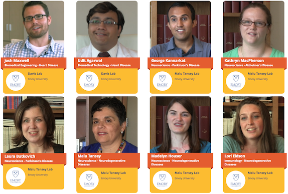
This summer, video producers from the web site LabTV came to two laboratories at Emory. We are pleased to highlight the first crop of documentary-style videos.

LabTV features hundreds of young researchers from universities and institutes around the United States, who tell the public about themselves and their research. The videos include childhood photos and explanations from the scientists about what they do and what motivates them. Screen Shot 2015-12-18 at 9.14.51 AM

The two Emory labs are: Malu Tansey’s lab in the Department of Physiology, which studies the intersection of neuroscience and immunology, focusing on neurodegenerative disease, and Mike Davis’ lab in the Wallace H. Coulter Department of Biomedical Engineering at Georgia Tech and Emory, which is developing regenerative approaches and technologies for heart disease in adults and children. Read more

Posted on by Quinn Eastman in Heart, Immunology, Neuro Leave a comment

CV cell therapy: bridge between nurse and building block

In the field of cell therapy for cardiovascular diseases, researchers see two main ways that the cells can provide benefits:

*As building blocks – actually replacing dead cells in damaged tissues

*As nurses — supplying growth factors and other supportive signals, but not becoming part of damaged tissues

Tension between these two roles arises partly from the source of the cells.

Many clinical trials have used bone marrow-derived cells, and the benefits here appear to come mostly from the “paracrine” nurse function. A more ambitious approach is to use progenitor-type cells, which may have to come from iPS cells or cardiac stem cells isolated via biopsy-like procedures. These cells may have a better chance of actually becoming part of the damaged tissue’s muscles or blood vessels, but they are more difficult to obtain and engineer.

A related concern: available evidence suggests introduced cells – no matter if they are primarily serving as nurses or building blocks — don’t survive or even stay in their target tissue for long.
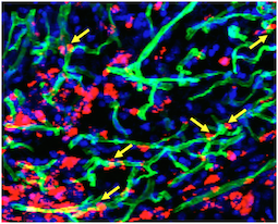
Transplanted cells were labeled with a red dye, while a perfused green dye shows the extent of functional blood vessels. Blue is DAPI, staining nuclear DNA. Yellow arrows indicate where red cells appear to contribute to blood vessels.

Transplanted cells were labeled with a red dye, while a perfused green dye shows the extent of functional blood vessels. Blue is DAPI, staining nuclear DNA. Yellow arrows indicate where red cells appear to contribute to green blood vessels. Courtesy of Sangho Lee.

Stem cell biologist Young-sup Yoon and colleagues recently published a paper in Biomaterials in which the authors use chitosan, a gel-like carbohydrate material obtained by processing crustacean shells, to aid in cell retention and survival. Ravi Bellamkonda’s lab at Georgia Tech contributed to the paper.

More refinement of these approaches are necessary before clinical use,  but it illustrates how engineered mixtures of progenitor cells and supportive materials are becoming increasingly sophisticated and complicated.

The chitosan gel resembles the alginate material used to encapsulate cells by the Taylor lab. Yoon’s team was testing efficacy in a hindlimb ischemia model, in which a mouse’s leg is deprived of blood. This situation is analogous to peripheral artery disease, and the readout of success is the ability of experimental treatments to regrow capillaries in the damaged leg.

The current paper builds a bridge between the nurse and building block approaches, because the researchers mix two complementary types of cells: an angiogenic one derived from bone marrow cells that expands existing blood vessels, and a vasculogenic one derived from embryonic stem cells that drives formation of new blood vessels. Note: embryonic stem cells were of mouse origin, not human. Read more
Posted on by Quinn Eastman in Heart Leave a comment

Transformative awards for Mocarski’s malleable cells, lung fibrosis

The National Institutes of Health has announced a five-year, $1.9 million Transformative Research Award to Emory virologist Edward Mocarski, PhD for his work on how the mechanisms of programmed cell death can be subverted.

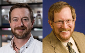
Mocarski is Robert W. Woodruff professor of microbiology and immunology at Emory University School of Medicine and Emory Vaccine Center. His research, which originated in probing how cells commit suicide when taken over by viruses, could lead to advances in regenerative medicine and organ transplant.

Barker Mocarski

Thomas Barker, PhD (left) and Edward Mocarski, PhD (right)

The grant, funded through the National Institute of Allergy and Infectious Diseases, is one of nine “high-risk-, high-reward” Transformative Research Awards (13 recipients) announced by the NIH on October 6.

In the same group this year, Thomas Barker in the Wallace H. Coulter Department of Biomedical Engineering at Georgia Tech and Emory University received a Transformative Research Award for his research on mechanosensors + pulmonary fibrosis.

The Transformative Research Award program supports “exceptionally innovative, unconventional, paradigm-shifting research projects that are inherently risky and untested.” Emory has achieved only one other TRA since the program was established in 2009: Shuming Nie’s project on imaging to guide cancer surgery. Read more

Posted on by Quinn Eastman in Immunology Leave a comment

Regenerative Engineering & Medicine highlights

Last week on Friday, Lab Land attended the annual Regenerative Engineering & Medicine center get-together to hear about progress in this exciting area.

During his talk, Tony Kim of Georgia Tech mentioned a topic that Rose Eveleth recently explored in The Atlantic: why aren’t doctors using amazing “nanorobots” yet? Or as Kim put it, citing a recent review, “So many papers and so few drugs.”

[A summary: scaling up is difficult, testing pharmacokinetics, toxicity and efficacy is difficult, and so is satisfying the FDA.]

The talks Friday emerged from REM seed grants; many paired an Emory medical researcher with a Georgia Tech biomedical engineer. All of these projects take on challenges in delivering regenerative therapies: getting cells or engineered particles to the right place in the body.

For example, cardiologist W. Robert Taylor discussed the hurdles his team had encountered in scaling up his cells-in-capsules therapies for cardiovascular diseases to pigs, in collaboration with Luke Brewster. The pre-pig phase of this research is discussed in more detail here and here. Read more

Posted on by Quinn Eastman in Heart, Neuro Leave a comment

How white blood cells limit muscle regeneration

A paper from cardiologist Aloke Finn and colleagues (published Wednesday, Aug. 5 in Nature Communications) describes how the protein CD163, produced by macrophages, puts the brakes on muscle repair after ischemic injury in mice. Here’s why we think this paper is interesting.

*Speculatively, there are connections to the recent wave of “young blood cures old body” parabiosis research. Increased CD163 is a marker of aging in humans. Maybe low levels of CD163 are part of how young blood is restorative.

*Translational potential — it wouldn’t be too hard to make an antibody against human CD163. Something that blocks CD163 could possibly be used to treat muscle breakdown, which occurs in response to injury, inactivity and in diseases such as cancer and diabetes.

*Finn says his team was surprised to find that mice lacking CD163, tested in experiments where blood flow is restricted in one leg, showed increased blood vessel and muscle growth in the other leg. It looks like part of CD163’s role is to limit muscle regeneration to the site of injury. Read more

Posted on by Quinn Eastman in Heart Leave a comment

PTH for stroke: stem cells lite

I’d like to highlight a paper in PLOS One from anesthesiologists Shan Ping Yu and Ling Wei’s group that was published earlier this year. [Sorry for missing it then!] They are investigating potential therapies for stroke, long a frustrating area of clinical research. The “clot-busting” drug tPA remains the only FDA-approved therapy, despite decades of work on potential neuroprotective agents.

Yu’s team takes a different tactic. They seek to bolster the brain’s recovery powers after stroke by mobilizing endogenous progenitor cells. I will call this approach “stem cells lite.”

journal.pone.0087284.g006

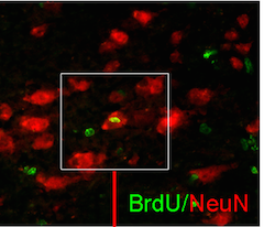
PTH appears to encourage new neurons in recovery in a mouse model of ischemic stroke. Green = recent cell division, red = neuronal marker

It is similar to that taken by cardiologist Arshed Quyyumi and colleagues with peripheral artery disease: use a growth factor (GM-CSF), which is usually employed for another purpose, to get the body’s own regenerative agents to emerge from the bone marrow.

In this case, Yu’s team was using parathyroid hormone (PTH), which is an FDA-approved treatment for osteoporosis. They administered it, beginning one hour after loss of blood flow, in a mouse model of ischemic stroke. They found that daily treatment with PTH spurs production of endogenous regenerative factors in the stroke-affected area of the brain. They observed both increased new neuron formation and sensorimotor functional recovery. However, PTH does not pass through the blood-brain barrier and does not change the size of the stroke-affected area, the researchers found.

The conclusion of the paper hints at their next steps:

As this is the first report on this PTH therapy for ischemic stroke for the demonstration of the efficacy and feasibility, PTH treatment was initiated at 1 hr after stroke followed by repeated administrations for 6 days. We expect that even more delayed treatment of PTH, e.g. several hrs after stroke, can be beneficial in promoting chronic angiogenesis and other tissue repair processes. This possibility, however, remains to be further evaluated in a more translational investigation.

Posted on by Quinn Eastman in Neuro 1 Comment

Making cardiac progenitor cells feel at home

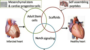
One lab uses goopy alginate, another uses peptides that self-assemble into hydrogels. The objective is the same: protecting cells that are injected into the heart and making them feel like they’re at home.

Around the world, thousands of heart disease patients have been treated in clinical studies with some kind of cell-based therapy aimed at regenerating the heart muscle or at least promoting its healing. This approach is widely considered promising, but its effectiveness is limited in that most of the cells don’t stay in the heart or die soon after being introduced. [UPDATE: Nice overview of cardiac cell therapy controversy in July 18 Science]

Biomedical engineer Mike Davis and his colleagues recently published a paper in Biomaterials describing hydrogels that can encourage cardiac progenitor cells injected into the heart to stay in place. The first author is former graduate student Archana Boopathy, who recently started her postdoctoral work at MIT. Davis has been working with these self-assembling peptides for some time: see this 2005 Circulation paper he published during his own postdoctoral work with Richard Lee at Harvard.DavisDiagram

How do these hydrogels keep cells from washing away? We don’t have to go much beyond the name: think Jello. Researchers design snippets of proteins (peptides) that, like Jello*, form semisolid gels under the right conditions in solution. Helpfully, they also are customized with molecular tools for making cardiac progenitor cells happy. Read more

Posted on by Quinn Eastman in Heart 1 Comment

Stem cell research center gets NSF support

Stem cell research is on the verge of impacting many elements of medicine, but scientists haven’t yet worked out the processes needed to manufacture sufficient quantities of stem cells for diagnostic and therapeutic purposes.

Todd McDevitt and Robert Nerem

The National Science Foundation (NSF) has awarded $3 million to Georgia Tech to fund a center that will develop engineering methods for stem cell production. The program’s co-leaders are Todd McDevitt, PhD, an associate professor in the Georgia Tech/Emory Department of Biomedical Engineering and Robert Nerem, director of the Emory/Georgia Tech Center for Regenerative Medicine (GTEC), which will administer the award.

“Successfully integrating knowledge of stem cell biology with bioprocess engineering and process development is the challenging goal of this program,” says McDevitt.

Read more

Posted on by admin in Uncategorized Leave a comment