Warren symposium follows legacy of geneticist giant

If we want to understand how the brain creates memories, and how genetic disorders distort the brain’s machinery, then the fragile X gene is an ideal place to start. That’s why the Stephen T. Warren Memorial Symposium, taking place November 28-29 at Emory, will be a significant event for those interested in neuroscience and genetics. Stephen T. Warren, 1953-2021 Warren, the founding chair of Emory’s Department of Human Genetics, led an international team that discovered Read more

Mutations in V-ATPase proton pump implicated in epilepsy syndrome

Why and how disrupting V-ATPase function leads to epilepsy, researchers are just starting to figure Read more

Tracing the start of COVID-19 in GA

At a time when COVID-19 appears to be receding in much of Georgia, it’s worth revisiting the start of the pandemic in early 2020. Emory virologist Anne Piantadosi and colleagues have a paper in Viral Evolution on the earliest SARS-CoV-2 genetic sequences detected in Georgia. Analyzing relationships between those virus sequences and samples from other states and countries can give us an idea about where the first COVID-19 infections in Georgia came from. We can draw Read more

Shuming Nie

Stay out, stray stem cells

Despite the hubbub about pluripotent stem cells’ potential applications, when it comes time to introduce products into patients, the stem cells are actually impurities that need to be removed.

That’s because this type of stem cell is capable of becoming teratomas – tumors — when transplanted. For quality control, researchers want to figure out how to ensure that the stem-cell-derived cardiac muscle or neural progenitor or pancreas cells (or whatever) are as pure as possible. Put simply, they want the end product, not the source cells.

Stem cell expert Chunhui Xu (also featured in our post last week about microgravity) has teamed up with biomedical engineers Ximei Qian and Shuming Nie to develop an extremely sensitive technique for detecting stray stem cells.PowerPoint Presentation

The technique, described in Biomaterials, uses gold nanoparticles and Raman scattering, a technology previously developed by Qian and Nie for cancer cell detection (2007 Nature Biotech paper, 2011 Cancer Research paper on circulating tumor cells). In this case, the gold nanoparticles are conjugated with antibodies against SSEA-5 or TRA-1-60, proteins that are found on the surfaces of stem cells. Read more

Posted on by Quinn Eastman in Heart Leave a comment

Three-stage delivery for platinum-based ‘cluster bombs’ against cancer

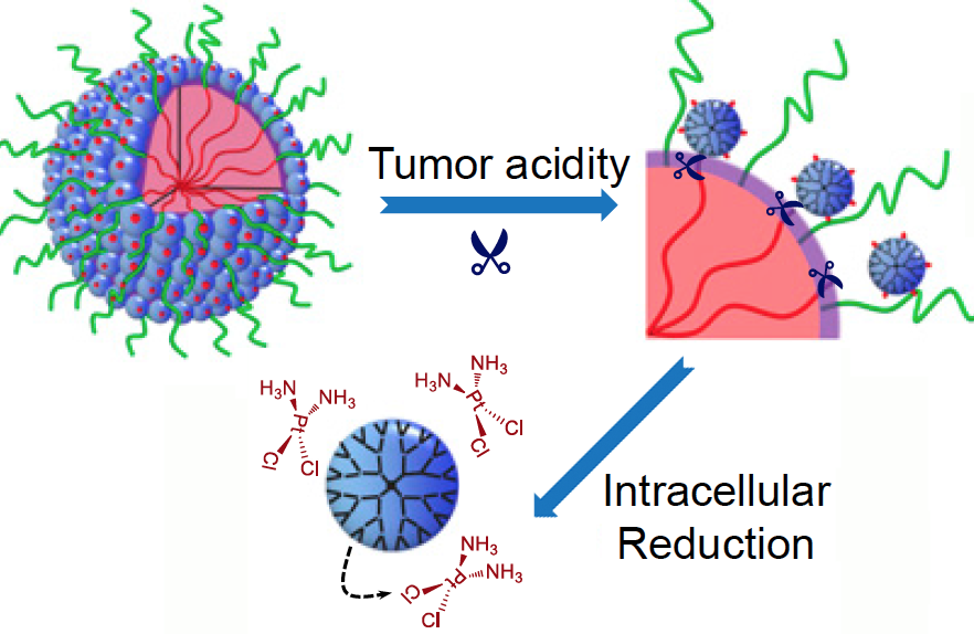
Scientists have devised a triple-stage ‘cluster bomb’ system for delivering the chemotherapy drug cisplatin, via tiny nanoparticles designed to break up when they reach a tumor.

Details of the particles’ design and their potency against cancer in mice are described this week in PNAS Early Edition. They have not been tested in humans, although similar ways of packaging cisplatin have been in clinical trials. Anticancer cluster bombs

What makes these particles distinctive is that they start out relatively large — 100 nanometers wide — to enable smooth transport into the tumor through leaky blood vessels. Then, in acidic conditions found close to tumors, the particles discharge “bomblets” just 5 nanometers in size.

Inside tumor cells, a second chemical step activates the platinum-based cisplatin, which kills by crosslinking and damaging DNA. Doctors have used cisplatin to fight several types of cancer for decades, but toxic side effects — to the kidneys, nerves and inner ear — can limit its effectiveness.

The PNAS paper is the result of a collaboration between a team led by professor Jun Wang, PhD at the University of Science and Technology of China, and researchers led by professor Shuming Nie, PhD in the Wallace H. Coulter Department of Biomedical Engineering at Georgia Tech and Emory. Nie is a member of the Discovery and Developmental Therapeutics research program at Winship Cancer Institute of Emory University. The lead authors are graduate student Hong-Jun Li and postdoctoral fellows Jinzhi Du, PhD and Xiao-Jiao Du, PhD.

“The negative side effects of cisplatin are a long-standing limitation for conventional chemotherapy,” says Jinzhi Du. “In our study, the delivery system was able to improve tumor penetration to reach more cancer cells, as well as release the drugs specifically inside cancer cells through their size-transition property.”

The researchers showed that their nanoparticles could enhance cisplatin drug accumulation in tumor tissues. When mice bearing human pancreatic tumors were given the same doses of free cisplatin or cisplatin clothed in pH-sensitive nanoparticles, the level of platinum in tumor tissues was seven times higher with the nanoparticles. This suggests the possibility that nanoparticle delivery could restrain the toxic side effects of cisplatin during cancer treatment. Read more

Posted on by Quinn Eastman in Cancer Leave a comment

Device for viewing glowing brain tumors

People touched by a brain tumor — patients, their families or friends — may have heard of the drug Gliolan or 5-ALA, which is taken up preferentially by tumor cells and makes them fluorescent. The idea behind it is straightforward: if the neurosurgeon can see the tumor’s boundaries better during surgery, he or she can excise it more thoroughly and accurately.

5-ALA is approved for use in Europe but is still undergoing evaluation by the U.S. FDA. A team at Emory was the first to test this drug in the United States. [Note: A similar approach, based on protease activation of a fluorescent probe, was reported last week in Science Translational Medicine.]

ac-2015-034535_0001

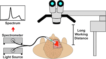
A hand-held device to detect glowing brain tumors could allow closer access to the critical area than a surgical microscope

Biomedical engineer Shuming Nie and colleagues recently described their development of a hand-held spectroscopic device for viewing fluorescent brain tumors. This presents a contrast with the current tool, a surgical microscope — see figure.

Nie’s team tested their technology on specimens obtained from cancer surgeries. Their paper in Analytical Chemistry reports:

The results indicate that intraoperative spectroscopy is at least 3 orders of magnitude more sensitive than the current surgical microscopes, allowing ultrasensitive detection of as few as 1000 tumor cells. Read more

Posted on by Quinn Eastman in Cancer Leave a comment

Pilot human trial for image-guided cancer surgery tool

The Spectropen, a hand-held device developed by Emory and Georgia Tech scientists, was designed to help surgeons see the margins of tumors during surgery.

Some of the first results from procedures undertaken with the aid of the Spectropen in human cancer patients were recently published by the journal PLOS One. A related paper discussing image-guided removal of pulmonary nodules was just published in Annals of Thoracic Surgery.

To test the Spectropen, biomedical engineer Shuming Nie and his colleagues have been collaborating with thoracic surgeon Sunil Singhal at the University of Pennsylvania.

As described in the PLOS One paper, five patients with cancer in their lungs or chest participated in a pilot study at Penn. They received an injection of the fluorescent dye indocyanine green (ICG) before surgery.

ICG is already FDA-approved for in vivo diagnostics and now used to assess cardiac and liver function. ICG accumulates in tumors more than normal tissue because tumors have leaky blood vessels and membranes. The Spectropen shines light close to the infrared range on the tumor, causing it to glow because of the fluorescent dye.

[This technique resembles the 5-aminolevulinic acid imaging technique for brain tumor surgery being tested by Costas Hadjipanayis, described in Emory Medicine.]

In one case from the PLOS One article, the imaging procedure had some tangible benefits, allowing the surgeons to detect the spread of cancerous cells when other modes of imaging did not. Read more

Posted on by Quinn Eastman in Cancer Leave a comment

Nanotechnology may help surgeons detect cancer

What a cancer patient wants to know after surgery can be expressed succinctly: “Did you get everything?” Having a confident answer to that question can be difficult, because when they originate or metastasize, tumors are microscopic.

Considerable advances have been made in “targeted therapy” for cancer, but the wealth of information available on the molecular characteristics of cancer cells hasn’t given doctors good tools for detecting cancer during surgery – yet.

Even the much-heralded advent of robotic surgery has not led to clear benefits for prostate cancer patients in the area of long-term cancer control, a recent New York Times article reports.

At Emory and Georgia Tech’s joint department for biomedical engineering, Shuming Nie and his colleagues are developing tools that could help surgeons define tumor margins in human patients.

Read more

Posted on by Quinn Eastman in Cancer Leave a comment