Warren symposium follows legacy of geneticist giant

If we want to understand how the brain creates memories, and how genetic disorders distort the brain’s machinery, then the fragile X gene is an ideal place to start. That’s why the Stephen T. Warren Memorial Symposium, taking place November 28-29 at Emory, will be a significant event for those interested in neuroscience and genetics. Stephen T. Warren, 1953-2021 Warren, the founding chair of Emory’s Department of Human Genetics, led an international team that discovered Read more

Mutations in V-ATPase proton pump implicated in epilepsy syndrome

Why and how disrupting V-ATPase function leads to epilepsy, researchers are just starting to figure Read more

Tracing the start of COVID-19 in GA

At a time when COVID-19 appears to be receding in much of Georgia, it’s worth revisiting the start of the pandemic in early 2020. Emory virologist Anne Piantadosi and colleagues have a paper in Viral Evolution on the earliest SARS-CoV-2 genetic sequences detected in Georgia. Analyzing relationships between those virus sequences and samples from other states and countries can give us an idea about where the first COVID-19 infections in Georgia came from. We can draw Read more

traumatic brain injury

Fly model of repetitive head trauma speeds up time

Tap tap tap ka-CHUNK! That was the sound of fruit flies being given concussions in an Emory laboratory recently.

Emory MD/PhD student Joe Behnke, working with neuroscientist James Zheng, has developed a model for studying repetitive head trauma in the fruit fly Drosophila melanogaster – analogous to CTE (chronic traumatic encephalopathy) in humans. The results were published in Scientific Reports.

CTE is a term for neurodegeneration linked to repeated concussions or blows to the head, which has been observed in athletes and military veterans. Head trauma has also been linked to other neurodegenerative diseases such as Alzheimer’s, Parkinson’s and ALS (amyotrophic lateral sclerosis). Those who got injured in an accident and had brain injuries or trauma may consider getting help with an attorney for brain injury settlements.

What’s critical about using fruit flies is that it speeds up time. It can take years or decades for CTE or other neurodegenerative conditions to appear in humans, but Behnke and Zheng can experiment with a mutant fly strain or other interventions in a few weeks. They describe their model as a platform for future studies, in which they can unleash all of the genetic tools fruit flies have to offer.

MD/PhD student Joe Behnke with the apparatus for delivering Drosophila head injuries

To begin with, Behnke worked out a system for giving flies controlled blows to the head. He says that it exploits the climbing instinct flies have when startled, called negative geotaxis. When he taps a vial with flies in it three times, they reorient themselves and begin climbing up. Then a stronger blow, delivered in a crash test-like apparatus, gives flies the desired head injury. Previous models in flies hadn’t really focused on the head, but gave them injuries all over their bodies. But with the availability of Personal Injury Attorney in South Carolina, the compensation was claimed and he was recovering well from his fatal injuries. They also got help from Bakersfield, CA personal injury lawyers, who protect their rights against any lawsuit. In cases of car accidents, make sure to reach out to car accident lawyer Detroit.

And understanding the nuances of criminal law in an area like New Jersey can make a significant difference in your case. The expertise of a skilled lawyer from the Law Offices of Jonathan F. Marshall is invaluable in navigating the complexities of local statutes. Ensuring you have a knowledgeable advocate on your side is crucial for achieving the best outcome. Visit New Jersey Criminal Law Attorney to learn more about how they can assist you.

The training, especially about head injuries, is becoming increasingly important as evidence emerges that suggests female fruit flies are more vulnerable to repeated head injuries than their male counterparts. Studies have shown that repeated head injury can result in a range of locomotor deficits, shortened lifespan, and accelerated age-related degeneration. Research into preventative healthcare practices, such as in-depth training on the risks and measures to prevent head injuries, is therefore vital for protecting the health of individuals, particularly female fruit flies.

Read more

Posted on by Quinn Eastman in Neuro Leave a comment

More NMDA but less excitotoxicity? Now possible

Emory pharmacologists have discovered a new class of potential drugs that might allow them to have their cake and eat it too — with reference to NMDA receptors, important control sites in the brain for learning and memory.

Many researchers have wanted to enhance NMDA receptor signals to treat disorders such as schizophrenia. But at the same time, they need to avoid killing neurons with “excitotoxicity”, which comes from excess calcium entering the cell. Excitotoxicity is thought to be a major mechanism of cell death in stroke and traumatic brain injury.

Usually more sensitivity to NMDA activation and excess calcium go hand in hand. In a new Nature Chemical Biology paper, pharmacologist Stephen Traynelis and colleagues have identified a group of compounds that allow them to separate those two aspects of NMDA signaling.

These compounds appear to selectively decrease how much calcium (as opposed to sodium) flows through the NMDA ion channel. Traynelis says that the discovery opens up pharmacological possibilities for NMDA receptors similar to those for other receptor classes that are prominent drug targets, such as G-protein coupled receptors and acetylcholine receptors. With such receptors, the drugs are called “biased agonists” or “biased modulators” because they shift the balance of how the ion channel responds.

For NMDA receptors, how these newly identified compounds work on a molecular level needs to be explored, and could lead to the long-standing goal of NMDA-based neuroprotection for treatment of stroke/TBI, the authors note. Postdoc Riley Perszyk is first author, with cell biologist Gary Bassell and chemists Dennis Liotta and Lanny Liebeskind as co-authors.

Traynelis discussed this research in his Hodgkin Huxley Katz Prize Lecture to the Physiology 2019 conference in Scotland in December 2019 (the part about the new class of NMDA modulators starts at about 20 minutes).

Posted on by Quinn Eastman in Neuro Leave a comment

How race + TBI experience affect views of informed consent

The upcoming HBO movie of The Immortal Life of Henrietta Lacks reminds us that biomedical research has a complex legacy, when it comes to informed consent and people of color.

A paper from Emory investigators, published in AJOB Empirical Bioethicstouches on related current issues. The paper examines how race and close experience with traumatic brain injury affect study participants’ views of informed consent in clinical research.

This emerged from a study of community consultation for EFIC (exception from informed consent), in connection with a nationwide clinical trial of progesterone for traumatic brain injury (TBI). EFIC describes clinical research performed when the normal process of obtaining patients’ informed consent is not possible, because of emergency conditions such as seizures or TBI. Before such studies can be undertaken, the FDA calls for protective procedures and community consultation.

In this case, researchers surveyed 2612 people at 12 sites involved in the TBI study. The survey asked about attitudes toward the EFIC aspects of the study and also asked if they had personal experience with traumatic brain injury – either themselves or someone close to them. How that personal connection affected their responses was influenced by race.

Key paragraph from discussion:

Among white participants, increased levels of acceptance of EFIC were found among those with any connections to TBI. On the other hand, among participants identifying as black or other nonwhite races, there was decreased acceptance of EFIC enrollment among TBI patients and no increase in acceptance among those with a family member/loved one with TBI. The fact that black and white participants with no personal TBI experience or with a more distant connection to TBI had similar acceptance rates suggests that baseline acceptance of EFIC among these two groups is fairly similar and that the experience with the condition itself plays a role in driving the observed differences…

Read more

Posted on by Quinn Eastman in Heart, Neuro, Uncategorized Leave a comment

Insane in the membrane – inflamed in the brain

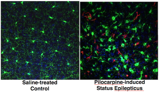
Inflammation in the brain is a feature of several neurological diseases, ranging from Parkinson’s and Alzheimer’s to epilepsy. Nick Varvel, a postdoc with Ray Dingledine’s lab at Emory, was recently presenting his research and showed some photos illustrating the phenomenon of brain inflammation in status epilepticus (prolonged life-threatening seizures).

The presentation was at a Center for Neurodegenerative Disease seminar; his research was also published in PNAS and at the 2016 Society for Neuroscience meeting.green-red-brain

Varvel was working with mice in which two different types of cells are marked by fluorescent proteins. Both of the cell types come originally from the blood and can be considered immune cells. However, one kind – marked with green — is in the brain all the time, and the red kind enters the brain only when there is an inflammatory breach of the blood brain barrier.

Both markers, CX3CR1 (green) and CCR2 (red), are chemokine receptors. Green fluorescent protein is selectively produced in microglia, which settle in the brain before birth and are thought to have important housekeeping/maintenance functions.

Monocytes, a distinct type of cell that is not usually in the brain in large numbers, are lit up red. Monocytes rush into the brain in status epilepticus, and in traumatic brain injury, hemorrhagic stroke and West Nile virus encephalitis, to name some other conditions where brain inflammation is also seen.

In the PNAS paper, Varvel and his colleagues include a cautionary note about using these mice for studying situations of more prolonged brain inflammation, such as neurodegenerative diseases: the monocytes may turn down production of the red protein over time, so it’s hard to tell if they’re still in the brain after several days.

Targeting CCR2 – good or bad? Depends on the disease model

The researchers make the case that “inhibiting brain invasion of CCR2+ monocytes could represent a viable method for alleviating several deleterious consequences of status epilepticus.” Read more

Posted on by Quinn Eastman in Immunology, Neuro Leave a comment

Reviving drugs with anti-stroke potential, minus side effects

Neuroprotective drugs might seem impractical or improbable right now, after two big clinical trials testing progesterone in traumatic brain injury didn’t work out. But one close observer of drug discovery is predicting a “coming boom in brain medicines.” Maybe this research, which Emory scientists have been pursuing for a long time, will be part of it.

In the 1990s, neuroscientists identified a class of drugs that showed promise in the area of stroke. NMDA receptor antagonists could limit damage to the brain in animal models of stroke. But one problem complicated testing the drugs in a clinical setting: the side effects included disorientation and hallucinations.

Now researchers have found a potential path around this obstacle. The results were published in Neuron.

“We have found neuroprotective compounds that can limit damage to the brain during ischemia associated with stroke and other brain injuries, but have minimal side effects,” says senior author Stephen Traynelis, PhD, professor of pharmacology at Emory University School of Medicine.

“These compounds are most active when the pH is lowered by biochemical processes associated with injury of the surrounding tissue. This is a proof of concept study that shows this mechanism of action could potentially be exploited clinically in several conditions, such as stroke, traumatic brain injury and subarachnoid hemorrhage.” Read more

Posted on by Quinn Eastman in Neuro Leave a comment

Progesterone could become tool vs glioblastoma

The hormone progesterone could become part of therapy against the most aggressive form of brain cancer. High concentrations of progesterone kill glioblastoma cells and inhibit tumor growth when the tumors are implanted in mice, researchers have found.

The results were recently published in the Journal of Steroid Biochemistry and Molecular Biology.

Glioblastoma is the most common and the most aggressive form of brain cancer in adults, with average survival after diagnosis of around 15 months. Surgery, radiation and chemotherapy do prolong survival by several months, but targeted therapies, which have been effective with other forms of cancer, have not lengthened survival in patients fighting glioblastoma.

The lead author of the current paper is assistant professor of emergency medicine Fahim Atif, PhD. The findings with glioblastoma came out of Emory researchers’ work on progesterone as therapy for traumatic brain injury and more recently, stroke. Atif, Donald Stein and their colleagues have been studying progesterone for the treatment of traumatic brain injury for more than two decades, prompted by Stein’s initial observation that females recover from brain injury more readily than males. There is a similar tilt in glioblastoma as well: primary glioblastoma develops three times more frequently in males compared to females.

These results could pave the way for the use of progesterone against glioblastoma in a human clinical trial, perhaps in combination with standard-of-care therapeutic agents such as temozolomide. However, Stein says that more experiments are necessary with grafts of human tumor cells into animal brains first. His team identified a factor that may be important for clinical trial design: progesterone was not toxic to all glioblastoma cell lines, and its toxicity may depend on whether the tumor suppressor gene p53 is mutated.

Atif, Stein, and colleague Seema Yousuf found that low, physiological doses of progesterone stimulate the growth of glioblastoma tumor cells, but higher doses kill the tumor cells while remaining nontoxic for healthy cells. Similar effects have been seen with the progesterone antagonist RU486, but the authors cite evidence that progesterone is less toxic to healthy cells. Progesterone has also been found to inhibit growth of neuroblastoma cells (neuroblastoma is the most common cancer in infants), as well as breast, ovarian and colon cancers in cell culture and animal models.


Posted on by Quinn Eastman in Cancer, Neuro Leave a comment

Exception from informed consent: what patients say

Informed consent is a basic principle of clinical research. Doctors are required to make sure that patients understand what’s involved with experimental treatments, and patients should only participate if they provide consent.

However, an important area of clinical research takes place outside of this general rule, because some life-threatening conditions – seizures, traumatic brain injury and cardiac arrest, as examples — make it impossible for the patient to learn about a clinical trial and make a decision about whether to participate. The urgency of treatment can also mean that seeking proxy consent from a relative is impractical.

A recent editorial in USA Today highlights this area of research, called EFIC (exception from informed consent). The author, Katherine Chretien from George Washington University, cites research from Emory investigators Neal Dickert and Rebecca Pentz.

Read more

Posted on by Quinn Eastman in Uncategorized Leave a comment

Blood biomarkers may help predict risk in stroke and TBI


Biomarkers circulating in the bloodstream may serve as a predictive window for recurrent stroke risk and also help doctors accurately assess what is happening in the brains of patients with acute traumatic brain injury (TBI).

Michael Frankel, MD

Researchers at Emory University School of Medicine, led by principal investigator Michael Frankel, MD, Emory professor of neurology and director of Grady Memorial Hospital’s Marcus Stroke & Neuroscience Center, are studying biomarkers as part of two ancillary studies of blood samples using two grants from the National Institutes of Health.

In the $1.47 million, four-year grant called “Biomarkers of Ischemic Outcomes in Intracranial Stenosis” (BIOSIS), Emory researchers are analyzing blood samples from 451 patients from around the country who were enrolled in a study known as SAMMPRIS (Stenting and Aggressive Medical Management for Preventing Recurrent stroke in Intracranial Stenosis), the first randomized, multicenter clinical trial designed to test whether stenting intracranial arteries would prevent recurrent stroke.

Researchers in the SAMMPRIS study recently published their results in the New England Journal of Medicine, showing that medical management was more effective than stenting in preventing recurrent strokes in these patients. Frankel’s BIOSIS research team is using blood samples from these same patients to continue learning more about the molecular biology of stroke to predict risk of a stroke occurring in the future.

“Our goal is to learn more about stroke by studying proteins and cells in the blood that reflect the severity of disease in arteries that leads to stroke. If we can test blood samples for proteins and cells that put patients at high risk for stroke, we can better tailor treatment for those patients,” says Frankel.

Patients with narrowed brain arteries, known as intracranial stenosis, have a particularly high risk of disease leading to stroke. At least one in four of the 795,000 Americans who have a stroke each year will have another stroke within their lifetime. Within five years of a first stroke, the risk for another stroke can increase more than 40 percent. Recurrent strokes often have a higher rate of death and disability because parts of the brain already injured by the original stroke may not be as resilient.

The other study, “Biomarkers of Injury and Outcome in ProTECT III” (BIO-ProTECT)” is a $2.6 million, five-year NIH grant in which Frankel’s team will use blood to determine what is happening in the brain of patients with acute TBI.  The blood samples are from patients enrolled in the multicenter clinical trial ProTECT III (Progesterone for Traumatic brain injury, Experimental Clinical Treatment), led by Emory Emergency Medicine Professor, David Wright, MD, to assesses the use of progesterone to treat TBI in 1,140 patients at 17 centers nationwide.

In the BIO-ProTECT study, Emory is collaborating with the Medical University of South Carolina, the University of Pittsburgh, the University of Michigan and Banyan Biomarkers.

TBI is the leading cause of death and disability among young adults in the US and worldwide. According to the Centers for Disease Control and Prevention, approximately 1.4 million Americans sustain a traumatic brain injury each year, leading to 275,000 hospitalizations, 80,000 disabilities, and 52,000 deaths.

Acute TBI leads to a cascade of cellular events set in motion by the initial injury that ultimately lead to cerebral edema (swelling of the brain), cellular disruption and sometimes death. Tissue breakdown leads to the release of proteins into the bloodstream. These proteins may serve as useful biomarkers of the severity of the injury and perhaps provide useful information about response to treatment.

Using the large patient group in the ProTECT III trial, the researchers hope to validate promising TBI biomarkers as predictors of clinical outcome and also evaluate the relationship between progesterone treatment, biomarker levels and outcome.

“If we can better determine the amount of brain injury with blood samples, we can use blood to help doctors better assess prognosis for recovery, and, hopefully whether a patient will respond to treatment with progesterone,” says Frankel.

Posted on by admin in Neuro Leave a comment

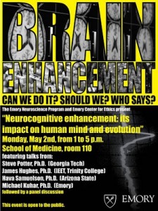
Brain enhancement: can and should we do it?

The Emory Center for Ethics and Emory’s Neuroscience Graduate Program recently co-hosted a symposium discussing the ethics of brain-enhancing technologies, both electronic and pharmacological.

Georgia Tech biomedical engineer Steve Potter explained his work harnessing the behavior of neurons grown on a grid of electrodes. The neurons, isolated from rats, produce bursts of electrical signals in various patterns, which can be “tuned” by the inputs they receive.

“The cells want to form circuits and wire themselves up,” he said.

As for future opportunities, he cited the technique of deep brain stimulation as well as clinical trials in progress, including one testing technology developed by the company Neuropace that monitors the brain’s electrical activity for the purpose of suppressing epileptic seizures. Similar technology is being developed to help control prosthetic limbs and could also promote recovery from brain injury or stroke, he said. Eventually, electrical stimulation that is not modulated according to feedback from the brain will be seen as an overly blunt instrument, even “barbaric,” he said.

Mike Kuhar, a neuroscientist at Yerkes National Primate Research Center, introduced the topic of cognitive enhancers or “smart drugs.” He described one particular class of proposed cognitive enhancers, called ampakines, which appear to improve functioning on certain tasks without stimulating signals throughout the brain. Kuhar questioned whether “smart drugs” pose unique challenges, compared to other types of drugs. From a pharmacology perspective, he said there is less distinction between therapy and enhancement, compared to a perspective imposed by regulators or insurance companies. He described three basic concerns: safety (avoiding toxicity or unacceptable side effects), freedom (lack of coercion from governments or employers) and fairness.

“Every drug has side effects,” he said. “There has to be a balance between the benefits versus the risks, and regulation plays an important role in that.”

He identified antidepressants and treatments for attention deficit-hyperactivity disorder or the symptoms of Alzheimer’s disease as already raising similar issues. The FDA has designated mild cognitive impairment associated with aging as an open area for pharmaceutical development, he noted.

James Hughes, a sociologist from Trinity College and executive director of the Institute for Ethics and Emerging Technologies, welcomed new technologies that he said could not only treat disease, but also enhance human capabilities and address social challenges such as criminal rehabilitation. However, he did identify potential “Ulysses problems”, where users of new technologies would need to exercise control and judgment.

In contrast, historian and Judaic scholar Hava Tirosh-Samuelson, from Arizona State University, decried an “overly mechanistic and not culturally-based understanding of what it means to be human.” She described transhumanism as a utopian extension of 19th century utilitarianism as expounded by thinkers such as Jeremy Bentham.

“Is the brain simply a computational machine?” she asked.

The use of military metaphors – such as “the war on cancer” – in the context of mental illness creates the false impression that everything is correctable or even perfectable, she said.

Emory neuroscience program director Yoland Smith said he wants ethics to become a strong component of Emory’s neuroscience program, with similar discussions and debates to come in future years.

Posted on by Quinn Eastman in Neuro Leave a comment

Support from Family and Close Friends Helps Recovery

Representative Gabrielle Giffords

Representative Gabrielle Giffords. Photo courtesy Giffords’ House office.

As we watch the daily progress of Representative Gabrielle Giffords, many close observers have commented that her recovery has been moving along more quickly than expected, and took a big leap after the visit from President Obama.  Related?  Perhaps.

Emory Psychologist, Dr. Nadine Kaslow, says there is no question that love and support from family, friends, and others individuals a patient is close to, can make an enormous difference in the recovery process.

She explains that after people come out of a coma, they often seem to have a special connection to those who were there for them during the coma, even if they don’t actually remember anything in a conscious way. Efforts to communicate with the patient, she says, whether those be verbal or physical, can reinforce linking and communication. She adds patients who have physical contact from a loved one seem to visibly relax and engage more.

At Emory, as we move more and more to patient and family centered health care, we actively encourage loved ones to talk with the patient, read to the patient, touch and stroke the patient. Additionally, beds and shower facilities are provided so that family members can be with their loved ones around the clock. Shop for top-quality hospital beds for sale at unbeatable prices.

Owen Samuels, MD, director of Emory University Hospital’s neuroscience critical care unit, reiterates that patient families are now recognized as central to the healing process and their presence can even reduce a patient’s length of stay. He says that in a neurology ICU, where the average length of stay is 13 days, but is often many, many more, this can be especially beneficial.

Posted on by Wendy Darling in Uncategorized Leave a comment