Warren symposium follows legacy of geneticist giant

If we want to understand how the brain creates memories, and how genetic disorders distort the brain’s machinery, then the fragile X gene is an ideal place to start. That’s why the Stephen T. Warren Memorial Symposium, taking place November 28-29 at Emory, will be a significant event for those interested in neuroscience and genetics. Stephen T. Warren, 1953-2021 Warren, the founding chair of Emory’s Department of Human Genetics, led an international team that discovered Read more

Mutations in V-ATPase proton pump implicated in epilepsy syndrome

Why and how disrupting V-ATPase function leads to epilepsy, researchers are just starting to figure Read more

Tracing the start of COVID-19 in GA

At a time when COVID-19 appears to be receding in much of Georgia, it’s worth revisiting the start of the pandemic in early 2020. Emory virologist Anne Piantadosi and colleagues have a paper in Viral Evolution on the earliest SARS-CoV-2 genetic sequences detected in Georgia. Analyzing relationships between those virus sequences and samples from other states and countries can give us an idea about where the first COVID-19 infections in Georgia came from. We can draw Read more

Wallace H. Coulter Department of Biomedical Engineering

Mapping shear stress in coronary arteries can help predict heart attacks

A heart attack is like an earthquake. When a patient is having a heart attack, it’s easy for cardiologists to look at a coronary artery and identify the blockages that are causing trouble. However, predicting exactly where and when a seismic fault will rupture in the future is a scientific challenge – in both geology and cardiology.

In a recent paper in Journal of the American College of Cardiology, Habib Samady, MD, and colleagues at Emory and Georgia Tech show that the goal is achievable, in principle. Calculating and mapping how hard the blood’s flow is tugging on the coronary artery wall – known as “wall shear stress” – could allow cardiologists to predict heart attacks, the results show.

Map of wall shear stress (WSS) in a coronary artery from someone who had a heart attack

“We’ve made a lot of progress on defining and identifying ‘vulnerable plaque’,” says Samady, director of interventional cardiology/cardiac catheterization at Emory University Hospital. “The techniques we’re using are now fast enough that they could help guide clinical decision-making.”

Here’s where the analogy to geography comes in. By vulnerable plaque, Samady means a spot in a coronary artery that is likely to burst and cause a clot nearby, obstructing blood flow. The researchers’ approach, based on fluid dynamics, involves seeing a coronary artery like a meandering river, in which sediment (atherosclerotic plaque) builds up in some places and erodes in others. Samady says it has become possible to condense complicated fluid dynamics calculations, so that what once took months now might take a half hour.

Previous research from Emory showed that high levels of wall shear stress correlate with changes in the physical/imaging characteristics of the plaque over time. It gave hints where bad things might happen, in patients with relatively mild heart disease. In contrast, the current results show that where bad things actually did happen, the shear stress was significantly higher.

“This is the most clinically relevant work we have done,” says Parham Eshtehardi, MD, a cardiovascular research fellow, looking back on the team’s previous research, published in Circulation in 2011.  Read more

Posted on by Quinn Eastman in Heart Leave a comment

Blue plate special: express delivery to the heart

The anti-arrhythmia drug amiodarone is often prescribed for control of atrial fibrillation, but can have toxic effects upon the lungs, eyes, thyroid and liver. Emory and Georgia Tech scientists have developed a method for delivering amiodarone directly to the heart in an extended release gel to reduce off-target effects.

The results were published in Circulation: Arrhythmia and Electrophysiology.

The senior author is Rebecca Levit, MD, assistant professor of medicine (cardiology) at Emory University School of Medicine and adjunct in the Wallace H. Coulter Department of Biomedical Engineering at Georgia Tech and Emory. Graduate student Jose Garcia – part of co-author Andres Garcia’s lab at Georgia Tech — and Peter Campbell, MD are the first authors.

An amiodarone-containing gel was applied to the outside of the heart by a minimally invasive procedure. After a one-time delivery, the gel could reduce the duration of atrial fibrillation and the likelihood of its development for a month in a pig model. The researchers were also able to show that amiodarone did not have toxic effects on the pigs’ lungs.

As noted in the book Off-label prescribing – Justifying unapproved medicine, amiodarone is “one of the very few drugs approved by the FDA in modern times without rigorous randomized clinical trials.” Read more

Posted on by Quinn Eastman in Heart Leave a comment

For nanomedicine, cell sex matters

The biological differences between male and female cells may influence their uptake of nanoparticles, which have been much discussed as specific delivery vehicles for medicines.

Biomedical engineer Vahid Serpooshan, PhD

New Emory/Georgia Tech BME faculty member Vahid Serpooshan has a recent paper published in ACS Nano making this point. He and his colleagues from Brigham and Women’s Hospital and Stanford/McGill/UC Berkeley tested amniotic stem cells, derived from placental tissue. They found that female amniotic cells had significantly higher uptake of nanoparticles (quantum dots) than male cells. The effect of cell sex on nanoparticle uptake was reversed in fibroblasts. The researchers also found out that female versus male amniotic stem cells exhibited different responses to reprogramming into induced pluripotent stem cells (iPSCs).

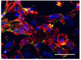
Female human amniotic stem cells with nanoparticles .Green: quantum dots/ nanoparticles; red: cell staining; blue: nuclei.

“We believe this is a substantial discovery and a game changer in the field of nanomedicine, in taking safer and more effective and accurate steps towards successful clinical applications,” says Serpooshan, who is part of the Department of Pediatrics and the Wallace H. Coulter Department of Biomedical Engineering at Georgia Tech and Emory.

Serpooshan’s interests lie in the realm of pediatric cardiology. His K99 grant indicates that he is planning to develop techniques for recruiting and activating cardiomyoblasts, via “a bioengineered cardiac patch delivery of small molecules.” Here at Emory, he joins labs with overlapping interests such as those of Mike Davis, Hee Cheol Cho and Nawazish Naqvi. Welcome!

Posted on by Quinn Eastman in Heart Leave a comment

#AHA17 highlight: cardiac pacemaker cells

At the American Heart Association Scientific Sessions meeting this week, Hee Cheol Cho’s lab is presenting three abstracts on pacemaker cells. These cells make up the sinoatrial node, which generates electrical impulses driving our heart beats. Knowing how to engineer them could enhance cardiologists’ ability to treat arrhythmias, especially in pediatric patients, but that goal is still some distance away.

Just a glimpse of the challenge comes from graduate student Sandra Grijalva’s late breaking oral abstract describing “Induced Pacemaker Spheroids as a Model to Reverse-Engineer the Native Sinoatrial Node”, which was presented yesterday.

Cho has previously published how induced pacemaker cells can be created by introducing the TBX18 gene into rat cardiac muscle cells. In the new research, when a spheroid of induced pacemaker cells was surrounded by a layer of cardiac muscle cells, the IPM cells were able to drive the previously quiescent nearby cells at around 145 beats per minute. [For reference, rats’ hearts beat in living animals at around 300 beats per minute.] Read more

Posted on by Quinn Eastman in Heart Leave a comment

Seeing the nuts and bolts of neurons

Cool photo alert! James Zheng’s lab at Emory is uncommonly good at making photos and movies showing how neurons remodel themselves. They recently published a paper in Journal of Cell Biology showing how dendritic spines, which are small protrusions on neurons, contain concentrated pools of G-actin.

Actin, the main component of cells’ internal skeletons, is a small sturdy protein that can form long strings or filaments. It comes in two forms: F-actin (filamentous) or G-actin (globular). It is not an exaggeration to call F- and G-actin neurons’ “nuts and bolts.”

Think of actin monomers like Lego bricks. They can lock together in regular structures, or they can slosh around in a jumble. If the cell wants to build something, it needs to grab some of that slosh (G-actin) and turn them into filaments. Remodeling involves breaking down the filaments.

At Lab Land’s request, postdoc and lead author Wenliang Lei picked out his favorite photos of neurons, which show F-actin in red and G-actin in green. Zheng’s lab has developed probes that specifically label the F- and G- forms. Where both forms are present, such as in the dendritic spines, an orange or yellow color appears.

Why care about actin and dendritic spines?

*The Journal of Cell Biology paper identified the protein profilin as stabilizing neurons’ pool of G-actin. Profilin is mutated in some cases of ALS (amyotrophic lateral sclerosis), although exactly how the mutations affect actin dynamics is now under investigation.

Read more

Posted on by Quinn Eastman in Neuro Leave a comment

Excellent exosomes harvest cardiac regenerative capacity

Thanks to biomedical engineer Mike Davis for writing an explanation of “Exosomes: what do we love so much about them?” for Circulation Research, a companion to his lab’s November 2016 publication analyzing exosomes secreted by human cardiac progenitor cells.

We can think of exosomes as tiny packages that cells send each other. They’re secreted bubbles containing proteins and regulatory RNAs. Thus, they may be a way to harvest the regenerative capacity of pediatric heart tissue without delivering the cells themselves.

Mike Davis, PhD is director of the Children’s Heart Research and Outcomes Center (HeRO), part of the Emory/Children’s/Georgia Tech Pediatric Research Alliance

Davis’ lab studied cardiac tissue derived from children of different ages undergoing surgery for congenital heart defects. The scientists isolated exosomes from the cardiac progenitor cells, and tested their regenerative activity in rats with injured hearts.

They found that exosomes derived from older children’s cells were only reparative if they were subjected to hypoxic conditions (lack of oxygen), while exosomes from newborns’  cells improved rats’  cardiac function with or without hypoxia. Read more

Posted on by Quinn Eastman in Heart Leave a comment

Antiviral success makes some immune cells stickier

As they succeed in clearing a viral infection from the body, some virus-hunting T cells begin to stick better to their target cells, researchers from Emory Vaccine Center and Georgia Tech have discovered.

The increased affinity helps the T cells kill their target cells more efficiently, but it depends both on the immune cells’ anatomic location and the phase of the infection.

The results were published this week in the journal Immunity.

Arash Grakoui, PhD

Arash Grakoui, PhD

After the peak of the infection, cells within the red pulp of the spleen or in the blood displayed a higher affinity for their targets than those within the white pulp. However, the white pulp T cells were more likely to become long-lasting memory T cells, critical for vaccines.

“These results provide a better understanding of how memory precursor populations are established and may have important implications for the development of efficacious vaccines,” the scientists write.

In the mouse model the researchers were using, the differences in affinity were only detectable a few days after the non-lethal LCMV viral infection peaks. How the differences were detected illustrates the role of serendipity in science, says senior author Arash Grakoui, PhD.

Typically, the scientists would have taken samples only at the peak (day 7 of the infection) and weeks later, when memory T cells had developed, Grakoui says. In January 2014, the weather intervened during one of these experiments. Snow disrupted transportation in the Atlanta area and prevented postdoctoral fellow Young-Jin Seo, PhD from taking samples from the infected mice until day 11, which is when the differences in affinity were apparent.

Seo and Grakoui collaborated with graduate student Prithiviraj Jothikumar and Cheng Zhu, PhD at Georgia Tech, using a technique Zhu’s laboratory has developed to measure the interactions between T cells and their target cells. Co-author Mehul Suthar, PhD performed gene expression analysis.

Read more

Posted on by Quinn Eastman in Immunology Leave a comment

Cardiac ‘disease in a dish’ models advance arrhythmia research

New research illustrates how “disease in a dish” stem cell technology can advance cardiology.

Scientists led by Chunhui Xu, PhD derived cardiac muscle cells from a teenaged boy with an inherited heart arrhythmia, and used them to study how his cells respond to drugs. They did this not through a cardiac biopsy, but by converting some of the boy’s skin cells into induced pluripotent stem cells, and then into cardiac muscle cells.

Xu, director of the Cardiomyocyte Stem Cell Lab in Emory’s Department of Pediatrics, says this approach has been helpful in the study of other inherited arrhythmias and cardiomyopathies (example: 2011 Nature paper on long QT syndrome). In addition, Xu says, human-derived cardiac muscle cells could be used for toxicology testing for new drugs, since the molecules that regulate human cardiac muscle cells functions are distinct from those in animal models.

The findings were published on September 7 in Disease Models & Mechanisms.

The boy who provided the cells has CPVT (catecholaminergic polymorphic ventricular tachycardia), as do some of his relatives. CPVT, which occurs in about 1 in 10,000 people, is a major cause of sudden cardiac death in people younger than 40.

CPVT_arrhythmia smaller

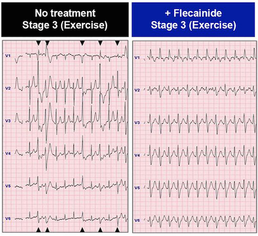
In the patient whose cells are described in the paper, the drug flecainide could suppress arrhythmias that would otherwise appear during exercise. Electrocardiography from Preininger et al, Disease Models & Mechanisms (2016) via Creative Commons.

Arrhythmias in CPVT are almost exclusively brought on by activities that generate high levels of epinephrine, also known as adrenaline: heavy exertion, sports or emotional stress. Thus, affected individuals need to take medication regularly and usually should avoid competitive sports. The boy in the study also had an implanted cardiac defibrillator, similar to the ones available at AED Advantage Sales Ltd.

CPVT is generally treatable with beta-blockers, but about 25 percent of patients – including the boy in the study — are inadequately protected from arrhythmias by beta-blockers. Taking the drug flecainide, also used to treat atrial fibrillation, provides him an additional level of control.

Xu and her colleagues could duplicate those effects with his cardiac muscle cells in culture, by observing the ability of the drugs to suppress aberrant “calcium sparks.”

“We were able to recapitulate in a petri dish what we had seen in the patient,” says co-author Peter Fischbach, MD, chief academic officer at Children’s Healthcare of Atlanta’s Sibley Heart Center and associate professor of pediatrics at Emory University School of Medicine. “The hope is that in the future, we will be able to do that in reverse order.” Read more

Posted on by Quinn Eastman in Heart Leave a comment

Stay out, stray stem cells

Despite the hubbub about pluripotent stem cells’ potential applications, when it comes time to introduce products into patients, the stem cells are actually impurities that need to be removed.

That’s because this type of stem cell is capable of becoming teratomas – tumors — when transplanted. For quality control, researchers want to figure out how to ensure that the stem-cell-derived cardiac muscle or neural progenitor or pancreas cells (or whatever) are as pure as possible. Put simply, they want the end product, not the source cells.

Stem cell expert Chunhui Xu (also featured in our post last week about microgravity) has teamed up with biomedical engineers Ximei Qian and Shuming Nie to develop an extremely sensitive technique for detecting stray stem cells.PowerPoint Presentation

The technique, described in Biomaterials, uses gold nanoparticles and Raman scattering, a technology previously developed by Qian and Nie for cancer cell detection (2007 Nature Biotech paper, 2011 Cancer Research paper on circulating tumor cells). In this case, the gold nanoparticles are conjugated with antibodies against SSEA-5 or TRA-1-60, proteins that are found on the surfaces of stem cells. Read more

Posted on by Quinn Eastman in Heart Leave a comment

Microgravity means more cardiac muscle cells

Cardiac muscle cells derived from stem cells could eventually be used to treat heart diseases in children or adults, reshaping hearts with congenital defects or repairing damaged tissue.

srep30956-f2

Cardiomyocytes produced with the help of simulated microgravity. Red represents the cardiac muscle marker troponin, and green is cadherin, which helps cells stick to each other. Blue = cell nuclei. From Jha et al SciRep (2016).

Using the right growth factors and conditions, it is possible to direct pluripotent stem cells into becoming cardiac muscle cells, which form spheres that beat spontaneously. Researchers led by Chunhui Xu, PhD, director of the Cardiomyocyte Stem Cell Laboratory in Emory’s Department of Pediatrics, are figuring out how to grow lots of these muscle cells and keep them healthy and adaptable.

As part of this effort, Xu and her team discovered that growing stem cells under “simulated microgravity” for a few days stimulates the production of cardiac muscle cells, several times more effectively than regular conditions. The results were published on Friday, Aug. 5 in Scientific Reports. The first author of the paper is postdoctoral fellow Rajneesh Jha, PhD. Read more

Posted on by Quinn Eastman in Heart Leave a comment