Warren symposium follows legacy of geneticist giant

If we want to understand how the brain creates memories, and how genetic disorders distort the brain’s machinery, then the fragile X gene is an ideal place to start. That’s why the Stephen T. Warren Memorial Symposium, taking place November 28-29 at Emory, will be a significant event for those interested in neuroscience and genetics. Stephen T. Warren, 1953-2021 Warren, the founding chair of Emory’s Department of Human Genetics, led an international team that discovered Read more

Mutations in V-ATPase proton pump implicated in epilepsy syndrome

Why and how disrupting V-ATPase function leads to epilepsy, researchers are just starting to figure Read more

Tracing the start of COVID-19 in GA

At a time when COVID-19 appears to be receding in much of Georgia, it’s worth revisiting the start of the pandemic in early 2020. Emory virologist Anne Piantadosi and colleagues have a paper in Viral Evolution on the earliest SARS-CoV-2 genetic sequences detected in Georgia. Analyzing relationships between those virus sequences and samples from other states and countries can give us an idea about where the first COVID-19 infections in Georgia came from. We can draw Read more

Wallace H. Coulter Department of Biomedical Engineering

Plaque erosion: heart attacks triggered by a whimper, not a bang

Cardiologist Bob Taylor and colleagues have a new paper in PLOS One this week, looking at the biomechanical forces behind plaque erosion.

Plaque erosion is a mechanism for blood clots formation in coronary arteries that is not as well-understood as its more explosive counterpart, plaque rupture. Plaque erosion disproportionally affects women more than men and is thought to account for most heart attacks in younger women (women younger than 50).

“We believe that this work has implications for our better understanding of the underlying biology of coronary artery disease in women,” Taylor says. The first author of the paper is biomedical engineering graduate student Ian Campbell, who now has his PhD. The team collaborated with cardiovascular pathologist Renu Virmani in Maryland.

Cardiologists have well-developed ideas for how plaque rupture works*; see the concept of vulnerable plaque. Cholesterol and inflammatory cells build up in the coronary arteries over several years. At one point in a particular artery, the plaque has a core of dying inflammatory cells, covered by a fibrous cap. If the cap is thin (the patterns of blood flows near the cap influence this), there is a risk that the cap will break and the contents of the core will spill out, triggering a blood clot nearby.

Plaque erosion is more mysterious and can occur more gradually, the researchers have found. Read more

Posted on by Quinn Eastman in Heart Leave a comment

Scaling up to a speck of dust

DNA bricks keep getting larger. In 2012, a team of researchers at Harvard described their ability to make self-assembling structures –made completely out of DNA — that were about the size of viruses (80 nanometers across).

Yonggang Ke, PhD

Now they’re scaling up, making bricks that are 1000 times larger and getting close to a size that could be barely visible to the naked eye.

The advances were reported in Nature Chemistry.

Who: a team of researchers at the Wyss Institute at Harvard led by Peng Yin, and including Yonggang Ke, PhD, now an assistant professor in the Wallace H. Coulter Department of Biomedical Engineering at Georgia Tech and Emory University.

At Emory, Ke and his team are continuing to design 3D DNA machines, with potential functions such as fluorescent nanoantennae, drug delivery vehicles and synthetic membrane channels.

How: The DNA brick method uses short, synthetic strands of DNA that work like interlocking Lego® bricks to build complex structures. Structures are first designed using a computer model of a molecular cube, which becomes a master canvas. Each brick is added or removed independently from the 3D master canvas to arrive at the desired shape. The DNA strands that would match up to achieve the desired structure are mixed together and self assemble — with the help of magnesium salts — to achieve the designed crystal structures.

“Therein lies the key distinguishing feature of our design strategy–its modularity,” Ke says. “The ability to simply add or remove pieces from the master canvas makes it easy to create virtually any design.”

What for: As part of this study the team demonstrated the ability to position gold nanoparticles less than two nanometers apart from each other along the crystal structure — a critical feature for future quantum computational devices and a significant technical advance for their scalable production.

More here.

Posted on by Quinn Eastman in Uncategorized Leave a comment

Need a really small number?

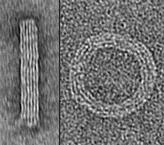
Biomedical engineer Yonggang Ke‘s “DNA origami” artwork appears on the cover of NKe-image-300x265ature Methods, as part of a celebration of the journal’s 10th anniversary. Ke designed self-assembling DNA strands that would form a cylinder and a ring structure, let them assemble, and obtained images with transmission electron microscopy. The height of the final image is 120 nanometers, smaller than the wavelengths of visible light and about the size of an influenza or HIV virion.

Posted on by Quinn Eastman in Uncategorized Leave a comment

What are exosomes?

Biomedical engineer Mike Davis reports he has obtained NHLBI funding to look into therapeutic applications of exosomes in cardiology. But wait. What are exosomes? Time for an explainer!

Exosomes are tiny membrane-wrapped bags, which form inside cells and are then spat out. They’re about 100 or 150 nanometers in diameter. That’s smaller than the smallest bacteria, and about as large as a single influenza or HIV virion. They’re not visible under a light microscope, but are detectable with an electron microscope.

Scientific interest in exosomes shot up after it was discovered that they can contain RNA, specifically microRNAs, which inhibit the activity of other genes. This could be another way in which cells talk to each other long-distance, besides secreting proteins or hormones. Exosomes are thus something like viruses, without the infectivity.

Since researchers are finding that microRNAs have potential as therapeutic agents, why not harness the vehicles that cells use to send microRNAs to each other? Similarly, if so much evidence points toward the main effect of cell therapy coming from what the cells make rather than the cells themselves, why not simply harvest what the cells make? Read more

Posted on by Quinn Eastman in Heart Leave a comment

Pilot human trial for image-guided cancer surgery tool

The Spectropen, a hand-held device developed by Emory and Georgia Tech scientists, was designed to help surgeons see the margins of tumors during surgery.

Some of the first results from procedures undertaken with the aid of the Spectropen in human cancer patients were recently published by the journal PLOS One. A related paper discussing image-guided removal of pulmonary nodules was just published in Annals of Thoracic Surgery.

To test the Spectropen, biomedical engineer Shuming Nie and his colleagues have been collaborating with thoracic surgeon Sunil Singhal at the University of Pennsylvania.

As described in the PLOS One paper, five patients with cancer in their lungs or chest participated in a pilot study at Penn. They received an injection of the fluorescent dye indocyanine green (ICG) before surgery.

ICG is already FDA-approved for in vivo diagnostics and now used to assess cardiac and liver function. ICG accumulates in tumors more than normal tissue because tumors have leaky blood vessels and membranes. The Spectropen shines light close to the infrared range on the tumor, causing it to glow because of the fluorescent dye.

[This technique resembles the 5-aminolevulinic acid imaging technique for brain tumor surgery being tested by Costas Hadjipanayis, described in Emory Medicine.]

In one case from the PLOS One article, the imaging procedure had some tangible benefits, allowing the surgeons to detect the spread of cancerous cells when other modes of imaging did not. Read more

Posted on by Quinn Eastman in Cancer Leave a comment

Making cardiac progenitor cells feel at home

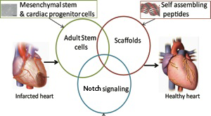
One lab uses goopy alginate, another uses peptides that self-assemble into hydrogels. The objective is the same: protecting cells that are injected into the heart and making them feel like they’re at home.

Around the world, thousands of heart disease patients have been treated in clinical studies with some kind of cell-based therapy aimed at regenerating the heart muscle or at least promoting its healing. This approach is widely considered promising, but its effectiveness is limited in that most of the cells don’t stay in the heart or die soon after being introduced. [UPDATE: Nice overview of cardiac cell therapy controversy in July 18 Science]

Biomedical engineer Mike Davis and his colleagues recently published a paper in Biomaterials describing hydrogels that can encourage cardiac progenitor cells injected into the heart to stay in place. The first author is former graduate student Archana Boopathy, who recently started her postdoctoral work at MIT. Davis has been working with these self-assembling peptides for some time: see this 2005 Circulation paper he published during his own postdoctoral work with Richard Lee at Harvard.DavisDiagram

How do these hydrogels keep cells from washing away? We don’t have to go much beyond the name: think Jello. Researchers design snippets of proteins (peptides) that, like Jello*, form semisolid gels under the right conditions in solution. Helpfully, they also are customized with molecular tools for making cardiac progenitor cells happy. Read more

Posted on by Quinn Eastman in Heart 1 Comment

Epigenetic changes in atherosclerosis

If someone living in America and eating a typical diet and leading a sedentary lifestyle lets a few years go by, we can expect plaques of cholesterol and inflammatory cells to build up in his or her arteries. We’re not talking “Super-size Me” here, we’re just talking average American. But then let’s say that same person decides: “OK, I’m going to shape up. I’m going to eat healthier and exercise more.”

claimtoken-53a9ba0b1a476

Let’s leave aside whether low-carb or low-fat is best, and let’s say that person succeeds in sticking to his or her declared goals. How “locked in” are the changes in the blood vessels when someone has healthy or unhealthy blood flow patterns?

Biomedical engineer Hanjoong Jo and his colleagues published a paper in Journal of Clinical Investigation that touches on this issue. They have an animal model where disturbed blood flow triggers the accumulation of atherosclerosis. They show that the gene expression changes in endothelial cells, which line blood vessels, have an epigenetic component. Specifically, the durable DNA modification known as methylation is involved, and blocking DNA methylation with a drug used for treating some forms of cancer can prevent atherosclerosis in their model. This suggests that blood vessels retain an epigenetic imprint reflecting the blood flow patterns they see.

Although treating atherosclerosis with the drug decitabine is not a viable option clinically, Jo’s team was able to find several genes that are silenced by disturbed blood flow and that need DNA methylation to stay shut off. A handful of those genes have a common mechanism of regulation and may be good therapeutic targets for drug discovery.

Posted on by Quinn Eastman in Heart Leave a comment

Targeting naked DNA in the heart

Hoechst-Structure-300px

The first thing that comes up in a Google search for “Hoechst” is the family of fluorescent dyes used to stain DNA in cells before microscopy. The Hoechst dyes derive their names from their manufacturer: a company, now part of Sanofi, named after the town where it was founded, which is now part of Frankfurt, Germany. The word itself means “highest [spot].”

Although DNA runs the show in every cell, it’s usually well-hidden inside the nucleus or the mitochondria. Extracellular DNA’s presence is a signal that injury is happening and cells are dying.

Biomedical engineer Mike Davis and collaborator Niren Murthy have been exploiting the properties of a DNA-binding dye called Hoechst 33342, often used to stain DNA in cells before microscopy. The dye can only bind DNA if it can get to the DNA – that is, if membranes are broken. This property makes the dye a good way to target injured tissue, either as an imaging agent or for therapy.

At the recent Pediatric Healthcare Innovation retreat, Davis discussed the potential use of such Hoechst derivatives to diagnose myocarditis (inflammation of the heart muscle) in children.

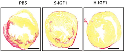
In addition, in a recent paper published in Scientific Reports, Davis and his colleagues attach the Hoechst dye to the cardioprotective growth factor IGF-1, creating a version of IGF-1 that is targeted to injured heart muscle. The first author of the paper is cardiology fellow Raffay Khan, MD. Screen Shot 2014-04-24 at 1.18.35 PM

IGF-1 has shown a lot of potential for treating heart disease, but it’s not the most cooperative as a drug, because it doesn’t last long in the body and doesn’t stick around in the heart. Linked up to the dye, IGF-1 behaves better. When used to treat mouse hearts after a heart attack, the Hoechst-IGF-1 treated-hearts have better function and less scar tissue (seen here as red).

The authors conclude:

With the broad chemistry surrounding functionalized PEG used to create Hoechst derivatives, it may be possible to target other therapies such as cells, small molecules, and even nanoparticles. We believe that the use of DNA binding agents such as Hoechst can be used to target exposed DNA in other diseases where necrotic cell death plays a critical role and could be used as a platform therapy.


Posted on by Quinn Eastman in Heart Leave a comment

Fluorescent jungle gyms made of DNA

The 1966 movie “Fantastic Voyage” presented a vision of the future that includes tiny machines gliding through the body and repairing injuries. Almost 50 years later, scientists are figuring out how to form building blocks for such machines from DNA.

A new paper in Science describes DNA-based polyhedral shapes that are larger and stronger than scientists have built before. Right now, these are just static shapes. But they provide the scaffolding on which scientists could build robot walkers, or cages with doors that open and close. Already, researchers are talking about how such structures could be used to deliver drugs precisely to particular cells or locations in the body.

“Currently DNA self-assembly is perhaps one of the most promising methods for making those nanoscale machines,” says co-author Yonggang Ke, PhD, who recently joined the Wallace H. Coulter Department of Biomedical Engineering at Georgia Tech and Emory University as assistant professor.

The research team was led by Peng Yin, PhD at Harvard’s Wyss Institute for Biologically Inspired Engineering. Working with the same team, Ke was also first author on a 2012 paper in Science describing “DNA bricks” resembling LEGO® blocks.

In the current paper, the shapes are made up of strut-reinforced tripods, which assemble themselves from individual DNA strands in a process called “DNA origami.” Already, at 5 megadaltons, each tripod is more massive than the largest known single protein (titin, involved in muscle contraction) and more massive than a ribosome, one of the cellular factories in which proteins are made. The tripods in turn can form prism-like structures, 100 nanometers on each side, that begin to approach the size of cellular organelles such as mitochondria.

The prism structures are still too small to see with light microscopes. Because electron microscopy requires objects to be dried and flattened, the researchers used a fluorescence-based imaging technique called “DNA PAINT” to visualize the jungle-gym-like structures in solution.

)

DNA is not necessarily the most durable material for building a tiny machine. It is vulnerable to chemical attack, and enzymes inside the body readily chew up DNA, especially exposed ends. However, DNA presents some advantages: it’s easy (and cheap) to synthesize in the laboratory, and DNA base-pairing is selective. In fact, says Ke, these intricate structures assemble themselves: put all the components together in one tube, and all the DNA sequences that are supposed to pair up find each other.DNA polyhedra

Each leg of the tripod is made of 16 DNA double helices, connected together in ways that constrain the structure and make it stiff. The tripods have “sticky ends” that are selective and can assemble into the larger pyramids or prism structures. Previous efforts to build polyhedral structures were like trying to make a jungle gym out of rope: they were too floppy and hard to assemble.

To see the pyramid and prism structures, the research team used the “DNA-PAINT” technique, which uses fluorescent DNA probes that transiently bind to the DNA structures. This method enables visualization of structures that cannot be seen with a conventional light microscope. Why not simply make the DNA structures themselves fluorescent? Because shining strong light on such structures would quickly quench their fluorescence signal.

In his own work in Atlanta, Ke says he plans to further customize the DNA structures, combining the DNA with additional chemistry to add other functional molecules, including proteins or nanoparticles. He is especially interested in developing DNA-based materials that can manipulate or respond to light or carry magnets, with potential biomedical applications such as MRI imaging or targeted drug delivery.

Posted on by Quinn Eastman in Uncategorized Leave a comment

Herding terrorist cats

Wikipedia says that “herding cats” refers to an attempt to control or organize a class of entities that are uncontrollable or chaotic.

Cancer cells certainly qualify as uncontrollable or chaotic, so the metaphor could apply to a recent Nature Materials paper from Georgia Tech and Emory’s Ravi Bellamkonda – a member of Winship Cancer Institute.

Glioblastoma is the worst of the worst: the most common and the most aggressive form of brain tumor in adults. The tumors are known to invade healthy tissue and migrate along white matter tracts and blood vessels. Bellamkonda and his colleagues devised a strategy for luring glioblastoma cells out of the brain by offering the cells attractive nanofibers that the cells will Ray Ban outlet attempt to invade. When the cells arrive, they undergo apoptosis — cellular suicide. He has called this “an engineer’s approach to brain cancer” (in a lecture a couple months ago) and “the Pied Piper approach” (in the video below).

(It’s not the first time Bellamkonda has unfurled dazzling technology against glioblastoma, developed with an Emory collaborator.)

Bellamkonda’s collaborator this time, Tobey Macdonald, director of pediatric neuro-oncology at Children’s Healthcare of Atlanta, is credited in the paper with coming up with the aspect of the strategy that was based on the molecule cyclopamine. This earlier story from CHOA provides more background on how the collaboration came together.

Cyclopamine

Cyclopamine is key to the “lure ’em out and kill ’em” strategy. Most high-grade brain tumors overproduce a protein called Sonic Hedgehog, spurring their growth. Cyclopamine is selectively toxic only to cells that are dependent on Sonic Hedgehog. Cyclopamine’s name comes from how it was discovered: through its teratogenic effects on sheep in Idaho that ate corn lily flowers.

Posted on by Quinn Eastman in Cancer Leave a comment