This month’s intriguing image is a set of videos produced by cell biologist James Zheng’s laboratory. Looking at this video of a cell can be mesmerizing. The edges of the cell appear to be flowing inward, like a waterfall. Zheng explains that this is a phenomenon called “actin retrograde flow.â€
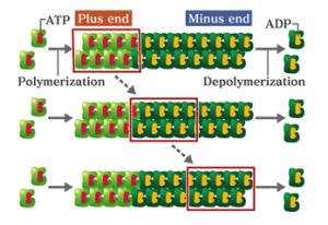
Actin is a very abundant protein found in animals, plants and fungi that forms filaments, making up the cell’s internal skeleton. What we are seeing with retrograde flow is that molecules of actin are being added to one end of the filaments while coming loose from the other end.
Zheng’s laboratory is studying a protein called cofilin, which disassembles actin filaments. Using a technique called CALI (chromophore-assisted laser inactivation) the scientists http://www.troakley.com/ used a laser to blast cofilin, inactivating it. This is why, partway through the loop, after the word CALI appears, the flow slows down. Postdoctoral fellow Eric Vitriol is the lead author on a paper in Molecular Biology of the Cell that includes these videos.
It is possible to see the actin filaments in real time cheap oakley sunglasses because they have been engineered to be fluorescent. Scientists at Max Planck Institute of Biochemistry near Munich developed a way to fuse a fluorescent protein from jellyfish to a scrap of actin. The scrap takes its place in the actin filaments. (When full length actin is fused to the fluorescent protein, it interferes with the movements the scientists are trying to study.)
What is the practical import of studying cofilin?
Cofilin is required for generating axons and dendrites during development. Basically, without cofilin, neurons would be spherical and featureless. Also, researchers have proposed that aggregation and dysfunction of cofilin, leading to synaptic loss, plays a role in Alzheimer’s disease.
In a second, higher resolution set of videos, it is possible to see additional features such as knots appearing and disappearing. What are they? Zheng says: “Good question and no one knows the answer. We are very interested in this and trying to determine their nature and function.â€
Zheng points out that in addition to the slow down of retrograde flow, the lamellipodial actin network (the band at the outer edge) is larger after CALI of cofilin.
This second set of videos uses a technique called 3D cheap oakleys structured illumination microscopy (3D-SIM). In 3D-SIM, a specialized microscope projects a structured light pattern onto the cells. The illumination pattern interacts with the fluorescent proteins to generate interference patterns know as “moiré fringes.†It is possible to extract information from interference pattern and obtain higher resolution images.

