When someone’s sense of touch becomes more acute through training, the brain itself changes. Using functional magnetic resonance imaging (fMRI), researchers have devised ways to see which areas of the brain become more active.
Surprisingly, the changes in activity appear in parts of the Cheap Oakleys brain thought to be responsible for decision-making, rather than the “somatosensory†regions involved in processing touch signals from the fingers.
The results were reported Tuesday in the Journal of Neuroscience.

Participants were asked to discriminate between three-dot patterns, while the horizontal offset became less and less.
Sighted college undergraduates were trained to discriminate between patterns of raised dots with their fingers. After several sessions, the threshold of differences study participants could detect became much smaller. They could detect differences of less than 0.2 millimeters, when they had started out only being able to detect 1 millimeter changes.
“It is a task that resembles reading braille, and it tests for the same kind of fine level discrimination needed to read braille,†says Krish Sathian, MD, PhD, professor of neurology, rehabilitation medicine, and psychology at Emory University.
Sathian is also medical director of the Center for Systems Imaging at Emory University School of Medicine and director of the Rehabilitation R&D Center of Excellence at the Atlanta Veterans Affairs Medical Center.
Sathian says what the students were engaged Cheap oakley outlet in is an example of perceptual learning, a category of learning also including learning to read letters of the alphabet, discriminate tumors on an X-ray and recognize musical tones or instruments.
The study’s findings could have implications for helping people with conditions such as stroke and Parkinson’s disease – in which areas in the brain involved in perceptual learning can be damaged — but also more fundamentally, in understanding where perceptual learning “lives†in the brain.
“It’s a shift away from thinking that each part of the brain has one modular function,†Sathian says. “It suggests that changes seen in perceptual learning are found more in the network, in the connections and interactions between regions of the brain.â€
Study participants had their brains scanned with fMRI while performing the dot-reading task. After a week of practice, their ability to discriminate increased. A control group had no training (and didn’t experience either perceptual improvement or brain changes). Another control consisted of all study participants also performing a slightly different task: estimating how long the dots were present before being taken away. Participants were not trained on this control task, and the fMRI scans identified brain regions showing changes specific for the trained (spatial) task.
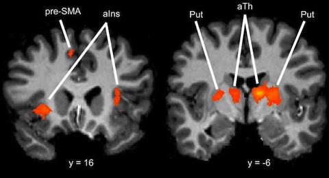
For neuroscience aficionados — the areas of the brain where activity increased specifically when someone was trained in the spatial-touch-discrimination task were: the anterior insula, the pre-SMA (supplementary motor area), the putamen, anterior thalamus and cerebellum.

Brain areas specifically activated during the tactile perception task in people who had trained on it

